O R I G I N A L AR T I C L E
JEADV (2003) 17, 291 – 295
Blackwell Publishing Ltd.
Lichen planus and hepatitis C virus infection
RGimenez-García,†*JLPérez-Castrillón‡
†Dermatology Section, ‡Internal Medicine Service, Hospital del Rio Hortega, Valladolid, Spain. *Corresponding author, Pago de la Barca 115, Boecillo, 47151 Valladolid, Spain, E-mail: [email protected]
AB S T R A C T
Background The association of lichen planus (LP) with liver diseases is well established. The reported prevalence rates of hepatitis C virus (HCV) antibodies in patients with LP tend to appear quite variable.
Objective The aim of this study was to assess the prevalence of HCV antibodies in a group of patients with LP and evaluate the clinical characteristics of the subgroup with LP associated with HCV.
Methods We studied 101 patients, 57 (56.4%) women and 44 (43.5%) men with a mean age of 48 years, consecutively diagnosed with cutaneous and/or mucosal LP between January 1992 and December 2000. We used 99 age- and sex-matched controls.
Results Anti-HCV antibodies were detected in nine cases (8.9%) of the LP group but only two (2.02%) of the controls. The odds ratio between the subjects with HCV positivity and those with negative HCV virus was 4.74, with a confidence interval at 95%, between 0.999 and 22.545. A statistically significant association was only demonstrated between erosive LP and infection by HCV.
Conclusions The possibility of liver disease caused by HCV should be ruled out in patients with LP, especially in the erosive form.
Key words: epidemiology, hepatitis C virus, lichen planus
Received: 7 March 2002, accepted 22 May 2002
Introduction
Lichen planus (LP) is a generally self-limiting disorder that usually appears in middle-aged adults and can affect the skin, mucosa, hair and nails. Various clinical forms, lichenoid eruptions (which are similar to LP and are normally drug induced) and association with other diseases have been
described,1,2 although in some cases it is not known if the
association is simply due to chance. It has been maintained since the 1980s that patients with LP have a greater proportion
of hepatopathies than the general population.3 Depending on
the various studies, the prevalence of chronic hepatic disease in LP patients oscillates between 0.1 and 35%, the majority of
which are based on the determination of aminotransaminases.4
In Spain hepatitis C antibody prevalence is surprisingly one of the highest in the world.
We describe a case – control study: (i) to determine the prev-alence of hepatitis C virus (HCV) infection in those patients diagnosed with LP in our studied population; (ii) to study the clinical characteristics of the subgroup with LP associated with hepatitis C; and (iii) to check for a possible relation between
some of the lesion localizations or some clinical form of LP and this viral infection.
Materials and methods
Patients
We studied 101 patients, 57 (56.43%) women and 44 (43.56%) men, consecutively diagnosed with cutaneous and /or mucosal LP between January 1992 and December 2000. The age range of the affected patients was 4–78 years with a mean age of 48 years.
Case definition
The diagnosis was based on the existence of typical clinical and histopathological findings of LP. Cases in which lichenoid reactions due to drugs were suspected were excluded.
292 Gimenez-García and Pérez-Castrillón
characteristic Wickham’s striae; (ii) appearance of the Koebner phenomenon; (iii) localization on the wrists, ankles, lumbar region and/or lesions of an annular morphology on the penis and/or hyperkeratotic lesions in the area of the palms and soles; (iv) evolution toward a long-lasting residual pigmentation; (v) off-white hyperkeratotic lesions of a reticular morphology in the buccal mucosa; and (vi) nail lesions as longitudinal striations, onychoschizia, onychomadesis or the characteristic formation of pterygium.
The histopathological diagnosis was based on the following findings: (i) orthohyperkeratosis, acanthosis and hypergranulosis (as changes in the epidermis); (ii) band-like lymphohistiocytic infiltrate in the dermoepidermal limit; (iii) the presence of hyaline or colloid bodies corresponding to degenerated epidermal basal cells in the lower epidermis layers; and (iv) melanin pigment incontinence.
Controls
We used a control group consisting of 99 patients treated in the same office who were matched by age and sex. Sixty-one were women (19–79 years old) consecutively diagnosed with chronic urticaria and 38 were men (13–77 years old) diagnosed with urticaria, warts, keratosis, eczema or psoriasis. The average age was 46.6.
Methods
A protocolized clinical history was prepared in which data corresponding to the following variables were collected: (i) sex and age; (ii) localization of LP lesions in the oral cavity, wrists, feet and ankles, back, upper extremities, genitalia, axillae, abdomen, nails, scalp, facial area and other localizations; (iii) determination of the clinical form of LP, such as lichen ruber planus, oral LP, lineal LP, erosive LP, lichen planopilaris, frontal fibrosing alopecia, annular LP, hypertrophic LP, bullous LP, follicular LP tumidus retroauricular, palmoplantar LP, actinic LP and follicular LP; and (iv) hepatic diseases previously diagnosed by other physicians.
The following laboratory tests were performed: (i) the patients’ serum was analysed to determine the analytic para-meters of alanine aminotransferase (GPT), aspartate
amino-transferase (GOT), bilirubin, alkaline phosphatase, γ-glutamyl
transpeptidase by automated methods in a BW/Hitachi 917 Roche Diagnostic Autoanalyser; (ii) antimitochondrial anti-bodies were determined by immunofluorescence with triple tissue portals; (iii) determination of anti-HCV antibodies was carried out using the second generation enzyme-linked immu-nosorbent assay immunoenzymatic method (Ortho Diagnostic Systems); and (iv) confirmation of enzyme-linked immuno-sorbent assay results was performed using second-generation four-antigen recombinant immunoblot assay (Ortho Diagnostic Systems).
Statistical analysis
The statistical analysis was applied as follows. In order to determine the possible associations among the variables of interest, the Chi-square test was used. The Fisher test was used in the study of dichotomic variables to detect possible
associations. A bilateral significance level was set at α = 0.05.
The data collection log was implemented in Microsoft Access 97. The software used in the statistical analysis was SPSS version 9.0 and Microsoft Excel 97. All these tools were used in a Pentium 133 MHz with 32 Mb of RAM.
Results
Prevalence of anti-hepatitis C virus antibodies
Anti-HCV antibodies were detected in nine cases (8.9%). In the control group we detected only two positive cases (both with chronic urticaria) for anti-HCV antibodies (2.02%) of the 99 studied.
From a statistical viewpoint, the association between cases/
controls and the hepatitis C is statistically significant (P = 0.05).
The odds ratio between the subjects with C virus positivity and those with negative C virus was 4.74, with a confidence interval at 95%, between 0.999 and 22.545.
Patients with lichen planus and hepatitis C
Age and localization
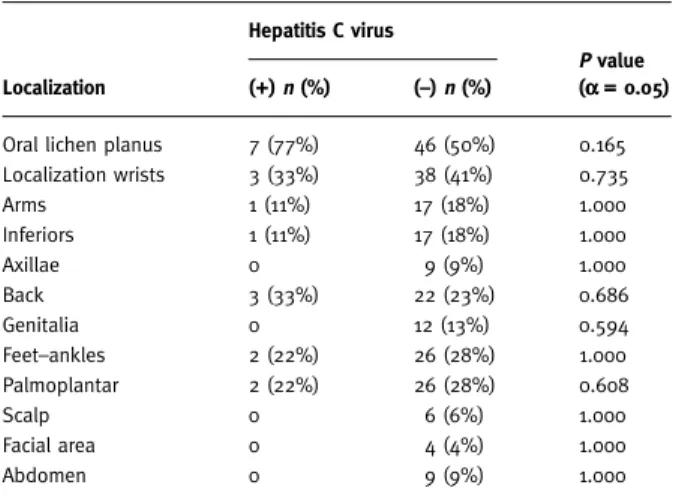
The characteristics of the nine-patient group with hepatitis C (six males and three females) from 19 to 78 years old (average age 55.5 years) were analysed. In this subgroup of patients with LP and hepatitis C, lesions localized in the buccal region, seven cases (77%), the back, three (33%), wrists, three (33%), feet and ankles, two (22%), palmo-plantar, two (22%), arms, one (11%) and other areas, two (22%), predominated.
In the group with HCV and LP, oral localization and on the wrists, back, feet and ankles and palmoplantar was frequently observed; nevertheless, when the Fisher test was applied no statistically significant differences were found between the lesion localization in the patients with LP and positive C virus and those that had LP and negative C virus (Table 1).
Clinical forms of lichen planus
In the group with anti-HCV antibodies, the diagnosed clinical forms of LP corresponded to lichen ruber planus in four cases (44.4%), to erosive LP in four (44.4%) and to hypertrophic LP in one case (11.1%). The clinical data of these nine patients is detailed in Table 2.
LP and HCV 293
Discussion
In recent years the association of hepatic diseases and especially hepatotropic virus with LP has been described. The first case of 0 in which a patient presented LP with hepatitis C following
10 months of treatment with interferon-α was later reported,6
along with other isolated cases of this association.7–12
Jubert et al.8 described six patients with LP (four had erosive
mucosal erosions) and a liver disease caused by HCV, demon-strated by the presence of viral RNA in serum. A 5% prevalence of LP in a series of 61 patients with active chronic hepatitis activated by HCV was detected. These data were confirmed in more in-depth studies. In Spain, numerous series with a variable prevalence of antibodies for C virus that oscillated between 10.5
and 47% as opposed to 2–5% for the control group13 –18 have
been published. In other countries, in the Mediterranean area19,20 as well as in other regions,21,22 similar data have been
found.
However, other epidemiological studies undertaken in France, Turkey and in Great Britain have not demonstrated a significant difference between the prevalence of HCV
anti-bodies in LP patients and control groups.23 –26 A British study of
180 cases of oral LP did not show an association with hepatic
disease.27
We have detected in our revision that 8.9% of the LP cases have anti-HCV antibodies, which signifies the lowest preval-ence found in the Spanish studies, where it oscillates between
10.5% and 47%.13 –18 One outstanding note is that in three of
them the diagnosis of hepatitis C was established based on the dermatological symptoms. These differences may be due to the fact that in some of them retrospective studies have been made, or that the diagnosed subjects were included after revising the histological diagnosis for hepatitis C, or that the sample was small. Another possible explanation is the different hepatitis C infection prevalence rate in our area compared with other areas
where it is higher.13,15,22,28,29
LP normally appears in persons between 50 and 70 years old. In our study the average global age was 55 for the group with hepatitis C, and males were more affected (six cases cf. three in
females). In the revision of Sánchez-Pérez et al.15 LP associated
with hepatitis C occurred more frequently in those subjects who were 70–90 years old; however, they demonstrated that females were doubly affected in comparison with males.
It is described that between 30 and 70% of patients with
classic LP have mucosal involvement.2 We found an oral
Table 1Lichen planus lesion localization
Localization
Hepatitis C virus
P value
(αααα = 0.05)
(+) n (%) (–) n (%)
Oral lichen planus 7 (77%) 46 (50%) 0.165 Localization wrists 3 (33%) 38 (41%) 0.735
Arms 1 (11%) 17 (18%) 1.000
Inferiors 1 (11%) 17 (18%) 1.000
Axillae 0 9 (9%) 1.000
Back 3 (33%) 22 (23%) 0.686
Genitalia 0 12 (13%) 0.594
Feet–ankles 2 (22%) 26 (28%) 1.000
Palmoplantar 2 (22%) 26 (28%) 0.608
Scalp 0 6 (6%) 1.000
Facial area 0 4 (4%) 1.000
Abdomen 0 9 (9%) 1.000
Table 2 Patients with lichen planus (LP) and hepatitis C (HCV)
Patient no. Age Sex LP clinical form
Diagnosed previously HCV
Induced or aggravated by interferon
1 55 Female Hypertrophic No
2 78 Male Lichen ruber planus Yes
3 54 Male Erosive No Yes
4 78 Male Lichen ruber planus Yes
5 62 Female Lichen ruber planus Yes
6 62 Male Erosive Yes
7 37 Male Erosive Yes Yes
8 19 Male Lichen ruber planus No
9 54 Female Erosive Yes Yes
Table 3 Relation of clinical form of lichen planus (LP) and infection by hepatitis C virus (HCV)
Clinical form HCV+ n (%) HCV– n (%)
Statistics Fisher P value (αααα = 0.05)
Odds ratio (Confidence interval 95%)
Erosive LP 4 (44.44%) 2 (2.2%) 0.001 36 (5.27, 245.91)
Lichen ruber planus 4 (44.44%) 52 (56.5%) 0.507 0.61 (0.15, 2.44)
294 Gimenez-García and Pérez-Castrillón
involvement in 52% of the patients with cutaneous LP that rose to 77% in the group with LP associated with HCV infection.
The clinical forms of presentation in the group of nine patients with positive HCV as opposed to those with negative C virus were four cases of lichen ruber planus (44.4% against 55.4%), four of erosive LP [44.4% against two (2.1%)] and one case (11.1%) of hypertrophic LP in contrast to 1% in the group of 92 patients with negative HCV. A statistically significant association between the erosive form of LP and HCV infection was demonstrated.
These results appear to confirm those suggested by other studies, which demonstrate that erosive LP is more frequent in patients with LP associated with hepatic disease or other
diseases;30 –33 a statistically significant association exists
between erosive LP and HCV infection.15,16 Miralles et al.13
found that of 13 patients with LP and positive HCV, eight had oral mucosal involvement, five of which had an erosive form.
Mignogna et al.,34 on the other hand, found that among 263
patients with oral LP the reticular form was more frequent in the carriers of HCV than in those who tested negative, while the plaque form was more prevalent in the latter. There were no sig-nificant differences in the erosive or atrophic forms. The asym-metrical localization in buccal mucosa and the localizations on the tongue, labial and gingival mucosa were more common in the cases with HCV, in which, furthermore, cutaneous involve-ment is less prevalent.
For some authors the association of LP and positive serology for HCV as well as for positive RNA is not a substantive enough reason to determine the role of HCV in the pathogenesis of LP. Nevertheless, the recent demonstration of RNA of HCV in epithelial cells of the oral mucosa of patients with LP would lead
to the theory that direct action of the virus is involved.35 HCV
could be involved, among other factors, in the pathogenesis of LP (acting as an exogenous antigen or due to the presence in the keratinocytes of common epitopes similar to that of the hepatocytes damaged by this hepatotropic virus). No relation between the different genotypes of HCV and the presence
or absence of LP lesions has been demonstrated.36,37 The
dif-ferences with respect to the results could be in relation to the different epidemiological methods and, perhaps, in aspects of a genetic type relative to the host, as has been recently
demonstrated.38
In conclusion, the possibility of liver disease caused by HCV should be ruled out in patients with LP (especially in the erosive form) if there is an alteration of the hepatic function tests or if there is no other apparent cause.
References
1 Boyd AS, Neldner KH. Lichen planus. J Am Acad Dermatol 1991;
25: 593 – 619.
2 Fellner MJ. Lichen planus. Int J Dermatol 1980; 19: 71–75.
3 Rebora A, Rongioletti F. Lichen et foie. Ann Dermatol Venereol 1994;
121: 533 –535.
4 Pawlotsky JM, Dhumeaux D, Bagot H. Hepatitis C virus in dermatology. A review. Arch Dermatol 1995; 131: 1185–1193. 5 Mokni M, Rybojad M, Puppin D Jr et al. Lichen planus and hepatitis
C virus. J Am Acad Dermatol 1991; 24: 792.
6 Agner T, Fogh H, Weismann K. The relation between lichen planus and hepatitis C: a case report. Acta Derm Venereol (Stockh) 1992; 72: 380. 7 Benchikhi H, Nejjam F, Habibeddine S et al. Lichen plan et hépatite
virale C. Ann Dermatol Venereol 1994; 121: 547–549.
8 Jubert C, Pawlotsky JM, Pouget F et al. Lichen planus and hepatitis C virus-related chronic active hepatitis. Arch Dermatol 1994; 130: 73–76.
9 Gomez Navarro E, Espinel Vazquez ML, Pique Duran E. Liquen plano y virus de la hepatitis C: descripción de cuatro casos. Actas Dermosifiliogr 1993; 84: 451– 453.
10 Espinel Ml Gomez E. Liquen plano y virus de la hepatitis C. Piel
1993; 8: 417– 419.
11 Revenga Arranz F, De Argila Fernandez-Duran D, Rivera Diaz R, Iglesias Diez L. Liquen plano e infección por el virus de la hepatitis C. Estudio de seis casos. Rev Clin Esp 1995; 195: 550 –552. 12 Mugoni MG, Montesu MA, Cottoni F. Lichen planus on the palms
and soles. J Eur Acad Dermatol Venereol 1994; 3: 535 –540. 13 Miralles J, Pujol RM, De Moragas JM. Liquen plano y hepatitis C.
Descripción de 13 casos. Actas Dermosifiliogr 1994; 85: 603 – 606. 14 Alayon Lopez C, Gimenez Arnau A, Gimenez Camarasa JM. Liquen
plano y hepatitis C. Actas Dermosifiliogr 1995; 86: 9–12. 15 Sanchez-Pérez J, De Castro M, Buezo G et al. Lichen planus and
hepatitis C virus: prevalence and clinical presentation of patients with lichen planus and hepatitis C virus infection. Br J Dermatol
1996; 134: 715 –719.
16 Olalquiaga Loewe J, Del Campo Hernandez I, Sendino Gómez R
et al. Infección por virus de la hepatitis C asociada a liquen plano. Estudio epidemiológico sobre una población del centro de España.
Actas Dermosifiliogr 1999; 90: 295 –299.
17 Santander C, De Castro M, García Monzón C. Prevalence of hepatitis C virus (HCV) and liver damage in patients with lichen planus (LP). Hepatology 1994; 20: 238A (Abstr.).
18 De Argila Fernandez-Duran D, Rovira Farré I, Alcalde Rubio MM, Pascasio Acevedo JM. Prevalencia de la infección por el virus de la hepatitis C en los pacientes con liquen plano del área de Badajoz.
Rev Clin Esp 1998; 198: 117–118.
19 Dupond AS, Lacour JPh, Laffont C et al. Lichen érosif buccal et hépatite C: étude rétrospective réalisée chez 29 patients. Ann Dermatol Venereol 1997 (Suppl.): S76 –S77.
20 Divano MC, Parodi A, Rebora A. Lichen planus: liver-kidney-microsomal (LKM1) antibodies and hepatitis C antibodies.
Dermatology 1992; 185: 132–133.
21 Bellman B, Reddy RJ, Falanga V. Lichen planus associated with hepatitis. Lancet 1995; 346: 1234.
LP and HCV 295
23 Cribier B, Garnier C, Laustriat D, Heid E. Lichen planus and hepatitis C virus infection: an epidemiologic study. J Am Acad Dermatol 1994; 31: 1070 –1072.
24 Dupin N, Chosidow O, Lunel F et al. Oral lichen planus and hepatitis C virus infection: a fortuitous association? Arch Dermatol
1997; 133: 1052–1053.
25 Ilter N, Senol E, Gürer MA, Altay Ö. Lichen planus and hepatitis C-virus infection in Turkish patients. J Eur Acad Dermatol Venereol
1998; 10: 192–193.
26 Tucker SC, Coulson IH. Lichen planus is not associated with hepatitis C virus infection in patients from North West England.
Acta Derm Venereol 1999; 79: 378 –379.
27 El Kabir M, Scully C, Porter S, Porter K, Macnamara E. Liver function in UK patients with oral lichen planus. Clin Exp Dermatol
1993; 18: 12–16.
28 Benchikhi H, Bastuji-Garin S, Pawlotsky Y et al. Lichen plan et infection par le virus de l′hépatite C: ëtude prospective de 40 cas.
Ann Dermatol Venereol 1994 (Suppl.): S53–C77.
29 Rebora A. Hepatitis viruses and lichen planus. Arch Dermatol 1994;
130: 138 –129.
30 Gandolfo S, Carbone M, Zulian P et al. Oral lichen planus and liver pathology. The clinico-statistical correlations between oral manifestations and liver damage. Minerva Stomatol 1992;
41: 209–213.
31 Monk BE, Pembroke AC. Skin problems in chronic active hepatitis.
Lancet 1981; 2: 1045.
32 Cottoni F, Solinas A, Piga MR et al. Lichen planus, chronic liver diseases, and immunologic involvement. Arch Dermatol Res 1988;
280 (Suppl.): S55–S60.
33 Rebora A, Rongioletti F. Lichen planus and chronic active hepatitis.
J Am Acad Dermatol 1984; 10: 840 – 841.
34 Mignogna MD, Lo Muzio L, Lo Russo L et al. Oral lichen planus: different clinical features in HCV-positive and HCV negative patients. Int J Dermatol 2000; 39: 134 –139.
35 Arrieta JJ, Rodriguez Inigo E, Casqueiro M et al. Detection of hepatitis C virus replication by in situ hybridization in epithelial cells of anti-hepatitis C virus-positive patients with and without oral lichen planus. Hepatology 2000; 32: 97–103.
36 Pawlotsky JM, Benkhiki H, Pellet C et al. Lichen planus and hepatitis C virus (HCV)-related chronic hepatitis: evaluation of HCV genotypes. Br J Dermatol 1995; 133: 666 – 667 (Letter). 37 Sánchez Perez J, Moreno Otero R, Borque MJ et al. Lichen planus
and hepatitis C virus infection: a clinical and virological study. Acta Derm Venereol (Stockh) 1998; 78: 305 –306.