PARK2 polymorphisms predict disease progression in
patients infected with hepatitis C virus
Ahmed A. Al-Qahtani,*,†,†† Mashael R. Al-Anazi,* Fahad A. Al-Zoghaibi,‡ Ayman A. Abdo,§,†† Faisal M. Sanai,||,†† Waleed K. Al-Hamoudi,§,†† Khalid A. Alswat,§,†† Hamad I. Al-Ashgar,¶ Mohammed Q. Khan,¶ Ali Albenmousa,** Hanif Khalak,‡‡ Mohammed N. Al-Ahdal*,†,§§
* Department of Infection and Immunity, Research Center, King Faisal Specialist Hospital & Research Center, Riyadh, Saudi Arabia.
† Department of Microbiology and Immunology, Alfaisal University School of Medicine, Riyadh, Saudi Arabia. ‡ Molecular BioMedicine Program, Research Center, King Faisal Specialist Hospital & Research Center, Riyadh, Saudi Arabia. § Section of Gastroenterology, Department of Medicine, College of Medicine, King Saud University, Riyadh, Saudi Arabia. || Gastroenterology Unit, Department of Medicine, King Abdulaziz Medical City, Jeddah, Saudi Arabia. ¶ Gastroenterology Unit, Department of Medicine, King Faisal Specialist Hospital & Research Center, Riyadh, Saudi Arabia.
** Department of Gastroenterology, Prince Sultan Medical Military City, Riyadh, Saudi Arabia.
†† Liver Disease Research Center, King Saud University, Riyadh, Saudi Arabia. ‡‡ Advanced Computing, Weill-Cornell Medical College, Doha, Qatar. §§ Department of Pathology and Laboratory Medicine, King Faisal Specialist Hospital and Research Center, Riyadh, Saudi Arabia.
A B S T R A C T A B S T R A C T A B S T R A C T A B S T R A C T A B S T R A C T
Background. Background.Background. Background.
Background. The protein encoded by PARK2 gene is a component of the ubiquitin-proteasome system that mediates targeting of proteins for the degradation pathway. Genetic variations at PARK2 gene were linked to various diseases including leprosy, typhoid and cancer. The present study investigated the association of single nucleotide polymorphisms (SNPs) in the PARK2 gene with the development of hepatitis C virus (HCV) infection and its progression to severe liver diseases. Material and methods.Material and methods.Material and methods.Material and methods. A total ofMaterial and methods. 800 subjects, including 400 normal healthy subjects and 400 HCV-infected patients, were analyzed in this study. The patients were classified as chronic HCV patients (group I), patients with cirrhosis (group II) and patients with hepatocellular carcinoma (HCC) in the context of cirrhosis (group III). DNA was extracted and was genotyped for the SNPs rs10945859, rs2803085, rs2276201 and rs1931223. Results.Results.Results.Results.Results. Among these SNPs, CT genotype of rs10945859 was found to have a significant association towards the clinical progression of chronic HCV infection to cirrhosis alone (OR = 1.850; 95% C. I. 1.115-3.069; p = 0.016) or cirrhosis and HCC (OR = 1.768; 95% C. I. 1.090-2.867; p value = 0.020). Conclusion. Conclusion. Conclusion. Conclusion. Conclusion. SNP rs10945859 in the PARK2 gene could prove useful in predicting the clinical outcome in HCV-infected patients.
Key words. Key words. Key words. Key words.
Key words. PARK2. Single nucleotide polymorphism. HCV. HCC. Liver cirrhosis.
November-December, Vol. 15 No. 6, 2016: 824-833
INTRODUCTION
Hepatitis C Virus (HCV) is an enveloped, positive-sense and single-stranded RNA virus classified within
Hepacivirus genus of the Flaviviridae family. It is considered as a major cause of chronic liver diseases and is considered as one of the most serious blood borne infection, with
nearly 170 million people infected worldwide.1 Although,
the majority of the HCV infections evolve to chronicity, nearly 15-25% of the cases clear the virus spontaneously
without any treatment intervention.2 Individuals who are
chronically infected with HCV could progress to fibrosis
and the infection could lead to cirrhosis with the risk of developing hepatocellular carcinoma (HCC). It has been reported that 4% of all HCV-infected cases show clinical
progression to HCC.3,4
Development of HCV-associated liver complications involves several abnormalities including genetic altera-tions, activation of cellular oncogenes, inactivation of tu-mor suppressor genes, and dysregulation of multiple signal transduction pathways.5,6 It is hypothesized that during liver
injury, hepatic stellate cells (HSCs) transform into myofi-broblast-like cells and this mechanism results in the initi-ation of hepatic fibrosis which could ultimately cause
The Official Journal of the Mexican Association of Hepatology, the Latin-American Association for Study of the Liver and
the Canadian Association for the Study of the Liver
Manuscript received: Manuscript received: Manuscript received: Manuscript received:
Manuscript received: October 27, 2015. Manuscript accepted:Manuscript accepted:Manuscript accepted:Manuscript accepted:Manuscript accepted: April 12, 2016.
cirrhosis. Activation and proliferation of HSCs precedes development of fibrosis, which in turn is followed by the
occurrence of HCC.7-10
It is well established that in addition to viral genotype, viral load and environmental factors, variations in host ge-netics strongly influence the disease outcome associated with HCV infection. Single nucleotide polymorphisms (SNPs) in host genes, e. g. genes involved in humoral and cellular immune responses and genes associated with acti-vation of antiviral mechanism, could determine the conse-quence of infection either towards viral clearance or progression to chronicity.11,12
Genetic polymorphisms in several human genes have strong association with HCV clearance and/or disease
pro-gression including human leukocyte antigen (HLA),13-15
CD24,16 IL23R17 and TNF-α.18 Also, polymorphisms
within DEPD5,19 IL1020 and chemokines21,22 have
been implicated in HCV infection and HCV-associated sequelae.
PARK2, which is encoded by a large gene located on chromosome 6q25.2-q27, is an E3 ubiquitin-protein ligase and is required for polyubiquitination of proteins before
degradation by the proteasome.23 PARK2 protein consists
of 465 amino acids with multiple distinct domains includ-ing an ubiquitin like domain, a unique Parkin-specific do-main, 2 RING domains and an in-between-RING
domain.24 It was originally discovered as a cause of
auto-somal recessive juvenile parkinsonism25 and since then it
has subsequently been linked to leprosy,26-28 autism,29 type
2 diabetes mellitus,30 Alzheimer disease31 and some types
of cancer.32,33 Further, heterozygous mutations in PARK2
gene also exist and are considered to be more controver-sial with regard to disease association.34 A study found that
variants in the shared PARK2 and PACRG regulatory re-gion act as a risk factor for infection by Mycobacterium leprae,
as a strong association was demonstrated between the
PARK2_e01(-2599) polymorphism and leprosy.28
In the present study, we investigated the role of poly-morphism in PARK2 gene as a host risk factor in the de-velopment of HCV infection and its progression to advanced stages of liver disease.
MATERIAL AND METHODS
Patients
Blood samples were procured from 400 HCV patients receiving treatment in three major hospitals in Riyadh city, namely King Faisal Specialist Hospital & Research Center, Riyadh Military Hospital and King Khalid Uni-versity Hospital.
The patients were classified on the basis of clinical manifestation of the disease into three groups. Group I
consisted of chronic HCV patients who were diagnosed on the basis of the presence of anti-HCV antibodies and persistent detection of HCV RNA for more than six months. Patients in this group exhibited normal, elevated and fluctuating levels of alanine aminotransferase (ALT). However, all subjects included in this group did not show clinical, radiological or histological evidence of cirrhosis. Group II included patients with HCV-induced liver cir-rhosis. Cirrhosis was diagnosed by the non-invasive as-sessment of hepatic fibrosis with ultrasound based
elastography (FibroScan)35 and/or the presence of
esopha-geal varices, platelet count < 100 x 109/L, imaging features
consistent with cirrhosis, albumin level less than 30 g/L, international normalized ratio (INR) more than 1.4 and
bilirubin (BIL) > 30 μmol/L. Group III is defined as
pa-tients who had HCV-induced HCC in the context of cir-rhosis. The diagnosis of HCC was made on the basis of the
recommended guidelines.36,37 Patients with significant
al-cohol ingestion, co-infection with HBV and/or HIV and other causes of liver disease such as Wilson’s disease, he-mochromatosis and autoimmune hepatitis were excluded from this study. Blood samples were also collected from 400 normal healthy subjects who volunteered to partici-pate in this study and set as control. They were character-ized by the absence of any known serological marker of HCV. The study was approved by the institutional review board of all participating hospitals, and conducted in ac-cordance with the Helsinki Declaration of 1975. Informed consents were obtained from all subjects enrolled in the study.
Genotyping of PARK2 SNPs
Genomic DNA from peripheral blood mononuclear cells was extracted using Gentra Pure Gene kit according to the manufacturer’s protocol (Qiagen, Hilden, Germa-ny). Blood samples from patients and controls were geno-typed for PARK2 SNPs.
PCR-based genotyping assay
anti-sense 5’-AACAAGGCAATACCTCTTACGC -3’. All PCR reactions were performed on Veriti 96-Well Thermal Cycler (Applied Biosystems, Foster City, CA, USA), un-der the following conditions: 2 min initial denaturation at 94°C; followed by 40 cycles of 94 °C for 1 min; 60 °C for 45 s; 72 °C for 1-min and a 5-min final extension at 72 °C.
DNA sequencing
The amplified PCR products were then electro-phoresed using a 2% agarose gel and visualized by
ethid-ium bromide (0.5 μg/mL). The DNA fragments obtained
were gel purified and sequenced using automated DNA
sequencing system (ABI 3100 and BigDye® Terminator
v3.1 cycle sequencing Kit (Applied Biosystems)) accord-ing to the manufacturer’s instructions.
The sequencing reaction mixture contained 5.6 μL
PCR products, 2 μL terminator ready reaction mix, 2 μL
sequencing buffer, 0.2 μM of each primer
(forward/re-verse primer specific for the target sequences), and
brought to a final volume of 10 μL. Sequenced products
were then purified using DyeEx spin column and eluted
into 25 μL ddH2O. Each sample was vacuum-dried,
re-suspended in 15 μL of Hi-Di formamide and analyzed by
ABI 3700 DNA Analyzer (Applied Biosystems).
Statistical analysis
Using the genotyping data, Haploview 4.2 software (Broad Institute of MIT and Harvard, Cambridge, MA) was used to calculate minor allele frequency (MAF), haplotype blocks and pairwise linkage disequilibrium (LD).
χ2 test and Fisher’s exact test were used to detect any
asso-ciation among categorical variables. While, One-way ANOVA and nonparametric test were used to analyze the continuous data. All statistical analyses were performed using SPSS software version 20 (SPSS Inc., Chicago, IL, USA). The link between the PARK2 SNPs and the disease outcome were expressed in odds ratio (OR) and their 95%
confidence intervals (C.I.). A p value ≤ 0.05 was
consid-ered to be statistically significant. The SNPs were tested for Hardy-Weinberg equilibrium (HWE) using the DeFi-netti program (http://ihg.gsf.de/cgi-bin/hw/hwa1.pl) with a cut-off p value of 0.01 and minor allele frequency (MAF) greater than 1%.
RESULTS
In the present study, samples from 400 control healthy subjects and 400 HCV-infected patients were assayed. The 400 HCV-infected patients included 309 chronic HCV pa-tients (group I), 80 cirrhotic papa-tients (group II) and 11
HCC patients (group III). The basic demographic and Table 1.
Baseline characteristics of all subjects included in this study.
Variable Chronic HCV Liver cirrhosis H C C Healthy Control p-value a Age (yrs) 50.9 ± 14.98 58.30 ± 11.86 63.23 ± 10.70 30.79 ± 8.90 < 0.0001 52.00 (41.00-62.00) 59.00 (50.00-67.00) 64.00 (55.75-71.25) 29.00 (24.00-36.00) < 0.0001 Sex
Male count (%)
1 6 3 (52.8%) 4 2 (52.5%) 4 (36.4%) 3 7 9 (94.75%) < 0.0001
Female count (%)
1 4 6 (47.2%) 3 8 (47.5%) 7 (63.6%) 2 1 (5.25%) BMI* 29.2 (25.67-33.39) 30.06 (26.22-33.06) 25.99 (21.89-31.39) 0.804
Platelet (per 10
9/L) *
230 (178.75-282.25) 163.5 (108.50-205.50) 104.00 (51.50-139.00) < 0.0001 ALT** 68.973 ± 57.99 78.66 ± 50.76 114.33 ± 31.78 0.738 AST** 43.54 ± 33.30 72.67 ± 48.81 91.33 ± 24.58 < 0.0001 ALP** 113.37 ± 78.91 133.66 ± 90.04 147.00 ± 96.70 0.072
HCV Load (Log10) *
5.85 (4.81-6.54) 5.63 (4.86-6.32) 5.98 (2.79-7.24) 0.552 HCV genotypes
Genotype 1 (1a +1d)
5 0 (58.8%) 3 1 (36.5%) 4 (4.7%) < 0.0001
Genotype 4 (4a +4d)
2 5 9 (82.2%) 4 9 (15.6%) 7 (2.2%)
*Values are expressed as median interquartile range (25th - 75th). **Values are expressed as Mean ± SD. p
a: nonparametric test and one way Anova for continuous data
and
χ
2 test for categorical data. BMI: body mass index. ALT: alanine aminotransferase. AST: aspartate aminotransferase. ALP: alkaline
some clinical data are shown in table 1. Significant
differ-ences were observed in age (p ≤ 0.0001), gender
distribu-tion (p ≤ 0.0001), platelet count (p ≤ 0.0001), and AST (p ≤
0.0001) across the different clinical groups. No significant differences were found in BMI, ALT, ALP and viral load between the groups. Also, the prevalence of steatosis (fatty liver) was 22.33% (69/309) in chronic patients (group I).
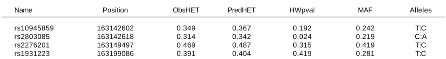
A total of seven SNPs were selected; rs76190375, rs10945859, rs2803085, rs163142645, rs2276201, rs1931223 and rs163877197. SNPs rs76190375, rs163142645 and
rs163877197 were excluded from the analysis as they devi-ated from HWE. Genotyping was performed on the re-maining four SNPs on all samples tested in this study. All SNPs analyzed were in HWE with a MAF more than 1% within this population (Table 2).
None of the SNPs showed any significant association when healthy control subjects were compared with HCV in-fected patients (i.e. groups I + II + III combined) (Table 3). Further, the frequency of these SNPs was analyzed and compared between the chronic HCV patients (group I)
Table 2. SNP marker information for PARK2 gene for all subjects observed (patients and healthy controls).
Name Position ObsHET PredHET HWpval MAF Alleles
rs10945859 163142602 0.349 0.367 0.192 0.242 T:C
rs2803085 163142618 0.314 0.342 0.024 0.219 C:A
rs2276201 163149497 0.469 0.487 0.315 0.419 T:C
rs1931223 163199086 0.391 0.404 0.419 0.281 T:C
Position: chromosomal position. ObsHET: observed heterozygosity. PredHET: predicted heterozygosity. HWpval: Hardy-Weinberg P-value. MAF: minor allele frequency.
Table 3. Genotypic distribution for PARK2 SNPs when healthy control subjects were compared to HCV-infected patients.
SNPs Genotype/ Healthy controls, HCV patients, OR (95% C.I.) χ² P-value Allele distribution n = 400 n = 400
rs10945859 CC 27 (6.75%) 27 (6.75%) 1.030 (0.587-1.810) 0.01 0.916
CT 136 (34%) 143 (35.75%) 1.083 (0.805-1.458) 0.28 0.596 TT 237 (59.25%) 230 (57.5%) Ref
CC+CT vs.TT 1.075 (0.811-1.424) 0.25 0.615
Allele
C 190 (23.75%) 197 (24.6%) 1.049 (0.834-1.319) 0.17 0.683 T 610 (76.25%) 603 (75.4%)
rs2803085 A A 24 (6%) 26 (6.5%) 1.096 (0.613-1.962) 0.1 0.756
AC 125 (31.25%) 126 (31.5%) 1.020 (0.753-1.382) 0.02 0.897
CC 251 (62.75%) 248 (62%) Ref
AA+AC vs.CC 1.032 (0.776-1.374) 0.05 0.826
Allele
A 173 (21.6%) 177 (22%) 1.037 (0.818-1.314) 0.09 0.763 C 627 (78.4%) 622 (78%)
rs1931223 CC 36 (9%) 32 (8%) 0.835 (0.500-1.396) 0.47 0.491
CT 161 (40.25%) 152 (38%) 0.887 (0.662-1.189) 0.64 0.423
TT 203 (50.75%) 216 (54%) Ref
CC+CT vs.TT 0.878 (0.665-1.159) 0.85 0.357
Allele
C 233 (29.1%) 216 (27%) 0.90 (0.724-1.120) 0.89 0.344 T 56 (70.9%) 584 (73%)
rs2276201 CC 68 (17%) 80 (20%) 1.350 (0.904-2.014) 2.16 0.141
CT 184 (46%) 191 (47.75%) 1.191 (0.873-1.625) 1.21 0.27
TT 148 (37%) 129 (32.25%) Ref
CC+CT vs.TT 1.234 (0.922-1.652) 1.99 0.158
Allele
C 320 (40%) 351 (43.9%) 1.173 (0.961-1.430) 2.47 0.116 T 480 (60%) 449 (56.1%)
and patients with cirrhosis (group II). For rs10945859, the frequency of CT genotype was significantly higher in cir-rhotic patients than chronic HCV patients (OR = 1.850; 95% C. I. 1.115-3.069; p = 0.016) (Table 4). This indicates that HCV-infected individuals who carry heterozygous CT genotype have higher likelihood of developing cir-rhosis than those with homozygous CC or TT genotype. None of the other SNPs showed any significant associa-tion with progression to liver cirrhosis in this analysis.
Next, the frequency of the SNPs in chronic HCV pa-tients (group I) was compared with papa-tients who devel-oped HCC (group III). None of the SNPs showed any significant association with the development of HCC (Ta-ble 5). However, when the two groups of patients with progressed liver disease (i.e. groups II and III) were com-bined and compared to group I (chronic HCV patients), rs10945859 CT genotype showed higher prevalence in pa-tients with liver complications than chronic HCV papa-tients (OR = 1.768; 95% C. I. 1.090-2.867; p = 0.020) (Table 5). This observation emphasizes the importance of CT geno-type of SNP rs10945859 as a predictor of liver
complica-tions in HCV-infected patients. Also, under the dominant
model, CC+CT vs. TT of rs10945859 showed association
close to significance with a p value of 0.077 (Table 5). However, SNPs rs2803085, rs1931223 and rs2276201 with p values of 0.179, 0.422 and 0.530, respectively, did not show any significant association with development of liver complications.
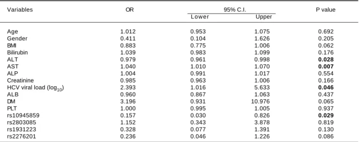
In addition, several independent factors contributing to cirrhosis were evaluated in the stepwise multivariate re-gression analysis (Table 6). The analysis showed that ALT (OR = 0.979; 95% C. I. = 0.961-0.998; p = 0.028), AST (OR = 1.040; 95% C. I. = 1.010-1.070; p = 0.007), viral load (OR = 2.393; 95% C. I. = 1.016-5.633; p = 0.046) and rs10945859 (OR = 0.157; 95% C. I. = 0.030-0.826; p = 0.029) were independently associated with HCV-associat-ed cirrhosis. Next, the multivariate regression analysis was performed on both cirrhotic and HCC patients combined (Table 7). The results revealed that BIL (OR = 1.057; 95% C. I. 1.001-1.117; p = 0.048), ALT (OR = 0.981; 95% C. I. = 0.964-0.998; p = 0.029), AST (OR = 1.039; 95% C. I. = 1.011-1.068; p = 0.005), HCV viral load (OR = 2.385; 95% Table 4. Genotypic distribution for PARK2 SNPs when chronic HCV patients (group I) were compared with cirrhotic patients (group II).
SNPs Genotype/ Group I Group II OR (95% C.I.) χ² P-value Allele distribution n = 309 n = 80
rs10945859 CC 24 (8%) 2 (2.5%) 0.395 (0.090-1.742) 1.60 0.205
CT 100 (32%) 39 (48.75%) 1.850 (1.115-3.069) 5.76 0.016
TT 185 (60%) 39 (48.75%) Ref
CC+CT vs.TT 1.568 (0.957-2.570) 3.22 0.073
Allele
C 148 (23.9%) 43 (26.9%) 1.167 (0.786-1.733) 0.59 0.443 T 470 (76.1%) 117 (73.1%)
rs2803085 A A 23 (7%) 2 (2.5%) 0.339 (0.077-1.487) 2.25 0.134
AC 95 (31%) 29 (36.25%) 1.190 (0.707-2.003) 0.43 0.513
CC 191 (62%) 49 (61.25%) Ref
AA+AC vs.CC 1.024 (0.618-1.697) 0.01 0.927
Allele
A 141 (22.8%) 33 (21%) 0.879 (0.574-1.347) 0.35 0.553 C 477 (77.2%) 127 (79%)
rs1931223 CC 23 (7%) 8 (10%) 1.400 (0.585-3.349) 0.57 0.449
CT 117 (38%) 30 (37.5%) 1.032 (0.611-1.743) 0.01 0.907
TT 169 (55%) 42 (52.5%) Ref
CC+CT vs.TT 1.092 (0.667-1.788) 0.12 0.726
Allele
C 163 (26.4%) 46 (28.75%) 1.126 (0.765-1.657) 0.36 0.546 T 455 (73.6%) 114 (71.25%)
rs2276201 CC 62 (20.1%) 18 (22.5%) 0.951 (0.487-1.858) 0.02 0.883
CT 152 (49.2%) 33 (41.25%) 0.711 (0.406-1.246) 1.43 0.233 TT 95 (30.7%) 29 (36.25%) Ref
CC+CT vs.TT 0.78 (0.466-1.308) 0.89 0.346
Allele
C 276 (44.7%) 69 (43.1%) 0.940 (0.662-1.334) 0.12 0.728 T 342 (55.3%) 91 (56.9%)
C. I. = 1.000-5.688; p = 0.050) and rs10945859 (OR = 0.196; 95% C. I. = 0.040-0.961; p = 0.044) were independ-ently associated with cirrhosis and HCC.
Haplotype analysis for these SNPs was performed to calculate the frequency of occurrence in HCV-infected patients (groups I, II and III) in comparison to the healthy controls. None of the eight haplotypes observed showed any significant association with increased susceptibility to HCV infection (Supplementary table 1). Other haplotype analyses were also performed including chronic HCV
pa-tients (group I) vs. cirrhosis patients (group II), chronic
HCV patients (group I) vs. HCC patients (group III) and
chronic HCV patients (group I) vs. cirrhotic and HCC
pa-tients (groups II and III). None of these analyses showed any statistically significant association (data not shown).
DISCUSSION
Although the number of newly infected HCV cases has declined in Saudi Arabia in the last few years, diseases as-sociated with the virus still pose a great financial and social
burden on the health system, especially among certain pa-tient population such as hemodialysis papa-tients and intrave-nous drug users. It is estimated that the prevalence of HCV in Saudi population is nearly 1.8 % and the most
dominant genotype is G4 followed by G1 and G3.38-40
Clinical outcome of HCV infection is determined by a network of interaction between host immune system, viral characteristics and environmental conditions. Several im-mune-related molecules, i.e. cytokines and chemokines, are hypothesized to play a crucial role in regulating anti-HCV immune responses that could ultimately determine
the outcome of HCV infection.41 The effect of such
mod-ulatory molecules depends largely on several factors in-cluding genetic variations and their expression activity profiles. Previous studies have shown that genetic variants that could confer a significant risk towards HCV infection are localized in genes involved in the activation of
im-mune response.42,43
We conducted this study to evaluate the effect of ge-netic variation in PARK2 gene area on the progression of HCV-related diseases. One of the most crucial steps
Table 5. Genotypic distribution for PARK2 SNPs when chronic HCV patients (group I) were compared with cirrhotic and HCC patients
(groups II and III).
SNPs Genotype/ Group I, Groups II + III, OR (95% C.I.) χ² P-value Allele distribution n = 309 n = 91
rs10945859 CC 24 (8%) 3 (3.3%) 0.514 (0.148-1.782) 1.14 0.286
CT 100 (32%) 43 (47.25%) 1.768 (1.090-2.867) 5.40 0.020
TT 185 (60%) 45 (49.45%) Ref
CC+CT vs.TT 1.525 (0.953-2.439) 3.12 0.077
Allele
C 148 (23.9%) 49 (27%) 1.170 (0.803-1.704) 0.67 0.413
T 470 (76.1%) 133 (73%)
rs2803085 A A 23 (7%) 3 (3.3%) 0.437 (0.127-1.509) 1.80 0.179
AC 95 (31%) 31 (34.1%) 1.093 (0.662-1.806) 0.12 0.727
CC 191 (62%) 57 (62.6%) Ref
AA+AC vs.CC 0.966 (0.596-1.565) 0.02 0.887
Allele
A 141 (22.8%) 37 (20.3%) 0.863 (0.575-1.297) 0.50 0.479 C 477 (77.2%) 145 (79.7%)
rs1931223 CC 23 (7%) 9 (9.9%) 1.407 (0.610-3.245) 0.65 0.422
CT 117 (38%) 35 (38.5%) 1.076 (0.654-1.768) 0.08 0.774
TT 169 (55%) 47 (51.6%) Ref
CC+CT vs.TT 1.130 (0.708-1.805) 0.26 0.609
Allele
C 163 (26.4%) 53 (29.1%) 1.147 (0.795-1.654) 0.54 0.463 T 455 (73.6%) 129 (70.9%)
rs2276201 CC 62 (20.1%) 18 (20%) 0.811 (0.421-1.561) 0.39 0.530
CT 152 (49.2%) 39 (43%) 0.717 (0.423-1.214) 1.54 0.214
TT 95 (30.7%) 34 (37%) Ref
CC+CT vs.TT 0.744 (0.457-1.213) 1.41 0.235
Allele
C 276 (44.7%) 75 (41.2%) 0.869 (0.621-1.214) 0.68 0.409 T 342 (55.3%) 107 (58.8%)
in conducting a candidate gene study is to identify a suitable gene that is likely to be involved in the disease under investigation. In this study, PARK2 was selected as it is part of the complex that is required for polyu-biquitination of proteins which are destined for
degra-dation by the proteasome.23 Strong evidence has
suggested that polyubiquitination is an important mechanism that regulates the interaction of host cells with HCV proteins. It has been shown that polyubiqui-tination of NS5A and NS5B of HCV accelerates the degradation of both proteins by proteasome-dependent pathway. Such mechanism is suggested to regulate HCV replication by controlling the rate of degradation of
NS5A and/or NS5B.44,45
The allele frequency of PARK2 SNPs in Saudi subjects included in this study was also determined. MAF values observed were 0.24, 0.22, 0.42 and 0.28 for rs10945859, rs2803085, rs2276201 and rs1931223, respectively.
In the present study, the possible association of four SNPs, rs10945859, rs2803085, rs2276201 and rs1931223 in PARK2 gene, with HCV infection and its progression to cirrhosis and HCC was investigated. The most important finding in this study is that the heterozygous CT genotype of SNP rs10945859 was found to have a significant associa-tion with HCV infecassocia-tion and its progression to cirrhosis and HCC, thereby, indicating the possibility of using it as a genetic marker to predict the progression of HCV-asso-ciated liver complications. However, this observation
Table 6. Multiple logistic regressions of PARK2 polymorphisms among chronic HCV patients and patients diagnosed with cirrhosis.
Variables OR 95% C.I. P value
Lower Upper
Age 1.012 0.953 1.075 0.692
Gender 0.411 0.104 1.626 0.205
BMI 0.883 0.775 1.006 0.062
Bilirubin 1.039 0.983 1.099 0.176
ALT 0.979 0.961 0.998 0.028
AST 1.040 1.010 1.070 0.007
ALP 1.004 0.991 1.017 0.554
Creatinine 0.985 0.963 1.006 0.166
HCV viral load (log10) 2.393 1.016 5.633 0.046
ALB 0.960 0.867 1.063 0.437
DM 3.196 0.931 10.976 0.065
PLT 1.000 0.995 1.005 0.937
rs10945859 0.157 0.030 0.826 0.029
rs2803085 1.152 0.343 3.878 0.819
rs1931223 0.328 0.077 1.391 0.130
rs2276201 0.236 0.046 1.226 0.086
Table 7. Multiple logistic regressions of PARK2 polymorphisms among chronic HCV patients and patients diagnosed with cirrhosis and
HCC.
Variables OR 95% C.I. P value
Lower Upper
AGE 1.004 0.946 1.067 0.890
Gender 0.663 0.187 2.347 0.524
BMI 0.889 0.781 1.013 0.077
PLT 0.999 0.994 1.005 0.824
BIL 1.057 1.001 1.117 0.048
ALT 0.981 0.964 0.998 0.029
AST 1.039 1.011 1.068 0.005
ALP 1.006 0.994 1.018 0.308
Creatinine 0.984 0.962 1.006 0.161
HCV Viral load (log10) 2.385 1.000 5.688 0.050
ALB 0.964 0.873 1.065 0.473
DM 2.897 0.903 9.298 0.074
rs10945859 0.196 0.040 0.961 0.044
rs2803085 1.153 0.355 3.742 0.813
rs1931223 0.463 0.117 1.824 0.271
needs to be confirmed in a larger group of patients and in different ethnic populations. Also, this genetic link needs to be substantiated by experimental evidence to identify functional significance and possible mechanism(s) through which this variant might contribute to liver ab-normalities in HCV-infected patients.
Further evidence that supports the observation that rs10945859 might modulate the progression of HCV-asso-ciated diseases was established from the logistic multivar-iate analysis.
The results in this study indicated that rs10945859 re-mained independently associated with cirrhosis and cir-rhosis/HCC in logistic regression analysis, that included several independent factors known to be important for HCV infection.
It is well-known that analysis using haplotypes of mul-tiple linked markers is more informative than single
markers.46 In the present study, analysis based on
haplo-types using all four SNPs did not show any evidence of as-sociation with HCV infection. This was also true when the analysis was performed involving any of the patient groups. Such results suggest that PARK2 haplotypes for the examined SNPs do not play a major role in HCV infec-tion and its related complicainfec-tions in Saudi patients ana-lyzed, emphasizing the need for additional investigations in a larger sample size from different populations.
Recently, a myriad of studies have been carried out to understand the role of various SNPs in HCV infection. The role of IL28B genetic variations in response to treatment and HCV clearance has been well established in various populations that are ethnically distinct.47-49 Other studies
have demonstrated that DRB1*01, DQB1*03, and DQB1*05 were associated with viral clearance in
Midwest-ern Americans.50 A large study was conducted on
Cauca-sians and Black Americans, in which HLA-A1101 and HLA-B57 was found to be significantly more frequent in in-dividuals with well-documented HCV clearance compared
to chronically-infected patients.51 More recently, DEPDC5
was found to have significant association with progression of liver-related diseases in HCV-infected patients.19
Genetic variations in PARK2 gene were identified as genetic risk factors for leprosy in several ethnically dis-tinct populations. PARK2_e01 (-2599) and rs1040079 SNPs were found to be the two most significantly associated
variants with leprosy.28 However, there was a significant
difference in the prevalence of the T allele of PARK2_e01 (-2599) between ethnically distinct populations and in its clinical significance as a genetic risk factor in leprosy in-fection.27 Other studies have suggested that the T allele of
the PARK2_e01(-2599) polymorphism is significantly as-sociated with typhoid and paratyphoid fever when patients were compared to randomly selected community unin-fected control individuals.52 Furthermore, in vitro studies
have suggested a possible role of PARK2 gene in Salmonella
pathogenesis and possible modulation of the intracellular
bacterial evasion mechanisms during the infection.53,54
In conclusion, significant association was found be-tween CT genotype of SNP rs10945859 of PARK2 gene and HCV-associated liver complications. This genetic var-iant could be considered as a predisposing genetic factor in progression of disease in HCV-infected patients. SNP rs10945859 of PARK2 gene can be a useful predictor of clinical outcome of HCV infection and help in identifying individuals at high risk of developing HCV-related liver complications. However, these findings need to be repli-cated in a larger sample of patients and the functional as-pects need to be substantiated to fully exploit the importance of PARK2 rs10945859 in HCV infection.
SUPPORT
This study was supported in part by a grant from King Abdulaziz City for Science and Technology, project no. ARP-27-18 and was approved by the Research Advisory Council (RAC) of King Faisal Specialist Hospital and Re-search Centre, RAC no. 2060040.
ACKNOWLEDGMENTS
The support of the Research Center administration at King Faisal Specialist Hospital and Research Center is highly appreciated. The authors are grateful to Hanan Shaarawi and Maureene Delos Reyes for secretarial and logistic assistance.
REFERENCES
1. Chisari FV. Unscrambling hepatitis C virus-host interactions. Nature 2005; 436: 930-2.
2. Westbrook RH, Dusheiko G. Natural history of hepatitis C. J Hepatol 2014; 61: S58-S68.
3. Croce CM. Causes and consequences of microRNA dysreg-ulation in cancer. Nat Rev Genet 2009; 10: 704-14.
4. Seeff LB. Natural history of chronic hepatitis C. Hepatology 2002; 36: S35-S46.
5. Lin MV, King LY, Chung RT. Hepatitis C virus-associated cancer. Annu Rev Pathol 2015; 10: 345-70.
6. Yamashita T, Honda M, Kaneko S. Molecular mechanisms of hepatocarcinogenesis in chronic hepatitis C virus infection. J Gastroenterol Hepatol 2011; 26: 960-4.
7. Jeong SW, Jang JY, Chung RT. Hepatitis C virus and hepato-carcinogenesis. Clin Mol Hepatol 2012; 18: 347-56. 8. Mikula M, Proell V, Fischer AN, Mikulits W. Activated hepatic
stellate cells induce tumor progression of neoplastic hepato-cytes in a TGF-beta dependent fashion. J Cell Physiol 2006; 209: 560-7.
10. Tsai WL, Chung RT. Viral hepatocarcinogenesis. Oncogene 2010; 29: 2309-24.
11. Hohler T, Kruger A, Gerken G, Schneider PM, Meyer zum Buschenfelde KH, Rittner C. Tumor necrosis factor alpha promoter polymorphism at position -238 is associated with chronic active hepatitis C infection. J Med Virol 1998; 54: 173-7.
12. Powell EE, Edwards-Smith CJ, Hay JL, Clouston AD, Craw-ford DH, Shorthouse C, Purdie DM, et al. Host genetic fac-tors influence disease progression in chronic hepatitis C. Hepatology 2000; 31: 828-33.
13. Harris RA, Sugimoto K, Kaplan DE, Ikeda F, Kamoun M, Chang KM. Human leukocyte antigen class II associations with hepatitis C virus clearance and virus-specific CD4 T cell response among Caucasians and African Americans. Hepatology 2008; 48: 70-9.
14. McKiernan SM, Hagan R, Curry M, McDonald GS, Kelly A, No-lan N, Walsh A, et al. Distinct MHC class I and II alleles are associated with hepatitis C viral clearance, originating from a single source. Hepatology 2004; 40: 108-14.
15. Neumann-Haefelin C, McKiernan S, Ward S, Viazov S, Span-genberg HC, Killinger T, Baumert TF, et al. Dominant influ-ence of an HLA-B27 restricted CD8+ T cell response in mediating HCV clearance and evolution. Hepatology 2006; 43: 563-72.
16. Sun H, Pan Y, Wu R, Lv J, Chi X, Wang X, Tu Z, et al. CD24 Ala57Val polymorphism is associated with spontaneous viral clearance in the HCV-infected Chinese population. Liver Int 2015; 35: 786-94.
17. Labib HA, Ahmed HS, Shalaby SM, Wahab EA, Hamed EF. Genetic polymorphism of IL-23R influences susceptibility to HCV-related hepatocellular carcinoma. Cell Immunol 2015; 294: 21-4.
18. Wei Y, Liu F, Li B, Chen X, Ma Y, Yan L, Wen T, et al. Poly-morphisms of tumor necrosis factor-alpha and hepatocellu-lar carcinoma risk: a HuGE systematic review and meta-analysis. Dig Dis Sci 2011; 56: 2227-36.
19. Al-Anazi MR, Matou-Nasri S, Abdo AA, Sanai FM, Khan MQ, Albenmousa A, Al-Ashgar, et al. Variations in DEPDC5 gene and its association with chronic hepatitis C virus infection in Saudi Arabia. BMC Infect Dis 2014; 14: 3839.
20. Mangia A, Santoro R, Piattelli M, Pazienza V, Grifa G, Iaco-bellis A, Andriulli A. IL-10 haplotypes as possible predictors of spontaneous clearance of HCV infection. Cytokine 2004; 25: 103-9.
21. Nischalke HD, Berger C, Luda C, Müller T, Berg T, Coenen M, Krämer B, et al. The CXCL1 rs4074 A allele is associated with enhanced CXCL1 responses to TLR2 ligands and pre-disposes to cirrhosis in HCV genotype 1-infected Caucasian patients. J Hepatol 2012; 56: 758-64.
22. Wasmuth HE, Zaldivar MM, Berres ML, Werth A, Scholten D, Hillebrandt S, Tacke F, et al. The fractalkine receptor CX3CR1 is involved in liver fibrosis due to chronic hepatitis C infection. J Hepatol 2008; 48: 208-15.
23. Asakawa S, Tsunematsu K, Takayanagi A, Sasaki T, Shimi-zu A, Shintani A, Kawasaki K, et al. The genomic structure and promoter region of the human parkin gene. Biochem Bi-ophys Res Commun 2001; 286: 863-8.
24. Beasley SA, Hristova VA, Shaw GS. Structure of the Parkin in-between-ring domain provides insights for E3-ligase dys-function in autosomal recessive Parkinson’s disease. Proc Natl Acad Sci U S A 2007; 104: 3095-100.
25. Kitada T, Asakawa S, Hattori N, Matsumine H, Yamamura Y, Minoshima S, Yokochi M, et al. Mutations in the parkin gene cause autosomal recessive juvenile parkinsonism. Nature 1998; 392: 605-8.
26. Chopra R, Ali S, Srivastava AK, Aggarwal S, Kumar B, Man-vati S, Kalaiarasan P, et al. Mapping of PARK2 and PACRG overlapping regulatory region reveals LD structure and func-tional variants in association with leprosy in unrelated indian population groups. PLoS Genet 2013; 9: e1003578.
27. Malhotra D, Darvishi K, Lohra M, Kumar H, Grover C, Sood S, Reddy BS, et al. Association study of major risk single nu-cleotide polymorphisms in the common regulatory region of PARK2 and PACRG genes with leprosy in an Indian popula-tion. Eur J Hum Genet 2006; 14: 438-42.
28. Mira MT, Alcais A, Nguyen VT, Moraes MO, Di Flumeri C, Vu HT, Mai CP, et al. Susceptibility to leprosy is associated with PARK2 and PACRG. Nature 2004; 427: 636-40.
29. Glessner JT, Wang K, Cai G, Korvatska O, Kim CE, Wood S, Zhang H, et al. Autism genome-wide copy number variation reveals ubiquitin and neuronal genes. Nature 2009; 459: 569-73.
30. Wongseree W, Assawamakin A, Piroonratana T, Sinsomros S, Limwongse C, Chaiyaratana N. Detecting purely epistatic multi-locus interactions by an omnibus permutation test on ensembles of two-locus analyses. BMC Bioinformatics 2009; 10: 294.
31. Burns MP, Zhang L, Rebeck GW, Querfurth HW, Moussa CE. Parkin promotes intracellular Abeta1-42 clearance. Hum Mol Genet 2009; 18: 3206-16.
32. Cesari R, Martin ES, Calin GA, Pentimalli F, Bichi R, McAdams H, Trapasso F, et al. Parkin, a gene implicated in autosomal recessive juvenile parkinsonism, is a candidate tumor sup-pressor gene on chromosome 6q25-q27. Proc Natl Acad Sci U S A 2003; 100: 5956-61.
33. Veeriah S, Taylor BS, Meng S, Fang F, Yilmaz E, Vivanco I, Janakiraman M, et al. Somatic mutations of the Parkinson’s disease-associated gene PARK2 in glioblastoma and other human malignancies. Nat Genet 2010; 42: 77-82.
34. Chaudhary S, Behari M, Dihana M, Swaminath PV, Govin-dappa ST, Jayaram S, Goyal V, et al. Parkin mutations in fa-milial and sporadic Parkinson’s disease among Indians. Parkinsonism Relat Disord 2006; 12: 239-45.
35. Bonder A, Afdhal N. Utilization of FibroScan in clinical prac-tice. Curr Gastroenterol Rep 2014; 16: 372.
36. Bruix J, Sherman M. Management of hepatocellular carcino-ma: an update. Hepatology 2011; 53: 1020-2.
37. Calvaruso V, Craxí A. 2011 European Association of the Study of the Liver hepatitis C virus clinical practice guide-lines. Liver International 2012; 32: 2-8.
38. Abdo AA, Sanai FM, Al-Faleh FZ. Epidemiology of viral hepa-titis in Saudi Arabia: are we off the hook? Saudi J Gastro-enterol 2012; 18: 349-57.
39. Abozaid SM, Shoukri M, Al-Qahtani A, Al-Ahdal MN. Prevail-ing genotypes of hepatitis C virus in Saudi Arabia: a system-atic analysis of evidence. Ann Saudi Med 2013; 33: 1-5. 40. WHO: World Health Organization Regional Committee for the
Eastern Mediterranean; The growing threats of hepatitis B and C in the Eastern Mediterranean Region: A call for action. 2009. Available from: http://applications.emro.who.int/docs/ EM_RC56_3_en.pdf
41. Li K, Lemon SM. Innate immune responses in hepatitis C vi-rus infection. Semin Immunopathol 2013; 35: 53-72. 42. Chuang WL, Yu ML. Host factors determining the efficacy of
hepatitis C treatment. J Gastroenterol 2013; 48: 22-30. 43. Slev P. Host genomics and HCV personalized medicine. Ann
Clin Lab Sci 2012; 42: 363-9.
45. Hou W, Tian Q, Zheng J, Bonkovsky HL. Zinc mesoporphyrin induces rapid proteasomal degradation of hepatitis C non-structural 5A protein in human hepatoma cells. Gastroenter-ology 2010; 138: 1909-19.
46. Zhang S, Sha Q, Chen HS, Dong J, Jiang R. Transmission/ disequilibrium test based on haplotype sharing for tightly linked markers. Am J Hum Genet 2003; 73: 566-79.
47. Ge D, Fellay J, Thompson AJ, Simon JS, Shianna KV, Urban TJ, Heinzen EL, et al. Genetic variation in IL28B predicts hep-atitis C treatment-induced viral clearance. Nature 2009; 461: 399-401.
48. Rao HY, Sun DG, Jiang D, Yang RF, Guo F, Wang JH, Liu F,
et al. IL28B genetic variants and gender are associated with spontaneous clearance of hepatitis C virus infection. J Viral Hepat 2012; 19: 173-81.
49. Tillmann HL, Thompson AJ, Patel K, Wiese M, Tenckhoff H, Nischalke HD, Lokhnygina Y, et al. A polymorphism near IL28B is associated with spontaneous clearance of acute hepatitis C virus and jaundice. Gastroenterology 2010; 139: 1586-92, 1592 e1.
50. Wang JH, Zheng X, Ke X, Dorak MT, Shen J, Boodram B, O’Gorman M, et al. Ethnic and geographical differences in HLA associations with the outcome of hepatitis C virus in-fection. Virol J 2009; 6: 46.
51. Thio CL, Gao X, Goedert JJ, Vlahov D, Nelson KE, Hilgartner MW, O’Brien SJ, et al. HLA-Cw*04 and hepatitis C virus per-sistence. J Virol 2002; 76: 4792-7.
52. Ali S, Vollaard AM, Widjaja S, Surjadi C, van de Vosse E, van Dissel JT. PARK2/PACRG polymorphisms and susceptibility to typhoid and paratyphoid fever. Clin Exp Immunol 2006; 144: 425-31.
53. Houde M, Bertholet S, Gagnon E, Brunet S, Goyette G, La-plante A, Princiotta MF, et al. Phagosomes are competent or-ganelles for antigen cross-presentation. Nature 2003; 425: 402-6.
54. Kubori T, Galan JE. Temporal regulation of salmonella viru-lence effector function by proteasome-dependent protein degradation. Cell 2003; 115: 333-42.
Correspondence and reprint request: Ahmed A. Al-Qahtani, Ph.D.
Department of Infection and Immunity, Research Center, King Faisal Specialist Hospital & Research Center, Riyadh,
Saudi Arabia
Tel.: 966114424550. Fax.: 966114424519 E-mail: [email protected]
Supplementary table 1. Haplotypes of PARK2 gene when control group was compared to HCV-infected patients (groups I, II and III).
Haplotype Freq. Chronic HCV patients, Chronic HCV patients, χ2 P value Healthy control Ratio Healthy control
Counts Frequencies