CopyrightqAmerican Museum of Natural History 2001 ISSN 0003-0082 / Price $2.10
Number 3333, 12 pp., 4 figures, 1 table
June 22, 2001
A New Species of Hisonotus (Siluriformes,
Loricariidae) of the Upper Rı´o Uruguay Basin
ADRIANA E. AQUINO,1 SCOTT A. SCHAEFER,1 AND
AMALIA M. MIQUELARENA2
ABSTRACT
A new species of the hypoptopomatine genus Hisonotus (Loricariidae) is described from a small tributary of the upper rı´o Uruguay basin near the border between Uruguay and Brazil. The new species can be distinguished from all other congeners by the following combination of characters: (1) presence of serrae along distal two thirds of posterior margin of pectoral-fin spine (versus serrae absent, posterior margin smooth); (2) odontodes along anterior margin of snout biserially arranged, dorsad and ventrad series separated by narrow odontode-free area covered by pad of soft tissue; (3) caudal peduncle short (27–34% SL, versus.34% SL) and deep (13–15 % SL, versus,13% SL); (4) eye large (15–19% HL, versus ,13% HL); and (5) caudal-fin pigmentation, when well defined, dark brown with a pair of whitish blotches on upper and lower lobes. The significance of the distribution of the new species is discussed relative to the degree of endemism of other fish groups in the Uruguay basin.
RESUMEN
Una nueva especie de Hypoptopomatinae del ge´nero Hisonotus (Loricariidae) es descripta para un pequen˜o tributario del rı´o Uruguay superior, cerca del lı´mite entre Uruguay y Brasil. La nueva especie puede distinguirse de todas las otras especies nominales del ge´nero por la siguiente combinacio´n de caracteres: (1) presencia de sierra a lo largo del margen posterior de los dos tercios distales de la espina pectoral (versus margen posterior liso), (2) odontodes del margen anterior del hocico ordenados biserialmente, las series dorsal y ventral separadas por una banda angosta libre de odontodes, cubierta por tejido blando; (3) pedu´ nculo caudal
1Division of Vertebrate Zoology (Ichthyology), American Museum of Natural History.
corto (27–34 % LE, versus usualmente.34) y alto (13–15 % LE, versus usualmente,13); (4) ojo grande (15–19 % in HL, versus usualmente ,13), y (5) patro´ n de coloracio´n de la aleta caudal, cuando se encuentra bien definido, marro´ n oscuro, con un par de manchas blan-quecinas sobre los lo´ bulos superior e inferior de la aleta. La distribucio´n geogra´fica de la nueva especie es discutida en relacio´n al grado de endemismo registrado en otros grupos de peces de la cuenca del rı´o Uruguay.
INTRODUCTION
As presently defined, the loricariid genus
Hisonotus Eigenmann and Eigenmann, 1889
consists of 12 nominal species (Reis and Schaefer, 1998; Schaefer, 1997, 1998), mostly occurring in Atlantic coastal streams of south-ern Brazil and the Paraguay-Parana´ system of southern South America. Hisonotus is a mem-ber of the loricariid subfamily Hypoptopoma-tinae, tribe Otothyrini (Reis and Schaefer, 1998; Schaefer, 1998), a monophyletic group diagnosed by the uniquely derived presence of a medially reflected ventral preopercle margin, forming a laminar shelf mesial to the canal-bearing cheek plate (Schaefer, 1998). The no-menclatural history of Hisonotus is intermin-gled with that of the genus Microlepidogaster Eigenmann and Eigenmann, 1889, which for most of the 20th century was considered a se-nior synonym of the former (e.g., Regan, 1904; Isbru¨cker, 1980). Under this classification,
Mi-crolepidogaster (including all nominal species
of both Microlepidogaster and Hisonotus) had been distinguished from other hypoptopoma-tines by a combination of plesiomorphic char-acter states, such as laterodorsal position of the eyes, arrector fossae open, presence of few pterotic fenestrae, and presence of an unplated region anterior to the nostrils (Britski, 1972; Buckup, 1981; Schaefer, 1991). Schaefer (1998) revalidated and diagnosed Hisonotus by the absence of plates anterior to the nostrils and the presence of robust rostral plates with enlarged odontodes, whereas
Microlepidogas-ter was distinguished by the posMicrolepidogas-terior position
of the dorsal fin and by having the rostrum composed of thin plates lacking enlarged odontodes. Revision of both Hisonotus and
Microlepidogaster are studies in progress by
the second author.
The new species is placed in the genus
Hi-sonotus on the basis of the diagnostic
char-acters mentioned above, and is diagnosed among congeners by a unique combination of characters. Specimens were collected by
Rau´l Ringuelet in the upper rı´o Uruguay ba-sin, a region of southeastern South America with endemic species of several groups of fishes (Buckup, 1981; Britski and Garavello, 1984; Reis and Schaefer, 1998).
METHODS
Measurements were taken following Buckup (1981) using a digital caliper to the nearest 0.1 mm, reported as proportions of standard length (SL) except where noted. Suborbital depth is defined as the distance in lateral view between the lower margin of the bony orbit and ventrolateral limit of the head. Meristic characters were obtained for right and left sides of each specimen. Nomencla-ture of body plates follows Schaefer (1997). Values for counts and measurements of the holotype are given in brackets. Bilateral counts are presented as left/right when asym-metric. Vertebral counts include five centra incorporated into the Weberian complex (Schaefer, 1987). In the text, ‘‘pectoral-fin spine’’ and ‘‘pelvic-fin spine’’ refer to the first lepidotrich of the pectoral and pelvic fins, respectively, which in siluriforms, though unbranched, are not true spines but rather highly ossified spine like segmented rays.
Osteological observations were made on specimens cleared and counter-stained for bone and cartilage following Taylor and Van Dyke (1985). Illustrations were prepared us-ing a Wild TYP stereomicroscope. In the list of material examined, cs denotes cleared and stained material.
Institutional abbreviations
AMNH American Museum of Natural History, New York
ANSP Academy of Natural Sciences of Phil-adelphia
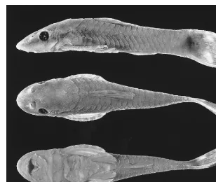
Fig. 1. Hisonotus ringueleti, holotype, ILPLA 886, female, 35.8 mm SL.
ILPLA Instituto de Limnologı´a ‘‘Dr. Rau´l A. Ringuelet’’, Buenos Aires
MCP Museo de Cieˆncias e Tecnologia, PUCRS, Porto Alegre, Brazil
MLP Museo de La Plata, La Plata, Argentina MZUSP Museu de Zoologia, Universidade de
Sa˜o Paulo, Brazil
UMMZ University of Michigan, Museum of Zoology, Ann Arbor
USNM National Museum of Natural History, Smithsonian Institution, Washington, DC
SYSTEMATIC ACCOUNT
Hisonotus ringueleti, new species Figure 1
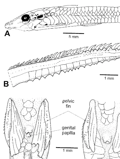
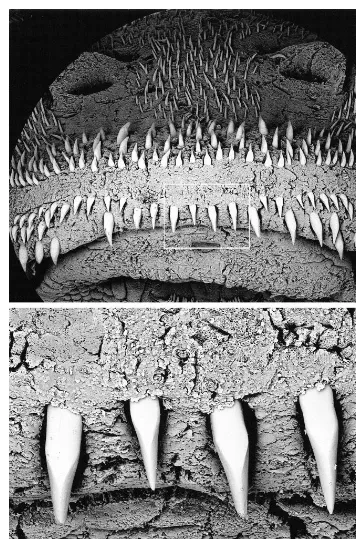
DIAGNOSIS: No autapomorphy was found for Hisonotus ringueleti. The new species can be distinguished from all other species of Hisonotus by the combination: (1) pres-ence of serrae along distal two-thirds of pos-terior margin of pectoral spine (versus serrae absent, posterior margin smooth) (fig. 2B); (2) odontodes along anterior margin of snout biserially arranged, dorsad and ventrad series separated by narrow odontode-free area (fig.
3, top); (3) caudal peduncle short (27–34% SL, versus . 31% SL) and deep (13–15 % SL, versus , 13% SL); (4) eye large (15– 19% HL, versus,13% HL); and (5) caudal-fin pigmentation, when well decaudal-fined, dark brown with pair of whitish blotches on upper and lower lobes (fig. 4, top).
REMARKS: Among nominal species of Hi-sonotus, the presence of serrae along the
pos-terior margin of the pectoral spine was also observed in Hisonotus taimensis Buckup, 1981, and H. nigricauda (Boulenger, 1891), which precludes this feature as autapomorph-ic for H. ringueleti among species of
Hison-otus. However, the consistency of certain
in-trinsic features of the serrae in H. ringueleti are noteworthy. Specifically, serrae of H.
rin-gueleti (1) are consistently present in
TABLE 1
Morphometric and Meristic Data for Hisonotus ringueleti
Holotype
Males (N510)
Min Max Mean SD
Females (N510)
Min Max Mean SD
Standard length 35.8 26.8 30.1 28.4 1.12 27.5 35.5 31.93 2.56 PERCENT OF STANDARD LENGTH
Predorsal length Head length Cleithral width Dorsal-fin spine length Trunk length
Pectoral-fin spine length Pelvic-fin spine length Abdominal length Caudal peduncle length Caudal peduncle depth Head depth
Snout length
Horizontal eye diameter Least interorbital diameter
45.5 33.8 23.3 25.9 16.2 25.6 14.6 17.2 31.2 14.1 17.6 10.5 5.6 13.9 46.4 34.9 22.1 26.0 15.2 23.9 19.6 17.5 28.8 13.0 18.0 9.3 5.7 13.5 48.0 37.5 23.8 31.2 19.0 27.9 23.7 21.4 32.0 14.9 19.4 11.5 6.6 16.4 46.9 36.2 23.0 27.9 17.0 26.3 21.2 19.6 30.9 13.7 8.4 10.5 6.2 15.0 0.47 0.89 0.64 1.75 1.07 1.41 1.41 1.19 1.08 0.58 0.39 0.60 0.29 0.99 46.8 35.2 21.8 25.4 15.1 25.9 14.9 17.4 27.3 13.0 17.7 10.5 5.6 14.2 49.5 39.2 25.4 29.0 18.6 28.2 19.1 20.1 33.8 14.9 19.6 11.7 6.8 17.0 47.63 36.80 23.47 27.30 16.61 27.09 17.56 19.02 30.63 13.65 18.67 11.08 6.08 15.19 0.94 1.15 1.09 1.09 1.10 0.70 1.36 0.83 1.71 0.56 0.65 0.44 0.33 0.83 PERCENT OF HEAD LENGTH
Head depth Snout length
Horizontal eye diameter Least interorbital diameter
52.1 31.1 16.7 41.0 49.5 26.7 15.8 38.2 53.4 30.7 18.8 44.9 50.9 28.9 17.1 41.5 1.61 1.29 0.97 2.41 48.1 28.7 15.3 38.7 53.0 32.8 17.5 44.4 50.76 30.12 16.52 41.28 1.89 1.32 0.73 1.71 COUNTS
Left lateral plates Right lateral plates Predorsal plates Left premaxillary teeth Right premaxillary teeth Left dentary teeth Right dentary teetha Dorsal-fin branched rays Pectoral-fin branched rays Pelvic-fin branched rays Anal-fin branched rays Caudal-fin branched rays
24 23 3 13 12 11 14 6 5 5 4 14 23 24 3 11 11 9 9 6 5 5 4 14 25 25 3 14 15 12 13 7 5 5 4 14 24.1 24.1 3.0 12.4 12.3 11.2 11.0 6.4 5.0 5.0 4.0 14.0 0.57 0.32 0 0.84 1.49 1.14 1.33 0.52 0 0 0 0 24 23 3 12 12 10 11 6 5 5 4 14 24 25 3 16 16 14 15 7 5 5 4 14 24.0 24.0 3.0 13.3 13.8 12.2 12.6 6.4 5.0 5.0 4.0 14.0 0 0.67 0 1.25 1.14 1.40 1.24 0.52 0 0 0 0 aN59 for females.
DESCRIPTION: Descriptive morphometric and meristic data are provided in table 1. Adult body size moderate (N 5 126; mean 28.3 mm SL, range 26–39). Body relatively stocky, greatest body depth at supraoccipital, 17.7–19.6 [17.6]% SL, slightly deeper than depth at dorsal-fin origin; caudal peduncle deep, 13.0–14.9 [14.1]% SL. Head moder-ately narrow, cleithral width 21.8–25.4 [23.3]% SL. Dorsal profile of head from snout tip to supraoccipital convex, anterior to nostrils slightly depressed, between eyes
slightly convex. Cross-sectional profile of su-praoccipital gently convex. Snout tip round-ed in dorsal view. Eyes placround-ed dorsolaterally, horizontal eye diameter 5.6–6.8 [5.6]% SL, larger than suborbital depth. Iris diverticulum present, large, its length two-thirds of pupil diameter.
(mode, 12) on dentary; accessory teeth (sen-su Reis and Schaefer, 1992) absent.
Body covered by dermal plates except for area around anus, skin covering lateral open-ing of swimbladder capsule, base of paired fins, area between pectoral girdle and lower lip, and snout anterior to nostrils. Lateral and anterior rostral plates reflected ventrally. Trunk plates arranged in five lateral series (fig. 2A): (1) dorsal series continuous; (2) mid-dorsal series discontinuous; (3) median series 23–24, incomplete, discontinuous, composed by anterior sector of 1–3 plates and posterior sector of 16–18 plates, sepa-rated by gap; (4) mid-ventral series incom-plete, continuous; and (5) ventral series complete, continuous. Lateral-line canal in-complete, discontinuous, with anterior field of 1–4 [4] canal-bearing plates along anterior sector of median series, and posterior field of 1–3 [3] plates along posterior sector of me-dian series. Abdomen partly covered by plates variable in size and shape, arranged in paired lateral series of 3–6 [6/4] plates each, and a median series of 3–6 [6] plates. Anal fin preceded by 4 paired lateral plates, vari-ably contacting antimeres at midline. Cora-coids and cleithra exposed ventrally, except for area at midline and surrounding arrector fossae.
Odontodes covering head, trunk, and fin rays. Head and trunk odontodes uniformly distributed, not arranged in distinct longitu-dinal lines or forming keels. Odontodes gen-erally small, except for enlarged odontodes on ventral aspect of pelvic and pectoral spines, anterior rostral margin of snout, and tuft at posterior supraoccipital tip, not ele-vated above level of plate posterior to supra-occipital. Odontodes along anterior margin of snout biserially arranged, dorsad and ven-trad series separated by narrow odontode-free area covered by pad of soft tissue; ven-trad series composed of a continuous row of enlarged and laterally faceted odontodes and paired lateral patch of smaller, conical odon-todes (fig. 3).
Dorsal-fin origin slightly posterior to ver-tical through pelvic-fin origin. Adipose fin absent. Pectoral fin, when depressed, over-lapping nearly two-thirds of pelvic-fin length; serrae along distal two-thirds of pos-terior margin of pectoral-fin spine, robust
(tooth height approximately 40 –50% of spine width at tip) (fig. 2B). Pelvic fin, when depressed, reaching beyond anal-fin origin only in males (see SEXUAL DIMORPHISM, be-low).
OSTEOLOGY: The following is not an ex-haustive description, but an account of char-acter states present in the new species for features that have been treated in recent phy-logenetic analyses (Schaefer, 1991, 1998). Mesethmoid tip bearing small, uncinate pro-cess directed ventrally; mesethmoid disk sep-arated from mesethmoid tip by one-quarter disk width. Parasphenoid shaft, posterior to lateral processes, laterally constricted. Pter-otic bone fenestrae relatively few in number, expanded and rounded, restricted to anter-oventral part of compound pterotic. Swim-bladder-capsule lateral opening wide.
Upper pharyngeal tooth- plate dentition with narrow extension anteriorly. Total ver-tebrae 27. Vertebral centra 10–15 with bifid neural spines, 15–18 with bifid hemal spines; distal portions of neural and hemal spines ta-pering distally, widely separated from one another. Seventh vertebral centrum not ex-panded anterior to dorsal-fin first proximal radial; anterior margin of seventh vertebral centrum simple.
Posterior margin of caudal-fin skeleton straight or with slight median notch. Dorsal-fin spinelet small, roughly triangular; dorsal-fin locking mechanism absent. Dorsal-dorsal-fin first three proximal radials with transverse process expanded.
vari-Fig. 2. Hisonotus ringueleti, ILPLA 883. A, Body lateral view, pattern of trunk lateral plates; B,
[image:6.612.52.456.86.620.2]Fig. 4. Hisonotus ringueleti, ILPLA 883,
cau-dal-fin pigmentation. Top, 27.4 mm SL, bottom, 31.5 mm SL.
able, ranging from a well-defined pattern of ground color dark brown and a pair of lighter blotches of moderate size placed symmetri-cally relative to longitudinal axis (fig. 4, top), to a pattern of ground color dark brown, with a series of small light blotches on dorsal and ventral lobes variably connected between lobes forming light transverse bars (fig. 4, bottom).
DISTRIBUTION: Known only from the type locality, a creek in the rı´o Quaraı´, a tributary of the upper rı´o Uruguay.
HABITAT: This species was collected from a small creek, ca. 0.5 m depth, with rapid current and clear water, bottom composed of rocks and sand, and with vegetated margins. Specimens of the new species were collected from around submerged rocks and aquatic plants (C. Rolda´n, personal commun.).
SEXUAL DIMORPHISM: Males smaller than females, mean standard length 26.9 (N558) versus 29.3 (N 5 66). Genital papilla of males pointed; fleshy flap along posterior margin of pelvic-fin spine of males. Males with longer pelvic fins (longest pelvic-fin ray length 19.6–23.7% SL, versus 14.9–19.1% SL); distance from anus to anal-fin origin shorter (16.3–18.6%, versus 19.8–22.8% SL
SL; fig. 2C, D). Pelvic fin not reaching anal fin origin in 85% of females (versus 7% of males); reaching first anal-fin ray in 15% of females (versus 35% of males); reaching be-yond first anal-fin rays in no females (versus 58% of males).
ETYMOLOGY: Named after Dr. Rau´l A. Rin-guelet (1914–1982), researcher and professor of the Museum of Natural Sciences of La Plata, Buenos Aires. Dr. Ringuelet’s vast ca-reer includes the publication of the book Los
Peces de Agua Dulce de la Repu´blica Ar-gentina (Ringuelet et al., 1967), which set
the standard for systematics research con-ducted during the last decades of the 20th century in the Austral region of the Neotrop-ics.
MATERIAL EXAMINED: Holotype: ILPLA 886 (35.8 mm, female), Uruguay, Rivera State, upper Uruguay River basin, Quaraı´ River drainage, creek at Km 18 of route join-ing Santana do Livramento, Brazil, and Ri-vera, Uruguay; close to border (ca. 318 009 S, 558 309W). Coll. R. A. Ringuelet and C. Rolda´n, 24 July 1981.
Paratypes: collected with holotype.
IL-PLA 883 (51/ 144?, 26.0–39.2 mm SL). AMNH 230702 (3 / 1 2 ? 1 3 cs, 23.3– 33.2 mm SL); ANSP 177878 (1 /1 2? 1 1 cs, 22.9–32.4 mm SL); FMNH 108806 (2 / 1 2 ?, 25.7–32.2 mm SL); MCP 26154 (2/11?11 cs, 26.4–31.3 mm SL); MLP 9536 (2 / 1 2 ?, 27.9–33.4 mm SL); MZUSP 62788 (1 / 1 2 ?, 23.3–31.1 mm SL); USNM 362665 (2 / 12 ?, 27.2–32.0 mm SL).
COMPARATIVE MATERIAL: Hisonotus sp.: FMNH 59635; USNM 206204, 297971, 235073, 300968, 235072, 345698, 345937.
Hisonotus laevior: USNM 235075, 285894,
326112. Hisonotus leucofrenatus: FMNH 59628. Hisonotus maculipinnis: UMMZ 206297; USNM 176024. Hisonotus
nigricau-da: USNM 181550, 177537 (2 cs). Hisonotus notatus: FMNH 59636. Hisonotus paulinus:
FMNH 59636. Hisonotus punctatus: MHNG 240825 (1 cs); UMMZ 206204 (1 cs).
Hi-sonotus taimensis: ANSP 168949 (1 cs);
USNM 235062. Microlepidogaster
perfora-tus: ANSP 174718 (1 cs).
DISCUSSION
The most distinctive feature of Hisonotus
posterior margin of the pectoral-fin spine, a character which had been previously reported also for three of six genera within the tribe Hypoptopomatini, subfamily Hypoptopoma-tinae (Acestridium Haseman, 1911,
Hypop-topoma Gu¨nther, 1868, and Oxyropsis Eigen-mann and EigenEigen-mann, 1889) (Schaefer, 1991). As far as we know, species of
Hison-otus are the only representatives of the tribe
Otothyrini having such pectoral-fin spine ser-rae. A more exhaustive examination of this feature revealed that the presence of serrae is more widespread among species of
Hisono-tus and not exclusive to H. ringueleti,
al-though Hisonotus remains the only genus of Otothyrini with species having serrated pec-toral spines. We observed this condition as variably present in H. taimensis and bilater-ally variable in one specimen of H.
nigricau-da. The condition observed in H. ringueleti
differs from that of both H. taimensis and H.
nigricauda in three respects. First, the
pres-ence of serrae appears to be fixed in H.
rin-gueleti, as it is observed consistently among
individuals, versus variably present among and within individuals of other Hisonotus species. Secondly, the serrated margin is composed of robust tooth like structures in
H. ringueleti, versus feeble and
inconspicu-ous serrae in the other species. Finally, in H.
ringueleti the serrae are more numerous and
occupy the distal two-thirds of the pectoral-fin spine, versus fewer in number and re-stricted to the distal quarter of the spine.
The particular odontode arrangement on the anterior margin of the snout of H.
rin-gueleti, composed of dorsad and ventrad
se-ries of odontodes separated by an odontode-free narrow gap, has not been previously re-ported for any other nominal species of
Hi-sonotus. Some species of the genus have a
similar arrangement of dorsally and ventrally directed odontode series on the rostral mar-gin (e.g., H. nigricauda), though without an associated odontode-free gap. Among other Otothyrini, the presence of a similar discon-tinuity in the odontode distribution on the snout was reported for species of
Pseudoto-cinclus Nichols, 1919 (Schaefer, 1991) and Otothyris Myers, 1927 (Garavello et al.,
1998). According to the phylogenetic scheme proposed by Schaefer (1998), the genera
Pseudotocinclus and Otothyris are both
rel-atively well nested within the Otothyrini and separated from the more basal position of
Hi-sonotus. Therefore, it is most parsimonious
to conclude that the presence of an odontode-free narrow gap between dorsad and ventrad series was independently derived in the aforementioned genera.
The presence of a pad of soft tissue on the snout tip has also been observed in H.
lae-vior Cope, 1884, H. nigricauda, and H. tai-mensis. However, the new species can be
dis-tinguished from those three by having the pad associated with an actual odontode-free area. The relatively deep caudal peduncle (greater than 13% SL) further distinguishes
H. ringueleti from other nominal species of Hisonotus.
Relative to other nominal species of
Hi-sonotus, the new species can be distinguished
from H. depressicauda (Ribeiro, 1918) by the absence of odontodes arranged as distinct keels on the head; from H. depressinotus (Ri-beiro, 1918) by the robust head and trunk (versus anterior region markedly depressed), from H. laevior, H. maculipinnis (Regan, 1912), and H. nigricauda by the presence of large dorsal and ventral light spots on the caudal fin (versus bar-pattern pigmentation), from H. taimensis by having fewer plates along the median lateral series (ca. 24, versus ca. 30), from H. leucofrenatus (Ribeiro, 1908) by having a shorter caudal peduncle (27.3–33.8% SL, versus ca. 40.5% SL) and abdominal plates comprising paired lateral series separated by a variably developed me-dian series (versus abdominal and preanal re-gion covered by few large irregularly ar-ranged plates), from H. notatus (Eigenmann and Eigenmann, 1889) by having fewer jaw teeth (premaxilla teeth 11–16, versus 24; dentary teeth 9–13, versus 19).
The geographic distribution of this spe-cies, being restricted to the upper rı´o Uru-guay, is congruent with an emerging pattern of enhanced species richness and endemism of fishes in the upper Uruguay and Jacui riv-er drainages, a phenomenon noted by othriv-er authors (e.g., Reis and Schaefer, 1998; Wim-berger et al., 1998). This region is one of the best sampled of the Neotropics. Neverthe-less, the rate of discovery of new endemic species for the region is still high (e.g.
Gym-nogeophagus Wimberger et al., 1998; Rine-loricaria Reis, 2000, unpubl., personal
com-mun.) which is perhaps a direct result of in-creased sampling effort in headwater portions of the rı´o Uruguay and its tributaries (Reis, 2000 unpubl., personal commun.).
A series of phylogeny-based biogeograph-ical analyses of species of del Plata basin (Curimatidae—Vari, 1988; Loricariidae— Schaefer, 1997; Cichlidae—Wimberger et al., 1998; Callichthyidae—Reis, 1998) provides support in favor of a hypothesis of early Ter-tiary hydrogeological isolation that prevent-ed dispersal between upper and lower reach-es of the Uruguay basin. Wimberger et al. (1998) provided evidence supporting such a hypothesis on the basis of a well supported clade of Gymnogeophagus species of the up-per Uruguay, relative to its sister clade in the lower Uruguay and Parana´ rivers. Known distributions of several other fish taxa (cich-lids—Reis and Malabarba, 1988; catfishes— Buckup, 1981; Britski and Garavello, 1984; Reis and Schaefer, 1998) provide further ev-idence in favor of a hypothesis of isolation.
As far as we can determine from available material, the distribution of Hisonotus
rin-gueleti is restricted to a single, small
tribu-tary of the rı´o Quaraı´, within the upper Uru-guay basin. Though it would be premature now to comment further on the significance of the distribution of H. ringueleti within the context of the biogeography of the genus as a whole, a number of emerging shared bio-geographic patterns involve monophyletic clades within the loricariid subfamily Hypop-topomatinae. Seven of nine genera described for the clade Otothyrini (Reis and Schaefer, 1998) have a distribution restricted to south-eastern Brazil (Epactionotus,
Eurycheili-chthys, Microlepidogaster, Otothyris, Pseu-dotocinclus, Pseudotothyris, Schizolecis).
The two exceptions are Hisonotus (sensu Schaefer, 1998) and Parotocinclus (sensu Schaefer, 1991), both of which are more widely distributed in cis-Andean drainages of South America. Species of Hisonotus also occur in the lower Parana´, Paraguay, and lower Uruguay River drainages, and
Paro-tocinclus species also occur in the Essequibo
River of Surinam, in the middle Amazon ba-sin, in the Atlantic coastal rivers of north-eastern Brazil, and in the Orinoco River
(Schaefer and Provenzano, 1993). In the most recent phylogenetic hypotheses, both
Hisonotus and Parotocinclus are relatively
basal taxa within the Otothyrini clade (Reis and Schaefer, 1998; Schaefer, 1998). In bio-geographical terms, this suggests the possi-bility of an ancient continent-wide distribu-tion of basal Otothyrini lineages, followed by subsequent isolation and speciation in more geographically restricted hydrogeographic regions of South America, a hypothesis that is congruent with the above-mentioned inter-pretation of Wimberger et al. (1998).
Among the Hypoptopomatini, Otocinclus and Hypoptopoma have the broadest distri-butions (Schaefer, 1991, 1997), with species present in the Paraguay, lower Parana´, Sa˜o Francisco, northeastern Brazil, and Amazon and Orinoco river basins (Schaefer, 1991, 1997). Considering the relatively extensive collecting effort in these regions, the absence of both genera from the upper Uruguay is not likely the result of sampling bias.
Based on a phylogenetic analysis of
Oto-cinclus Cope, 1872, Schaefer (1997)
pre-sented a hypothesis of area relationships in-volving many of the same areas of endemism shared by other hypoptopomine genera, such as Hisonotus and Hypoptopoma. Further evaluation of congruence among biogeo-graphical patterns involving genera and su-praspecific clades of Hypoptopomatinae must await the results of ongoing revisionary and phylogenetic analyses.
ACKNOWLEDGMENTS
the field trip to collect the specimens, pro-vided helpful information about the type lo-cality and collecting methods. Adriana Aqui-no was supported by a Starr Fellowship and an Axelrod Fund Grant of the American Mu-seum of Natural History and by a CONICET (Argentina) fellowship while in residence at the AMNH.
REFERENCES
Britski, H. A.
1972. Peixes de a´gua doce do Estado de Sa˜o Paulo; sistema´tica. In: Comissa˜o Inter-estadual da Bacia Parana´-Uruguai. Po-luic¸a˜o e Piscicultura. Sa˜o Paulo, Facul-dade de Sau´de Pu´blica da USP, Instituto de Pesca, pp. 79–108.
Britski, H. A., and J. C. Garavello
1984. Two new southeastern Brazilian genera of Hypoptopomatinae and redescription of Pseudotocinclus Nichols, 1919 (Os-tariophysi, Loricariidae). Pap. Avulsos Zool. 35: 225–241.
Buckup, P. A.
1981. Microlepidogaster taimensis sp.n., novo Hypoptopomatinae da Estac¸a˜o Ecolo´gica do Taim, Rio Grande do Sul, Brasil (Ostariophysi, Loricariidae). Iheringia, Se´rie Zoologia 60: 19–31. Eigenmann, C. H., and R. S. Eigenmann
1889. Preliminary notes on South American Nematognathi. Proc. California Acad. Sci. (ser. 2) 1: 119–172.
Garavello, J. C., H. A. Britski, and S. A. Schaefer
1998. Systematics of the genus Otothyris My-ers 1927, with comments on geograph-ic distribution (Siluriformes, Lorgeograph-icari- Loricari-idae, Hypoptopomatinae). Am. Mus. Novitates 3222: 19 pp.
Isbru¨ cker, I.J.H.
1980. Classification and catalogue of the mailed Loricariidae. Versl. Tech. Ge-gevens, Inst. Taxon. Zool. Univ. Am-sterdam 22: 1–180.
Regan, C. T.
1904. A monograph of the fishes of the fam-ily Loricariidae. Trans. Zool. Soc. Lon-don 17: 191–350.
Reis, R. E.
1998. Anatomy and phylogenetic analysis of the neotropical callichthyid catfishes (Ostariophysi, Siluriformes). Zool. J. Linn. Soc. 124: 105–168.
2000 (unpubl.). Revision of the cascudinhos of the genus Eurycheilichthys
(Siluri-formes, Loricariidae, Hypoptopomati-nae): high species richness in a limited area. 80th Annual Meeting of the American Society of Ichthyologists and Herpetologists, Abst. p. 304, oral com-mun.
Reis, R. E., and L. R. Malabarba
1988. Revision of the neotropical cichlid ge-nus Gymnogeophagus Ribeiro, 1918, with description of two new species (Pisces, Perciformes). Rev. Brasil. Zool. 4: 259–305.
Reis, R. E., and S. A. Schaefer
1992. Eurycheilus pantherinus (Siluroidei:
Loricariidae), a new genus and species of Hypoptopomatinae from southern Brazil. Copeia 1992: 215–223. 1998. New cascudinhos from southern Brazil:
Systematics, endemism, and relation-ships (Siluriformes, Loricariidae, Hy-poptopomatinae). Am. Mus. Novitates 3254: 25 pp.
Ringuelet, R. A., R. H. Ara´mburu, and A. Alonso de Ara´mburu
1967. Los peces argentinos de agua dulce. Comisio´n de Investigaciones Cientı´fi-cas de la Provincia de Buenos Aires, La Plata, 602 pp.
Schaefer, S. A.
1987. Osteology of Hypostomus plecostomus (Linnaeus), with a phylogenetic analy-sis of the loricariid subfamilies (Pisces: Siluroidei). Contrib. Sci. Nat. Hist. Mus. Los Angeles Cty. 394: 1–31. 1991. Phylogenetic analysis of the loricariid
subfamily Hypoptopomatinae (Pisces: Siluroidei: Loricariidae), with com-ments on generic diagnoses and geo-graphic distribution. Zool. J. Linn. Soc. 102: 1–41.
1997. The Neotropical Cascudinhos: System-atics and biogeography of the
Otocin-clus catfishes (Siluriformes:
Loricari-idae). Proc. Acad. Nat. Sci. Philadel-phia 148: 1–120.
1998. Conflict and resolution: Impact of new taxa on phylogenetic studies of the neo-tropical cascudinhos (Siluriformes: Loricariidae). In: L. R. Malabarba, R. E. Reis, R. P. Vari, C. A. S. Lucena, and Z. M. S. Lucena (eds.), Phylogeny and Classification of Neotropical Fish-es, EDIPUCRS, Porto Alegre, pp. 375– 400.
Schaefer, S. A., and F. Provenzano R.
(Siluro-idei: Loricariidae). Ichthyol. Explor. Freshwaters 4: 39–56.
Taylor, W. R., and G. C. Van Dyke
1985. Revised procedures for staining and clearing small fishes and other verte-brates for bone and cartilage study. Cy-bium 9: 107–119.
Vari, R. P.
1988. The Curimatidae, a lowland neotropical fish family (Pisces: Characiformes); distribution, endemism, and phyloge-netic biogeography. In: W. R. Heyer and P. E. Vanzolini (eds.), Proceedings
of a Workshop on Neotropical Distri-butions Patterns, Academia Brasileira de Ciencias, Rio de Janeiro, pp. 343– 377.
Wimberger, P. H., R. E. Reis, and K. R. Thornton 1998. Mitochondrial phylogenetics,
biogeog-raphy, and evolution of parental care and mating system in
Gymnogeopha-gus (Perciformes: Cichlidae). In: L. R.
Malabarba, R. E. Reis, R. P. Vari, C. A. S. Lucena, and Z. M. S. Lucena (eds.), Phylogeny and Classification of Neo-tropical Fishes, EDIPUCRS, Porto Ale-gre, pp. 309–318.
Recent issues of the Novitates may be purchased from the Museum. Lists of back issues of the
Novitates and Bulletin published during the last five years are available at World Wide Web site
http://nimidi.amnh.org. Or address mail orders to: American Museum of Natural History Library, Central Park West at 79th St., New York, NY 10024. TEL: (212) 5545. FAX: (212) 769-5009. E-MAIL: [email protected]