Video-based assistance system for training in minimally invasive
surgery
J U A N A . S Á N C H E Z - M A R G A L L O1, F R A N C I S C O M . S Á N C H E Z - M A R G A L L O1, J O S É B. P A G A D O R1, E N R I Q U E J. G Ó M E Z2, P A T R I C I A S Á N C H E Z - G O N Z Á L E Z2, J E S Ú S U S Ó N1, J O S É M O R E N O3
Minimally Invasive Surgery Centre Jesús Usón, Cáceres, Spain, Biomedical Engineering and Telemedicine Centre, Polytechnic University of Madrid, Madrid, Spain, and Laboratory of Robotics and Artificial Vision, University of Extremadura, Cáceres, Spain
Abstract
In this paper, the development of an assisting system for laparoscopic surgical training is presented. With this system, we expect to facilitate the training process at the first stages of training in laparoscopic surgery and to contribute to an objective evaluation of surgical skills. To achieve this, we propose the insertion of multimedia contents and outlines of work adapted to the level of experience of trainees and the detection of the movements of the laparoscopic instrument into the monitored image. A module to track the instrument is implemented focusing on the tip of the laparoscopic tool. This tracking method does not need the presence of artificial marks or special colours to distinguish the instruments. Similarly, the system has another method based on visual tracking to localize support multimedia content in a stable position of the field of vision. Therefore, this position of the support content is adapted to the movements of the camera or the working area. Experimental results are presented to show the feasibility of the proposed system for assisting in laparoscopic surgical training.
training, visual tracking, video-based system Key words: Laparoscopy, laparoscopic instrument tracking,
I n t r o d u c t i o n
T h e introduction of laparoscopic surgery constitutes one of the m o s t important advances in the field of surgery in the last twenty years, and it still has a great impact on surgical practice. T h i s surgical p r o c e d u r e has modified m a n y well-established surgical concepts, such as the reduction of surgical stress (1,2), less parietal problems (3), reduction of post-operative pain (4), better aesthetic results (5), a n d faster rein-corporation into the working life ( 6 - 8 ) , and as con-sequence the sanitary costs are r e d u c e d . T h e s e benefits have led to m a n y surgeons being trained in this surgical p r o c e d u r e (9).
I n order to carry out an adequate performance of laparoscopic surgery it is important to progress from the basic principles to advanced skills t h r o u g h a steep learning curve (10). However, future advances should
improve these practical aspects of training in laparos-copy (11).
I n laparoscopic surgery the video images acquired from the laparoscopic camera are displayed in real time on a m o n i t o r to provide visual feedback to the surgeon (12), thus minimally invasive p r o c e d u r e s guided by imaging involve a new paradigm in surgical p r o c e d u r e s and surgical training.
Regarding the training process in laparoscopic sur-gery, especially d u r i n g the early stages, teachers have some difficulties such as paying attention to all trai-nees simultaneously, repeating the same contents of explanation and making a subjective assessment. W e hypothesize that this training process in laparoscopic surgery could be improved by providing video-based assistance d u r i n g the performance of surgical tasks and a real-time tracking m e t h o d applied to laparoscopic instruments. Therefore, this tracking
method allows us to carry out an objective assessment of surgical skills.
To make the training in laparoscopic surgery during the early stages easier, there are currently several hybrid simulation systems. Most of them are focused on the objective assessment of surgical skills (12-15). Others focus on providing an image more similar to a real surgical scenario. Yet others include the insertion of virtual contents into the training process (16).
Regarding the tracking of the laparoscopic instru-ments, several approaches have been proposed to deal with this problem. We think that addressing the problem by using image processing techniques to track the instrument is an interesting alternative to others based on the use of sensors located on the instrument (optical, electromagnetic or mechanical) which can be very uncomfortable and unmanageable for surgeons. In the scientific literature, some of these approaches analyze the colour of the instrument as Wang et al. (17), who carry out a statistical analysis to distinguish between pixels belonging to a laparoscopic instrument or environment of the abdominal cavity. Other authors (18), however, perform statistical anal-ysis using Bayes theorem. On the other hand, Doignon et al. (19) use an image segmentation to find the instrument using the hue and saturation of the image with a HSI (Hue, Saturation, Intensity) colour model and region growing techniques.
In works that use artificial marks on the laparo-scopic instruments, these marks are often located next to the tip and then are identified by image segmen-tation. The use of an unusual colour in surgical images, as artificial mark, is performed in other cases (20,21). Zhang and Payandeh (22) use a pattern formed by three black lines with the same width and space. Tonet et al. (23) propose a heuristic approach to estimate the depth of the instrument by means of the knowledge of the instrument width and the orientation of its edges. Others propose the use of a LED placed in the tip of the instrument that projects laser dots onto the surface of organs to allow them to localize the instrument with respect to the scene (24).
Another possibility is not using artificial marks or analyzing the colour of the instrument. For instance, Climent and Mares (25) and Voros et al. (26) employ the Hough transform to find the straight lines belong-ing to the edges of the instrument. Besides usbelong-ing the Hough transform, Voros et al. (26) also use the Otsu thresholding and the position of the insertion point of the instrument to restrict the search range of the tool. On the other hand, other authors (27) propose a geometrical optical model to determine the tip posi-tion and the orientaposi-tion of a laparoscopic instrument with respect to the camera coordinates.
In this paper, a new way to improve the laparo-scopic training process for new trainees is presented. This system supports the surgical training with video-based contents (image and video) and carries out an automatic vision-based tracking of the movement performed by the laparoscopic instruments. This tracking method could be useful, in future works, to analyze the movements performed by trainees and make an objective assessment of surgical skills during the surgical training process.
Material and methods
To standardize the performance within a controllable and reproducible scenario, we use the physical lapa-roscopic simulator SIMULAP-IC05® (CCMIJU, Cáceres, Spain) (9,28,29), mainly dedicated to the learning of laparoscopic maneuvers which involve some technical difficulty. This training device simu-lates the abdominal cavity and presents a transparent cover made of plastic, which allows the insertion of the trocars for the handling of the laparoscopic instru-ments. The surgeon follows his maneuvers performed through the displayed images on the screens.
For the implementation of the system, we use the C++ programming language and the OpenCV 1.0.1 graphic library (30), all under a Linux-based operating system.
The video-based system is implemented on a com-puter with an Intel® Core™ 2 processor to 2.4 GHz and 3GB RAM. The system has a standard video capture card, a NVidia GeForce GTX 260 896MB DDR3 graphic card and a T F T monitor which has 17 inches, 1024x768 resolution, 500 cd/m2
bright-ness, 400:1 contrast, 0.297 mm of pixel size and 16 ms response time.
The image capture from the camera of the physical simulator is taken by a RCA video connector con-nected to the video capture card of the computer.
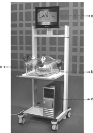
The hardware components of the system are installed in a tower (Figure 1), which integrates the physical simulator, the corresponding camera, a screen to display the information and a computer which captures the video image and runs the pro-cesses of the assisting system.
Visual tracking for the viewing support content
Figure 1. The complete system consists of a monitor (a), the physical laparoscopic simulator (b), a camera (c), and a computer (d).
This visual tracking method performs an analysis of the optical flow via the Lucas-Kanade algorithm with Gaussian Pyramids (31,32). This algorithm accom-plishes the extraction of the x and j ; components of the velocity vector, in a local neighborhood around the analyzed point. Therefore, an estimation of the optical flow in blocks of pixels is established, and thus the movement vector for this block is considered.
The pyramidal Gaussian representation of an image consists of creating a series of images, so that each image is the Gaussian distribution of its previous image. Thus, the representation of the image is divided into different hierarchical levels. The first level is the original image, the second one is the image after applying it to a Gaussian filter, and so on.
The aim of this visual tracking method is to deter-mine the position of a certain point of an image in the next frame of the video sequence. The method keeps the superimposed visual content in a stable position of the working area.
A pyramidal representation (n levels) is obtained from two consecutive images of the sequence; being n the level with more Gaussian smoothing, and 0 the initial level (original image). In our particular case, we use five levels for the pyramidal representation of the Gaussian image.
For each level, starting from the nl level backwards,
using the two consecutive images of the sequence, the velocity vector of the selected point is calculated by the Lucas-Kanade optical flow method. For one point of the ith level (1 < i < ri) corresponds to a block of
points of level i-1 of the pyramidal representation. Therefore, if we have the velocity vector of the i level point (calculated in the previous step), we will obtain the velocity vector of a block of points in the level i-1. When we reach the last level, we have the motion estimation of the marked point in the original image (level 0).
Once we track the marked point in the current frame, the next step is to establish the region of interest (ROI) superimposed on this point, so the support visual content is inserted within this ROI. We define the rectangular structure of the ROI with the same width and height as the visual content and with the same coordinates of its centre as the tracked point in the image. Now, we can insert a static image, outlines of the laparoscopic task or a video tutorial into this region.
This visual tracking method updates the position of the visual content each time there are changes in the sequence of video which affect the position of the tracked point. Therefore, the visual content is always located in a stable position of the working area in the field of vision.
For assessment, we take three 25-second video sequences, each one of them with a marked point to which is applied the tracking algorithm. For each video sequence, we carry out different movements of the camera to analyze the behavior of this method: Horizontal, vertical and diagonal movements.
For each 25-second video sequence, we take one frame per second (25 frames in total) and an expert evaluates manually the location of the marked point within the limits in a neighborhood of 24 pixels, which is considered unnoticeable for the user. This thresh-old was empirically determined, so that 24 pixels of neighborhood were ideal for the given images sequences.
Laparoscopic instrument tracking method
An automatic classifier system is developed, previ-ously trained, to be able to distinguish among images where our object of interest appears and images where it does not, and its position in the image.
The classifier is developed in four phases: Image acquisition, creation of samples, training, and testing.
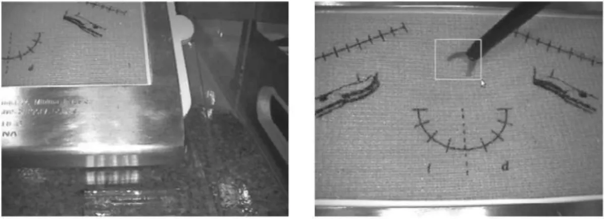
Figure 2. Negative image (left). Positive image (right), selection of the coordinates and dimensions of the object of interest (tip of the laparoscopic scissors).
are taken in an arbitrary way, and they do not contain the object to be identified. The positive images con-tain the object to be identified, and it is necessary to indicate the coordinates and dimensions of this object. In the positive images, the object must appear in a clear way, in different angles, in different back-grounds, and with different illumination settings. The positive and negative images are exclusively acquired inside of the physical simulator. To facilitate the task of selecting the coordinates and the dimensions of the objects, we develop a marking application tool which allows users to use the mouse for this purpose.
To proceed to the creation of samples, the vector which contains the sample images (standardized, grayscale and proper-sized) is created.
The following stage is the training of the classifier. We use the OpenCV graphic library to carry out a training based on AdaBoost algorithm (33). AdaBoost is a learning algorithm focused on two important aspects: Selection of a set of features which represent the object of interest, and training the classifier with a lineal combination of the best features.
When the training stage is over, we have a cascade classifier (34); this classifier points out if there are any tips of laparoscopic instruments in the input image. In
case the algorithm gives a positive answer, we will know in real time the position of the tip of the laparoscopic tool in the image.
Regarding assessment, the tests are tried on three 25-second video sequences. Conditions of these sequences vary in terms of illumination, speed of movement and position of the instrument in the field of vision.
For each sequence, we take one frame per second (25 frames in total) and an expert manually assesses the result of the automatic method for detecting the tip of the analyzed laparoscopic instrument, using laparoscopic scissors.
Results
Measuring of the effectiveness of the methods allows us to determine the validity of the system for assis-tance during the training of basic laparoscopic tasks.
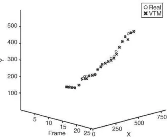
The results of the reliability of the visual tracking method (Figures 3 and 4) are presented in three different graphs with regard to horizontal (Figure 5), vertical (Figure 6) and diagonal (Figure 7) move-ments of the camera.
Figure 4. Three sequences of laparoscopic tasks which include several support visual contents, such as a video sequence (left and center images) and an outline of the task (right image). The support content remains in a stable position with respect to the movements of the camera and working area.
The result will be considered wrong if the marked point is located outside a neighbourhood of 24 pixels from its real position. Therefore, the results show that the horizontal movements of the camera (Figure 5) provide better results (84% success) for the visual tracking method than vertical (Figure 6) (72% success) and diagonal movements (Figure 7) (64% success). We consider that results with success rates near to 100% provide greater robustness to the system under different work-ing conditions. Takwork-ing into account the results obtained, they show that diagonal movements of the camera make it difficult to predict the correct position of the marked point by this visual tracking method. The percentage of success is calculated by dividing by the number of success cases and the number of analyzed frames.
The results of the tracking method of the tip of the laparoscopic scissors (Figure 8) are summarized in
the Table I. Three sequences of images with different working conditions are analyzed: For Sequence 1, the speed of movements is increased and the usual positions of the scissors in the working area are avoided. For Sequence 2, a usual laparoscopic suturing task is carried out under usual working conditions (illumination, speed of movements and position of the scissors). Finally, for Sequence 3, the usual light conditions in the laparoscopic simu-lator are decreased.
As observed, in Sequence 3, the lack of illumination can worsen the process of recognition of the object. On the other hand, for Sequence 1, the movements of the scissors are faster and are mainly located in areas of the field of vision with texture and colour similar to the tip of the laparoscopic instrument, such as the metallic edge of the working sheets holder. Conse-quently, both circumstances complicate the identifi-cation of the tip of the scissors.
500 j
400 \
> 300
200
100
O Real XVTM
** • » • • • „ • * • • • " • " A * s s * *
750
500
400 J
>- 300
200 j
100
O Real XVTM
c#"
o
***
750
Figure 5. Recorded position (x, y) of the marked point along 25 frames of a video sequence (frame) in which a horizontal movement with the camera is performed. The graph shows the position obtained by the visual tracking method (VTM) and the real position which should be obtained (real).
procedure. However, future advances should improve some practical aspects of training in laparoscopy.
Due to the surgeon's need for visual feedback to perform the surgical training tasks, we hypothesize that this training process could be improved by pro-viding video-based assistance using support visual contents (video tutorial or outline of the task) during surgical tasks and adapted ones to the level of expe-rience of trainees. Similarly, analyses of path and movements travelled by laparoscopic instruments and their speed are important sources to carry out an objective evaluation of surgical skills (35).
This paper presents the first steps in developing a new hybrid simulation system. This system has the advantages of the physical laparoscopic simulator SIMULAP-IC05® (CCMIJU, Cáceres, Spain) and combines the performance of laparoscopic tasks with the use of computer vision techniques.
Preliminary results show satisfactory percentages of success (84%, 72% and 64%) (Figure 5,6 and 7) with regard to the assessment of the visual tracking method and prove the effectiveness of the system for locali-zation of support video-based content. Likewise, the favorable results (success rates of 72%, 88% and 76%) (Table I) with respect to the detection of the tip of the laparoscopic scissors show that the system is appropriate for the analysis of the movement of this instrument and for later use with another type of laparoscopic instrument.
Figure 8. Example of execution of the automatic tracking method of the tip of the laparoscopic scissors. The sequence of six images shows the bounding boxes of the tracking method of the tip of the laparoscopic instrument. This sequence of images corresponds to a task of cutting a sheet of latex with laparoscopic scissors.
O Real XVTM
500
400
>-300 J
200 J
100
o o O x £> x
• »
°x
» x *
Frame 2 0 25 2 0° X
Figure 7. Recorded position (x, y) of the marked point along 25 frames of a video sequence (frame) in which a diagonal move-ment with the camera is performed. The graph shows the position obtained by the visual tracking method (VTM) and the real position which should be obtained (real).
Discussion
Table I. Results of the tracking of the laparoscopic scissors tip in three sequences of images.
Images (frames) No. Successes No. Mistakes % Successes Sequence 1 25 18 7 72% Sequence 2 25 22 3 88% Sequence 3 25 19 6 76%
Nowadays, there are several hybrid systems for training in laparoscopic surgery. One of these systems is the laparoscopic simulator CELTS (Center for Integration of Medicine and Innovative Technology -CIMIT, Cambridge, MA, USA) (12), which incor-porates two haptics mechanisms to analyze the movements of each of the laparoscopic instruments used. Other types of hybrid simulators are the LTS3e (RealSim Systems, Albuquerque, N M , USA) (13) and SurgicalSim-LTS® (METI, Sarasota, FL, USA) (14), which implement a series of tests using a set of sensors for measuring the efficacy during the performance of laparoscopic training tasks. The laparoscopic simulator with SimuVisio (SimuLab Corporation, Seattle, WA, United States) only focuses on improving the quality of the displayed image to convert it into a more real one. On the other hand, an ultrasound system for locating laparoscopic instruments is used by Zebris (Zebris Medical GmbH, Isny, Germany) (15). Finally, somewhat more advanced is the laparoscopic simulator ProMIS® (Haptica Inc., Boston, MA, USA) (16), which makes use of augmented reality techniques and performs the insertion of virtual contents together with real objects.
Besides sharing some of the features of the men-tioned hybrid systems, the proposed system offers the possibility of inserting support visual contents adapted to the level of experience of the trainees and allows them to determine the localization of these contents on the field of vision in a stable way. As a result, by employing visual tracking techniques for points or areas of the image, the contents are visual-ized and localvisual-ized within these areas, and suffer no displacement due to possible movements of the cam-era or objects of the working area. In this way, possible interruptions in the field of vision of the working area are avoided.
The proposed system uses a cascade classifier (34), previously trained, to automatically identify the posi-tion of the tip of the laparoscopic instrument in a standardized surgical environment with different settings.
Regarding the results of the behavior analysis of the visual tracking method, it is possible that the increase
in the number of mistakes is mainly due to the kind of movements developed by the camera. Diagonal movements of the camera are more complex than horizontal and vertical movements, and make the calculation of the motion estimation of the marked point in the image difficult.
As regards the results of the laparoscopic instru-ment tracking method, rapid moveinstru-ments of laparo-scopic scissors, lack of illumination, and the location of the instrument in areas with background texture and color similar to its tip increase the number of mistakes during the tracking process.
One way to decrease the rate of mistakes is to include a larger set of training cases for the classifier, increasing the number of cases with lack of illumina-tion and situaillumina-tions where the tip of the laparoscopic instrument is localized in areas which present diffi-culties for its identification.
The method used for tracking the laparoscopic scissors has permitted the system to work without colour restrictions or special marks on the instrument, in contrast to many reports in the scientific literature. The system is not affected by the specular reflections as other approaches implement some methods to solve this problem (17,22,26).
Total occlusion of the tip of the laparoscopic instru-ment complicates the detection process of this image-guided system. Nevertheless, some studies partially solve this problem by estimating the instrument posi-tion in successive frames.
This system allows real-time execution to assist training in laparoscopic surgery. Consequently, the system may be used together with real-time objective methods to assess a surgeon's skills, with automatic positioning methods of the laparoscopic camera or with augmented reality tools. Other studies leave open the possibility of the reduction of the speed of calculation of their methods, in order to achieve real-time execution (26,27).
Like other studies, the tracking method of this system uses the x and y values to describe the tip of the laparoscopic instrument, therefore the next step of this work is to calculate the depth of the tip of the instrument. We consider that depth value is important to complete the required information to carry out a proper objective assessment. To obtain this depth value, we propose the use of stereoscopic techniques
(36) or geometrical optical models. In the same way, the widening of possible laparoscopic instruments that can be detected by the system is something to have in mind for future works.
and the level of experience of the trainees. This support content is located in a stable position of the field of vision. For extracting the movement of the laparoscopic instrument, a robust visual-based tracking method of the instrument was imple-mented and presented. This method does not need the presence of artificial marks, or special colours to distinguish the tip of the instrument used. Similarly, this tracking method could be useful, in future works, to analyze the movements performed by trainees and to make an objective assessment of surgical skills during the surgical training process.
Acknowledgements
This study was supported in part by grant PRI07B132 from Plan Regional de Investigación, Consejería de Economía, Comercio e Innovación, Junta de Extremadura and the European Social Fund. The authors are especially grateful to Ms Elena Crespo for her technical assistance.
Declaration of interest: The authors report no
conflicts of interest. The authors alone are responsible for the content and writing of the paper.
References
1. Schietroma M, Carlei F, Cappelli S, Pescosolido A, Lygidakis NJ, Amicucci G. Effects of cholecystectomy (lapa-roscopic versus open) on PMN-elastase. Hepatogastro-enterology 2007;54:342-5.
2. Buunen M, Gholghesaei M, Veldkamp R, Meijer DW, Bonjer HJ, Bouvy N D . Stress response to laparoscopic surgery - A review. Surg Endose. 2004;18:1022-8.
3. Beldi G, Ipaktchi R, Wagner M, Gloor B, Candínas D. Laparoscopic ventral hernia repair is safe and cost effective. Surg Endose. 2006;20:92-5.
4. Cordera F, Long KH, Nagorney DM, McMurtry EK, Schleck C, Ilstrup D, et al. Open versus laparoscopic sple-nectomy for idiopathic thrombocytopenic purpura: Clinical and economic analysis. Surgery 2003;134:45-52.
5. Seitz G, Seitz EM, Kasparek MS, Konigsrainer A, Kreis ME. Long-term quality-of-life after open and laparo-scopic sigmoid colectomy. Surg Endose. 2008;18:162-7. 6. Delaney CP, Chang E, Senagore AJ, Broder M. Clinical
out-comes and resource utilization associated with laparoscopic and open colectomy using a large national database. Ann Surg. 2008;247:819-24.
7. Roumm AR, Pizzi L, Goldfarb NI, Cohn H. Minimally inva-sive: minimally reimbursed? An examination of six laparo-scopic surgical procedures. Surg Innov. 2005;12:261-87. 8. Nguyen N T , Zainabadi K, Mavandadi S, Paya M,
Stevens CM, Root J, et al. Trends in utilization and outcomes of laparoscopic versus open appendectomy. Am J Surg. 2004; 188:813-18.
9. Usón J, Sánchez FM, Pascual S, Climent S. Formación en Cirugía Laparoscópica Paso a Paso. 3rd ed. Minimally Inva-sive Surgery Centre Jesús Usón, editor. Cáceres, Spain; 2007.
10. Sánchez-Margallo FM, Asencio JM, Tejonero MC, Sánchez MA, Pérez FJ, Usón J, et al. Training design and improvement of technical skills in the transvaginal cholecys-tectomy (NOTES). Cir Esp. 2009;85:307-13.
11. Van Velthoven RF, Piechaud PT. Training centers: an essential step to developing skills in urolaparoscopy. Curr Urol Rep. 2009;10:93-6.
12. Stylopoulos N , Cotin S, Dawson S, Ottensmeyer M, Neumann P, Bardsley R, et al. CELTS: a clinically-based Computer Enhanced Laparoscopic Training System. Stud Health Technol Inform. 2003;94:336-42.
13. Soyinka A, Schollmeyer T, Meinhold-Heerlein I, Gopalghare D, Hasson H, Mettler L. Enhancing laparoscopic performance with the LTS3e: a computerized hybrid physical reality simulator. Fértil Steril. 2008;90:1988-94.
14. Mathis KL, Wiegmann DA. Construct validation of a lapa-roscopic surgical simulator. Simul Healthc. 2007;2:178-82. 15. SokollikC, Gross J, BuessG. New model for skills assessment
and training progress in minimally invasive surgery. Surg Endose. 2004;18:495-500.
16. Van Sickle KR, McClusky DA, Gallagher AG, Smith CD. Construct validation of the ProMIS simulator using a novel laparoscopic suturing task. Surg Endose. 2007;19:1227-31.
17. Wang YF, Uecker DR, Wang YL. A new framework for vision-enabled and robotically assisted minimally invasive surgery. Comput Med Imaging Graph. 1998;22:429-37. 18. McKenna S, Charif H, Frank T, editors. Towards Video
Understanding of Laparoscopic Surgery: Instrument Track-ing. Proceedings of Image and Vision Computing; 2005; New Zealand.
19. Doignon C, Graebling P, Mathelin M. Real-time segmenta-tion of surgical instruments inside the abdominal cavity using a joint hue saturation color feature. Real-Time Imaging 2005; 11:429-42.
20. Wei GQ, Arbter K, Hirzinger G, editors. Automatic tracking of laparoscopic instruments by color coding. Proceedings of the first international joint conference CRVMed-MRCAS'97; Grenoble, France; 1997.
21. Nishikawa A, Asano S, Fujita R, Yohda T, Miyazaki F, Sekimoto M, et al., editors. Robust visual tracking of multiple surgical instruments for laparoscopic surgery. Proceedings of Computer Assisted Radiology and Surgery; London, UK; 2003.
22. Zhang XL, Payandeh S. Application of visual tracking for robot-assisted laparoscopic surgery. J Robot Syst. 2002;19: 315-28.
^ 2 3 . Tonet O, Thoranaghatte RU, Megali G, Dario P. Tracking endoscopic instruments without a localizer: A shape-analysis-based approach. Comput Aided Surg. 2007;12:35-42. 24. Doignon C, Nageotte F, Maurin B, Krupa A. Pose estimation
and feature tracking for robot assisted surgery with medical imaging. In: Kragic D, Kyrki V, editors. Unifying Perspectives in Computational and Robot Vision: Springer Verlag; 2007. 25. Climent J, Mares P. Automatic instrument localization in
laparoscopic surgery. Electronic Letters on Computer Vision and Image Analysis. 2004;4:21-31.
26. Voros S, Long JA, Cinquin P. Automatic detection of instru-ments in laparoscopic images: A first step towards high-level command of robotic endoscopic holders. Int J Rob Res. 2007; 26:1173-90.
28. Sánchez-Margallo FM, Díaz-Guemes I, Pérez FJ, Sánchez MA, Loscertales B, Usón J. Preliminary results with a training program for thoracoscopic atrial fibrillation therapy. Surg Endose. 2009;23:1882-6.
29. Usón J, Sánchez FM, Díaz-Guemes I, Loscertales B, Soria F, Pascual S. Animal models in urological laparoscopic training. Actas Urol Esp. 2006;30:443-50.
30. Bradski G, Kaehler A. Learning OpenCV. Computer Vision with the OpenCV Library. Loukides M, editor. Sebastopol, CA, USA: O'Reilly; 2008.
31. Lucas B, Kanade T, editors. An Iterative Image Registration Technique with an Application to Stereo Vision. Proceedings of IJCAI; Vancouver, BC, Canada; 1981.
32. Bouguet J. Pyramidal implementation of the Lucas Kanade feature tracker. Intel Corporation, Microprocessor Research Labs. 1999.
33. Freund Y, Schapire RE. A Decision-Theoretic Generaliztion of On-Line Learning and an Application to Boosting. Journal of Computer and System Sciences. 1997;55:119-39. 34. Viola P, Jones M. Robust real-time face detection. Int J
ComputVis. 2004;57:137-54.
35. Judkins T N , Oleynikov D, Stergiou N . Objective evaluation of expert and novice performance during robotic surgical train-ing tasks. Surg Endose. 2009;23:590-7.