USING NEURAL NETWORKS TO SIMULATE THE ALZHEIMER'S DISEASE
JÚLIO L. R. MONTEIRO, University of São Paulo, Brazil, [email protected] MARCIO LOBO NETTO, University of São Paulo, Brazil, [email protected] DIEGO ANDINA, Technical University of Madrid, Spain, [email protected]
JAVIER ROPERO PELÁEZ, University of São Paulo, Brazil, [email protected]
ABSTRACT
Making use of biologically plausible artificial neural networks that implement Grossberg’s presynaptic learning rule, we simulate the possible effects of calcium dysregulation in the neuron’s activation function, to represent the most accepted model of Alzheimer's Disease: the calcium dysregulation hypothesis. According to Cudmore and Turrigiano calcium dysregulation alters the shifting dynamic of the neuron’s activation function (intrinsic plasticity). We propose that this alteration might affect the stability of synaptic weights in which memories are stored. The results of the simulation supported the theoretical hypothesis, implying that the emergence of Alzheimer's disease's symptoms such as memory loss and learning problems might be correlated to intrinsic neuronal plasticity impairment due to calcium dysregulation.
KEYWORDS: Artificial Neural Networks, Computer Simulation, Alzheimer's Disease,
Calcium Hypothesis, Neural Plasticity, MatLAB.1. INTRODUCTION
Although the Alzheimer's Disease (AD) was first documented by Alois Alzheimer[3] one hundred years ago, the understanding of its ultimate cause still represents a mystery to neurobiology. There are many theories for the disease mechanism, including the cholinergic hypothesis [4], the Tau hypothesis [5], the Amyloid hypothesis [6] and the recent Calcium dysregulation hypothesis [7]. The calcium dysregulation hypothesis (CDH) of brain aging and AD [1],[8],[9],[10], suggests that aging alters brain Ca2+ regulation, impairing neuronal function and leading to neurodegeneration. In this article, we present a formal method of simulating the effects of the dysregulation of calcium in an artificial neural network to allow a better understanding on how the AD takes place. We do not intend, however, to give an extensive account of the processes in which calcium is involved, as this can be found elsewhere [1][2][6][7] [8][9]. We merely will focus our study in the effect produced by calcium dysregulation on the dynamic of the neuron’s activation function and in the consequences of this effect on memory and learning.
In the following section (section 2) we present an introduction to the regulatory mechanisms of neuronal plasticity, to allow the understanding of the biological process involved in the CDH (section 3). In section 4 we introduce the neural network computational model that makes use of the neuronal plasticity properties explained in section 3. The experimental setup of the simulation utilizing MathWorks's MatLAB is also explained. Section 5 presents the results obtained when intrinsic neuronal plasticity is hampered, according to the calcium dysregulation hypothesis. Finally section 6 compares experimental and expected results, concluding that the results of the simulation are consistent with the memory loss and learning impairments in AD.
2. HOMEOSTATIC MECHANISMS OF NEURONAL PLASTICITY
The regulation of an internal environment so as to maintain a stable, constant condition is also called homeostasis, which was a term coined by Walter Bradford Cannon in 1932.
In this paper two relevant regulatory or homeostatic mechanisms of neuronal activity will be presented: metaplasticity and intrinsic plasticity. For a correct understanding of these two mechanisms we will start first with an introduction to synaptic plasticity.
2.1 Synaptic plasticity
The transmission of information between neurons is mediated by molecules known as neurotransmitters that act on synapses, and interact with ionic channels residing in the synaptic membrane, thereby allowing the inflow or outflow of positive and negative ions such as potassium, sodium, chlorine and calcium. Synaptic plasticity, refers to the modulation of the efficacy of information transmission between neurons, being related to the regulation of the number of ionic channels in synapses.
The first model of synaptic plasticity was postulated by Hebb and is known as the Hebb rule[11] that is stated as follows: when two neurons fire together wire together or, in other words, the synaptic strength between neurons with a correlated firing tends to increase. Mathematically the change in the synaptic strength (synaptic weight) between neurons i and j is calculated by the product of the output of neuron i and the input Ij (which corresponds to the output of neuron j) multiplied by a learning constant.
(1)
Some authors proposed revised versions of the Hebb's rule, taking into account more recent biological studies [12]. The formulation that was adopted for our simulation of synaptic plasticity, due to its biological plausibility, is the Grossberg's presynaptic learning rule [13] either in its incremental or in its probabilistic version.
The incremental version of the pre-synaptic rule is as follows:
(2)
According to Minai [14], this incremental version of the pre-synaptic rule is asymptotically equivalent to the following probabilistic version (Eq.3), where the synaptic weight between two neurons is the conditional probability of the output neuron’s firing, given that the input neuron have previously fired.
(3)
2.2 Synaptic metaplasticity
One of the important biological characteristics of the presynaptic rule is that it also models metaplasticity, which is an important homeostatic mechanism of neurons. It slows down the process of weight increment or decrement, making more difficult for the neuron to become either inactive or saturated.
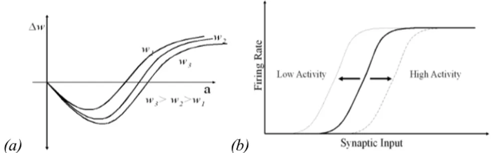
The property of metaplasticity [15] is shown in Fig.1a representing a family of curves in which each curve yields the variation of weight given the neuron’s activation. The parameter that defines what curve must be used is the value of the synaptic weight. According to this figure, for higher values of the weight the curves are more elongated to the right.
This means that in synapses with higher weights, the interval in which the variation of weight is negative is broader, thereby favoring synaptic depression. The opposite takes place in the lower weight curves.
j i
ij
O
I
w
=
ε
∆
(
i j)
ij PO I w = /
)
( i ij
j
ij I O w
w = −
Synaptic metaplasticity is a homeostatic mechanism because it regulates weight variation, down-regulating weight increment in synapses with initial high weights and up-down-regulating weight increment in synapses with initial low weights.
2.3 Intrinsic plasticity
Although synaptic metaplasticity makes it difficult for synaptic weights to become either null or saturated, it does not totally preclude either of these two extreme situations. For totally precluding the possibility of either weight annihilation or saturation, another important homeostatic property of real neurons should be taken into account: the so-called intrinsic plasticity.
Intrinsic plasticity, demonstrated in Fig. 1b, regulates the position (rightward shift) of the neuron’s activation function, usually modeled as a sigmoidal function, according to previous levels of activity:
(4)
In which O is the output probability of the neuron and “a” is the activation function given by the sum of synaptic contributions.
Intrinsic plasticity (Fig. 1b) was modeled according to the following equation that yields the position of the sigmoid in terms of the previous position shift-1 and the previous sigmoid output.
(5)
Parameter
ξ
determines the velocity of the sigmoid shifting. This equation means that the more the neuron keeps firing, the higher will be the rightward shift of the activation function, leading to a moderation of the neurons’ firing probability in the future. Conversely, if the firing probability is low, the sigmoid will move leftwards, thereby increasing the probability of the neuron’s firing in the future.We simulate the effects of the calcium dysregulation by setting
ξ
in Eq. (4) to zero, so that the shifting dynamics of the sigmoid is stopped, destroying the effect of intrinsic plasticity. In thisξ
ξ
+
+
=
− −1
1
1 t
t
t
O
shift
shift
(
a
shift
)
e
O
−
−
+
=
25
1
1
(a) (b)
way, without the compensatory mechanism of intrinsic plasticity, weights are free to increase or decrease without bound.
3. THE CALCIUM DYSREGULATION HYPOTHESIS
The calcium dysregulation hypothesis (CDH) is referred to a broad category of calcium-dependent neural processes related to the gradual impairment of cognitive abilities in AD, especially those related to memorizing and learning. Thibault and colleagues [1] enumerate some of these calcium-dependent processes like the increase in slow after-hyperpolarization (AHP), reduction of long-term potentiation (LTP), enhancement of long-term depotentiation (LTD) and impairment of short-term frequency facilitation (FF). In most of these cases, the role of Ca2+ is correlated to changes in plasticity, although the exact mechanism explaining this correlation remains uncertain. However, in the case of intrinsic plasticity, Cudmore and Turrigiano [19] showed that there is a clear relationship between intrinsic plasticity and Ca2+ influx. Reducing extracellular calcium, prevents the leftward shifting of the activation function curve. In this way, if calcium influx is altered, a dysregulation in the shifting dynamic of the activation function takes place. In the following simulations we will show that the dysregulation of the shifting dynamic of the activation function leads to the alteration of synaptic weights, where memories are encoded. Synaptic weight destabilization might be an explanation of the impairment in memory retrieval and in learning in AD.
The same equations used in this section were described in [15] and used successfully to describe and simulate other type of neural disorders[16][17][18].
4. COMPUTER SIMULATION
For allowing a better understanding of the relationship between calcium dysregulation and cognitive loss, a computational simulation was performed.
In this simulation, a small artificial neural network of 30 sparsely connected neurons was studied. The synaptic weights of each one of these neurons were modified according to the presynaptic learning rule Eq. (3). The shift of the sigmoidal activation functions of these neurons is calculated according to Eq. (5). The neuronal network is exposed repetitively to a series of 5 patterns that were presented to 5 neurons considered as “input” neurons. The whole presentation of these 5 patterns was considered an “epoch” of patterns, according to the neural networks terminology. After 150 epochs, the network weights reached either static or dynamic equilibrium in a predictable behavior. For simulating the starting point of the Alzheimer Disease, the shift variations of all neurons in the network are suddenly hampered by setting the shifting velocity
ξ
to zero. In this way, we simulate the cessation of intrinsic plasticity dynamics caused by calcium dysregulation.5. RESULTS
The simulation was written in MathWorks's MatLAB. The network is implemented as a 30x30 sparse matrix of weights, with the probability of having positive weights equal to 0.10 and a probability of having negative weights (representing inhibitory neurons) equal to 0.30. According to biology, neurons in the model are either excitatory or inhibitory, without any neuron being excitatory and inhibitory at the same time.
All neurons have the same sigmoid function given by Eq. (4) for calculating the action potential probability from incoming synaptic activation. The shift of the sigmoid is initially set to 0.5 whereas
ξ
is set to 0.01 during the first 150 epochs of the simulation.The results are a set of graphs showing the outputs of each of the 30 neurons in the network as the simulation progresses.
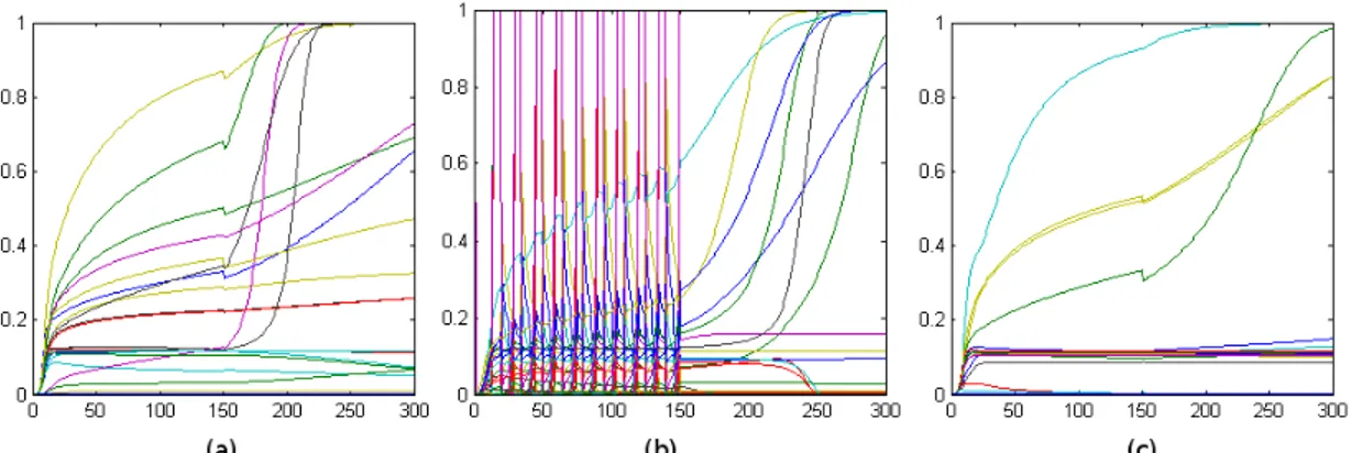
The simulation yielded three different categories of results that are exemplified by the following pair of graphs.
Each graph represents the variation of the outputs and weights in the network along the different epochs, indicating the outputs of each of the 30 neurons during the presentation of the last pattern of each epoch.
In 80% of the random simulated networks, synaptic weight lose their stability after the sigmoid stall, resulting in graphs similar to Fig. 2a.
Around 25% of the random simulated networks exhibits an oscillatory, burst-like behavior during the first phase of the simulation. This behavior is lost after the sigmoid stall in the second phase of the simulation, as is exemplified in Fig. 2b. No network exhibiting burst behaviour continues bursting after the stall of the sigmoid dynamics.
However, 20% of the randomly initialized networks continue behaving as in the initial phase, so that even after the sigmoid stall, no significant change in the network is perceived, as exemplified in Fig. 2c. This is consistent with the fact that a reduce number of patients with Azheimer disease do not exhibit memory or learning impairments.
6. CONCLUSIONS
In this paper we have shown that calcium dysregulation might be the starting point of a cascade of events leading to the lose of synaptic weight stability and, thereby, to the lose of the memories stored in synaptic weights. This might explain memory deficits in Alzheimer’s disease patients.
According to Cudmore and Turrigiano [19], the inflow of calcium is determinant in intrinsic plasticity. Intrinsic plasticity is a homeostatic mechanism that shifts the activation function to the right or to the left depending on the degree of synaptic activation of the neuron, so that the neuron is never zeroed or saturated. At the same time intrinsic plasticity leads to a stability of weights. When the compensatory mechanism of intrinsic plasticity is disrupted synaptic weights may lose their stable values.
We have tested this hypothesis by training a neural network until weights reached a stable situation. Afterwards, the shifting dynamics of the activation function is hampered. The computational model shows that, although in some cases synaptic weights remain stable, in most of the cases they enter in a situation of instability. These results are confirmed with networks of different sizes (networks of 30, 50 and 100 neurons were tested with similar results) with
different architecture of connections (in each simulation synaptic weights were initialized by a randomly generated sparse matrix in which we define the percentage of excitatory or inhibitory connections) and with different input patterns (that were also randomly initialized at the beginning of each simulation).
These results show that calcium dysregulation that is correlated to the impairment of intrinsic plasticity, leads to synaptic instability, which is consistent with cognitive deficits in AD.
7. REFERENCES
[1] Thibault O, Hadley R, Landfield PW, Elevated Postsynaptic [Ca2+]
i and L-Type Calcium Channel Activity in Aged Hippocampal Neurons: Relationship to Impaired Synaptic Plasticity, The Journal of Neuroscience, Vol.21, No.24, 2001, pp. 9744-9756.
[2] Desai NS, Homeostatic plasticity in the CNS: synaptic and intrinsic forms, Journal of Physiology, Vol.97, 2003, pp.391-402.
[3] Alzheimer A, Über eine eigenartige Erkrankung der Hirnrinde, Allgemeine Zeitschrift fur Psychiatrie Vol.64, 1907, pp.146-148.
[4] Shen ZX, Brain cholinesterases: II. The molecular and cellular basis of Alzheimer's disease,
Med. Hypotheses 63 (2), 2004, pp. 308-21.
[5] Schmitz C, Rutten BP, Pielen A, et al, Hippocampal neuron loss exceeds amyloid plaque load in a transgenic mouse model of Alzheimer's disease, American Journal of Pathology, Vol. 164, No.4, 2004, pp.1495-1502.
[6] Iijima K, Liu HP, Chiang AS, et al, Dissecting the pathological effects of human Abeta40 and Abeta42 in Drosophila: a potential model for Alzheimer's disease, Proc. National Academy of Sciences U.S.A. 101 (17), 2004, pp.6623-6628.
[7] O’Day, Danton H. and Michael A. Myre, Calmodulin Binding Domains in Alzheimer’s Disease Proteins: Extending the Calcium Hypothesis, Biochemical Biophysical Research
Communications 320, 2004, pp.1051-1054.
[8] Khachaturian ZS, Towards theories of brain aging, Handbook of studies on psychiatry and old age (Kay D, Burrows GD, eds), Amsterdam: Elsevier, 1984, pp 7–30.
[9] Landfield PW, Pitler TA, Prolonged Ca 2+ dependent afterhyperpolarizations in hippocampal neurons of aged rats. Science 226, 1984, pp. 1089–1092.
[10] Michaelis ML, Johe K, Kitos TE, Age-dependent alterations in synaptic membrane systems for Ca2+ regulation. Mech Aging Dev 25, 1984, pp. 215–225.
[11] Hebb DO, The organization of behavior. New York: Wiley, 1949.
[12] Brown TH, Kairiss EW, Keenan CL, Hebbian Synapses: Biophysical Mechanisms and Algorithms, Annual Review in Neurosciences, Vol. 13, 1990, pp. 475-511.
[13] Grossberg S, Adaptive pattern classification and universal recoding: I. Parallel development and coding of neural feature detectors. Biological Cybernetics, Vol. 23, 1976, pp.121-134. [14] Minai AA, Levy WB, The Dynamics of Sparse Random Networks, Biological Cybernetics,
Vol. 70(2), 1993, pp.177-187.
[15] Pelaez JR, Simoes MG, A Computational Model of Synaptic Metaplasticity. Proceedings of the International Joint Conference of Neural Networks 1999, Vol.1, 1999, pp. 6-11. [16] Nascimento AN, Andina D, Peláez FJR, Towards a Neural-Networks Based Therapy for
Limbs Spasticity, Bio-inspired Modeling of Cognitive Tasks, Lecture Notes in Computer Science, Vol. 4527/2007, 2007, pp. 124-131.
[17] Peláez FJR, Towards a neural network based therapy for hallucinatory disorders. Neural Networks, 2000 Special Issue 13(2000), 2000, pp. 1047-1061.
[18] Granado LC, Ranvaud R, Peláez FJR, A Spiderless Arachnophobia Therapy: Comparison between Placebo and Treatment Groups and Six-Month Follow-Up Study, Neural Plasticity, Vol. 2007, Article ID 10241, 2007, 11p, doi:10.1155/2007/10241.