897 J. Gen. Physiol. © The Rockefeller University Press • 0022-1295/99/06/897/11 $2.00 Volume 113 June 1999 897–907
Kv
b
1.2 Subunit Coexpression in HEK293 Cells Confers O
2Sensitivity
to Kv4.2 but not to
Shaker
Channels
María Teresa Pérez-García, José Ramón López-López, and Constancio González
From the *Departamento de Bioquímica y Biología Molecular y Fisiología, Facultad de Medicina, Universidad de Valladolid, 47005 Valladolid, Spain
a b s t r a c t Voltage-gated K1 (KV) channels are protein complexes composed of ion-conducting integral
mem-brane a subunits and cytoplasmic modulatory b subunits. The differential expression and association of a and b subunits seems to contribute significantly to the complexity and heterogeneity of KV channels in excitable cells, and their functional expression in heterologous systems provides a tool to study their regulation at a molecular level. Here, we have studied the effects of Kvb1.2 coexpression on the properties of Shaker and Kv4.2 KV channel a subunits, which encode rapidly inactivating A-type K1 currents, in transfected HEK293 cells. We found that Kvb1.2 functionally associates with these two a subunits, as well as with the endogenous KV channels of HEK293 cells, to modulate different properties of the heteromultimers. Kvb1.2 accelerates the rate of inactivation of the Shaker currents, as previously described, increases significantly the amplitude of the endogenous currents, and confers sensitivity to redox modulation and hypoxia to Kv4.2 channels. Upon association with Kvb1.2, Kv4.2 can be modi-fied by DTT (1,4 dithiothreitol) and DTDP (2,29-dithiodipyridine), which also modulate the low pO2 response of the Kv4.21b channels. However, the physiological reducing agent GSH (reduced glutathione) did not mimic the effects of DTT. Finally, hypoxic inhibition of Kv4.21b currents can be reverted by 70% in the presence of carbon monoxide and remains in cell-free patches, suggesting the presence of a hemoproteic O2 sensor in HEK293 cells and a membrane-delimited mechanism at the origin of hypoxic responses. We conclude that b subunits can mod-ulate different properties upon association with different KV channel subfamilies; of potential relevance to under-standing the molecular basis of low pO2 sensitivity in native tissues is the here described acquisition of the ability of Kv4.21b channels to respond to hypoxia.
k e y w o r d s : potassium channels • b subunit • hypoxia
i n t r o d u c t i o n
Voltage-gated K1 (KV)1 channels help establish the rest-ing membrane potential and modulate the frequency and duration of the action potentials in excitable cells. Molecular biology techniques have identified several mammalian genes encoding the pore-forming a sub-units of KV channels that can give rise to delayed recti-fier or A-type currents upon expression in heterolo-gous systems (Chandy and Gutman, 1995). The func-tional and structural diversity of the KV channels’ a subunits is further increased by their capacity to form functional heterotetrameric structures and to associate with modulatory b subunits (for review see Pongs, 1995; Jan and Jan, 1997). For example, association of b subunits with some members of the Shaker subfamily
re-sults in ab heteromultimers with inactivation kinetics more rapid than those of the corresponding a homo-multimers (Rettig et al., 1994; Chouinard et al., 1995; Heinemann et al., 1995; Majumder et al., 1995; McCor-mack et al., 1995; Morales et al., 1995), and, even fur-ther, some of these b subunits can convert a delayed rectifier into a rapidly inactivating channel (Rettig et al., 1994; England et al., 1995; Majumder et al., 1995; Mo-rales et al., 1995; Heinemann et al., 1996).
In some tissues, K1 currents exhibit specific proper-ties, such as regulation by oxygen levels (Lopez-Barneo et al., 1988; Post et al., 1992; Youngson et al., 1993). It has been hypothesized that O2 sensitivity of K1 currents could be intrinsic to the channels themselves (Ruppers-berg et al., 1991; Duprat et al., 1995; Weir and Archer, 1995) or, alternatively, that a membrane-bound O2 sen-sor or a regulatory subunit of the K1 channels confers the observed sensitivity (Gonzalez et al., 1992; Lopez-Barneo, 1994; Patel et al., 1997).
In the present work, we have used an heterologous expression system to study the association of the auxil-iary subunit Kvb1.2 (formerly Kvb3) with some cloned KV channels and its possible contribution to the hy-poxic sensitivity of the heteromultimers. The KV chan-nels used (Shaker B and Kv4.2) express rapidly inactivat-Portions of this work have previously appeared in abstract form
(Pérez-García, M.T., J.R. López-López, and C. Gonzalez. 1998. J. Phys-iol. 509P:36P).
Address correspondence to Constancio González, Departamento de Fisiología, Facultad de Medicina, c/ Ramón y Cajal s/n, 47005 Valladolid, Spain. Fax: 34 83 423588; E-mail: [email protected]
1
ing currents comparable to the oxygen-sensitive K1 cur-rents described in some preparations (Lopez-Barneo et al., 1988; Gonzalez et al., 1992). We found subfamily-specific functional interactions between Kvb1.2 and the different KV channels studied, so that Kvb1.2 coexpres-sion is able to regulate the amplitude of the endoge-nous HEK293 KV currents, the rate of inactivation of the Shaker currents, and the redox and oxygen sensitiv-ity of the Kv4.2 currents. The hypoxic response of the Kv4.21Kvb1.2 heteromultimers was unaffected by ap-plication of reduced glutathione (GSH) in the pipette solution or in the bath, but was prevented by treatment with DTT (1,4 dithiothreitol) and restored with DTDP (2,29-dithiodipyridine), suggesting that reduction of some, but not all, of the residues susceptible to redox modulation can disrupt the mechanism underlying the low pO2 regulation of these channels. Hypoxic inhibi-tion was reverted by carbon monoxide (suggesting the presence of an hemoproteic O2 sensor in HEK cells) and remains in excised membrane patches, indicating that the mechanism of low pO2 inhibition is restricted to the plasma membrane.
m a t e r i a l s a n d m e t h o d s
HEK293 Cell Maintenance and Transfections
HEK293 cells were maintained in DMEM supplemented with 10% fetal calf serum (GIBCO BRL), 100 U/ml penicillin, 100
mg/ml streptomycin, and 2 mM l-glutamine. Cells were grown as a monolayer and plated on squared coverslips (24 3 24 mm) placed in the bottom of 35-mm Petri dishes at a density of 2–4 3 105 cells/dish the day before transfection. Transient transfections
were performed using the calcium-phosphate method (Wigler et al., 1978) with 1 mg of plasmid DNA encoding the drosophila Shaker B (H4) K1 channel a subunit (into pRcRSV; Invitrogen Corp.), or the Kv4.2 K1 channel a subunit (into E42c) alone or in combination with 2 mg of plasmid DNA encoding the Kvb1.2 subunit into pREP4. In a group of experiments, the cells were only transfected with 2 mg of Kvb1.2 subunit. In all cases, 0.2 mg of green fluorescent protein (GFP) in a CMV-promoter expres-sion plasmid (GFPPRK5), was included to permit transfection ef-ficiency estimates (10–40%) and to identify cells for voltage-clamp analysis (Marshall et al., 1995). Voltage-voltage-clamp recordings revealed typical inactivating currents in 100% of the cells ex-pressing GFP. A group of control cells was obtained by analyzing the currents present in cells transfected with GFP alone or in un-transfected cells. All plasmids used in this study were generously provided by Drs. E. Marban and G.F. Tomaselli (John Hopkins University, Baltimore, MD).
Electrophysiological Recordings
K1 currents were studied using either the whole-cell or the out-side-out configuration of the patch-clamp technique. The hold-ing potential was 260 or 280 mV, respectively. Isolated HEK cells were studied 1–3 d after transfection. The coverslips with the at-tached cells were transferred to a small recording chamber (0.2 ml) placed in the stage of an inverted microscope and perfused by gravity with (mM): 141 NaCl, 4.7 KCl, 1.2 MgCl2, 1.8 CaCl2, 10
glucose, 10 HEPES, pH 7.4 with NaOH. The bath solution was connected to ground via a 3 M KCl agar bridge and a Ag-AgCl
electrode. Patch pipettes were double pulled (PP-83; Narishige Co.) and heat polished (MF-83; Narishige Co.) to resistances ranging from 1.5–3 MV for whole-cell experiments to 10–15 MV for cell-free recordings when filled with a internal solution con-taining (mM): 125 KCl, 4 MgCl2, 10 HEPES, 10 EGTA, 5 MgATP,
pH 7.2 with KOH. Hypoxia was achieved by bubbling the reser-voir that fed the perfusion chamber with 100% N2. The final pO2
level in the perfusion chamber was below 10 mmHg. The time course of the fall in the pO2 was complete within 1 min of
solu-tion exchange. In selected experiments, the control solusolu-tions were also bubbled with air to exclude potential artifactual effects due to the bubbling of the solutions. Whole-cell currents were re-corded using an Axopatch 200 patch-clamp amplifier, sampled at 10 and filtered at 2 kHz (23 dB, four-pole Bessel filter). The se-ries resistance (ranging from 4 to 10 MV) was routinely compen-sated by 60–80%. Data were leak subtracted on line by a P/4 pro-tocol. K1 currents from macropatches in the outside-out configu-ration were registered several minutes after excision and were taken as the difference between the current recorded in a 50-ms depolarizing pulse to 140 mV from a holding potential of 280 mV and the average current obtained applying four pulses to
140 mV after inactivating the K1 channels with 200-ms prepulses to the same potential. To facilitate the subtraction of capacitative transients, the potential was held at 280 mV during 1 ms be-tween prepulse and pulse. Currents were sampled at 5 and fil-tered at 1 kHz. Records were digitized with a Digidata-1200 A/D converter (Axon Instruments), and stored on disk using PCLAMP version 6.02 software. All the experiments were done at room temperature (20–228C).
Data Analysis
Analysis of the data was performed with the CLAMPFIT subrou-tines of the PCLAMP software and ORIGIN 4.0 software (Micro-cal Software, Inc.). Pooled data are expressed as mean 6 SEM. Statistical comparisons between groups of data were carried out with the two-tailed Student’s t test for paired or unpaired data, and values of P , 0.05 were considered statistically significant. The analysis of the differences between two groups of data when comparing more than one variable was done with a fully factorial analysis of variance [(M)ANOVA] using commercial software (SYSTAT; Systat Inc.).
Materials
DTT, DTDP, and GSH were obtained from Sigma Chemical Co. DTT and GSH were prepared fresh and dissolved in the bath or in the pipette solution, and DTDP was first dissolved in ethanol to a concentration of 500 mM, and then diluted in bath solution to a final concentration of 100mM.
r e s u l t s
Effects of Kvb1.2 on the Amplitude of Shaker, Kv4.2, and HEK293 Endogenous KV Currents
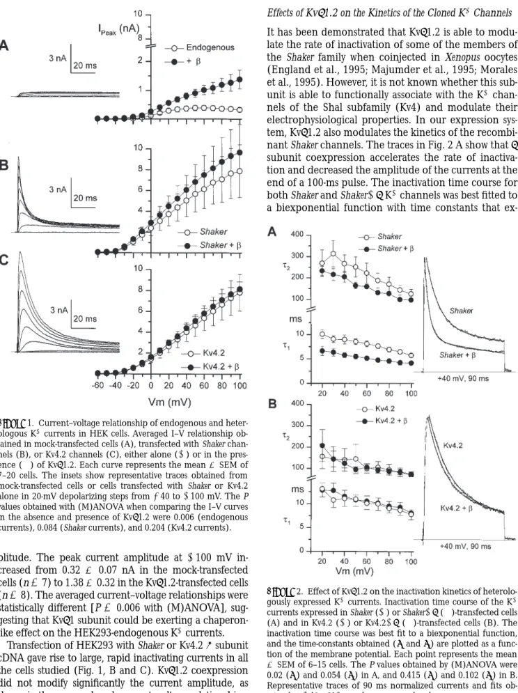
Untransfected or mock-transfected (GFP alone) HEK293 cells show KV currents of variable size, ranging from 100 to 600 pA at 160 mV. As shown in Fig. 1 A, over the length of a 100-ms pulse, this endogenous current exhib-ited almost no inactivation. Outward current was ob-served at potentials above 220 mV, and peak current typ-ically showed a plateau at potentials more positive than
am-plitude. The peak current amplitude at 1100 mV in-creased from 0.32 6 0.07 nA in the mock-transfected cells (n 5 7) to 1.38 6 0.32 in the Kvb1.2-transfected cells (n 5 8). The averaged current–voltage relationships were statistically different [P 5 0.006 with (M)ANOVA], sug-gesting that Kvb1 subunit could be exerting a chaperon-like effect on the HEK293-endogenous K1 currents.
Transfection of HEK293 with Shaker or Kv4.2 a subunit cDNA gave rise to large, rapid inactivating currents in all the cells studied (Fig. 1, B and C). Kvb1.2 coexpression did not modify significantly the current amplitude, as shown in the averaged peak current–voltage relationships.
Effects of Kvb1.2 on the Kinetics of the Cloned K1 Channels
It has been demonstrated that Kvb1.2 is able to modu-late the rate of inactivation of some of the members of the Shaker family when coinjected in Xenopus oocytes (England et al., 1995; Majumder et al., 1995; Morales et al., 1995). However, it is not known whether this sub-unit is able to functionally associate with the K1 chan-nels of the Shal subfamily (Kv4) and modulate their electrophysiological properties. In our expression sys-tem, Kvb1.2 also modulates the kinetics of the recombi-nant Shaker channels. The traces in Fig. 2 A show that b subunit coexpression accelerates the rate of inactiva-tion and decreased the amplitude of the currents at the end of a 100-ms pulse. The inactivation time course for both Shaker and Shaker1b K1 channels was best fitted to a biexponential function with time constants that
ex-Figure 1. Current–voltage relationship of endogenous and heter-ologous K1 currents in HEK cells. Averaged I–V relationship ob-tained in mock-transfected cells (A), transfected with Shaker chan-nels (B), or Kv4.2 chanchan-nels (C), either alone (s) or in the pres-ence (d) of Kvb1.2. Each curve represents the mean 6 SEM of 7–20 cells. The insets show representative traces obtained from mock-transfected cells or cells transfected with Shaker or Kv4.2 alone in 20-mV depolarizing steps from 240 to 1100 mV. The P values obtained with (M)ANOVA when comparing the I–V curves in the absence and presence of Kvb1.2 were 0.006 (endogenous currents), 0.084 (Shaker currents), and 0.204 (Kv4.2 currents).
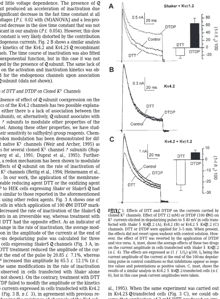
Figure 2. Effect of Kvb1.2 on the inactivation kinetics of heterolo-gously expressed K1 currents. Inactivation time course of the K1 currents expressed in Shaker (s) or Shaker1b (d)-transfected cells (A) and in Kv4.2 (s) or Kv4.21b (d)-transfected cells (B). The inactivation time course was best fit to a biexponential function, and the time-constants obtained (t1 and t2) are plotted as a
func-tion of the membrane potential. Each point represents the mean
6 SEM of 6–15 cells. The P values obtained by (M)ANOVA were 0.02 (t1) and 0.054 (t2) in A, and 0.415 (t1) and 0.102 (t2) in B.
1
hibited little voltage dependence. The presence of b subunit produced an acceleration of inactivation due to a significant decrease in the fast time constant at all the voltages [P 5 0.02 with (M)ANOVA] and a less-pro-nounced decrease in the slow time constant that was not significant in our analysis (P 5 0.054). However, this slow time constant is very likely distorted by the contribution of endogenous currents. Fig. 2 B shows a similar analysis of the kinetics of the Kv4.2 and Kv4.21b recombinant channels. The time course of inactivation was also fitted to a biexponential function, but in this case it was not changed by the presence of b subunit. The same lack of effect on the activation and inactivation kinetics was ob-served for the endogenous channels upon association with b subunit (data not shown).
Effects of DTT and DTDP on Cloned K1 Channels
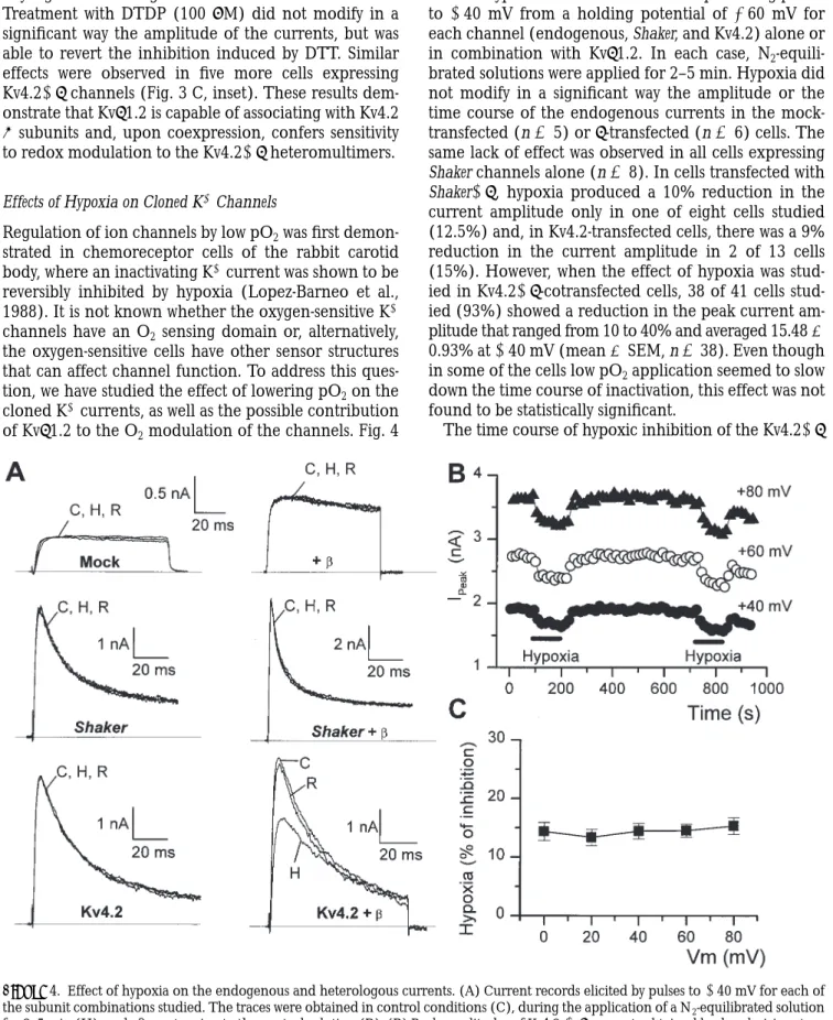
The absence of effect of b subunit coexpression on the kinetics of the Kv4.2 channels has two possible explana-tions: either there is a lack of association between the two subunits, or, alternatively, b subunit associates with Kv4.2 a subunits to modulate other properties of the channel. Among these other properties, we have stud-ied their sensitivity to sulfhydryl group reagents. Chem-ical redox modulation has been demonstrated for dif-ferent native K1 channels (Weir and Archer, 1995) as well as for several cloned K1 channel a subunits (Rup-persberg et al., 1991; Duprat et al., 1995). Further-more, a redox mechanism has been shown to modulate the effects of b subunit on the rate of inactivation of Kv1.4 K1 channels (Rettig et al., 1994; Heinemann et al., 1995). In our work, the application of the membrane-permeable reducing agent DTT or the oxidizing agent DTDP to HEK cells expressing Shaker or Shaker1b had effects similar to those reported in the aforementioned works using other redox agents. Fig. 3 A shows one of four cells in which application of 100 mM DTDP mark-edly decreased the rate of inactivation of the Shaker1b currents in an irreversible way, whereas treatment with 2 mM DTT had the opposite effect. As an indicator of the change in the rate of inactivation, the average mod-ification in the amplitude of the currents at the end of a 100-ms depolarizing pulse was calculated for three more cells expressing Shaker1b channels (Fig. 3 A, in-set). DTT treatment reduced the amplitude of the cur-rent at the end of the pulse by 20.85 6 7.1%, whereas DTDP increased this amplitude by 65.5 6 12.1% (n 5 4). The same modifications in the rate of inactivation were observed in cells transfected with Shaker alone (data not shown). On the contrary, treatment with DTT or DTDP failed to modify the amplitude or the kinetics of the currents expressed in cells transfected with Kv4.2 alone (Fig. 3 B, n 5 3), in agreement with previous re-sults showing the resistance of channels of the Kv4 sub-family to treatment with other redox agents (Duprat et
al., 1995). When the same experiment was carried out in Kv4.21b-transfected cells (Fig. 3 C), we could ob-serve that application of 2 mM DTT produced an irre-versible reduction of the current amplitude without Figure 3. Effects of DTT and DTDP on the currents carried by cloned K1 channels. Effect of DTT (2 mM) or DTDP (100 mM) on K1 currents elicited in depolarizing pulses to 140 mV in cells trans-fected with Shaker 1 Kvb1.2 (A), Kv4.2 (B) or Kv4.2 1 Kvb1.2 (C) channels. DTT or DTDP were applied for 3–5 min. When present, the effects did not revert upon washout with control solution. How-ever, the effect of DTT was reverted by the application of DTDP and vice versa. A, inset, shows the average effects of these two drugs on the current amplitude in cells transfected with Shaker 1 Kvb1.2 (n 5 4). The effects are expressed as (1 2 I/I0) ? 100, I0 being the
any significant change in the kinetics of the currents. Treatment with DTDP (100 mM) did not modify in a significant way the amplitude of the currents, but was able to revert the inhibition induced by DTT. Similar effects were observed in five more cells expressing Kv4.21b channels (Fig. 3 C, inset). These results dem-onstrate that Kvb1.2 is capable of associating with Kv4.2
a subunits and, upon coexpression, confers sensitivity to redox modulation to the Kv4.21b heteromultimers.
Effects of Hypoxia on Cloned K1 Channels
Regulation of ion channels by low pO2 was first demon-strated in chemoreceptor cells of the rabbit carotid body, where an inactivating K1 current was shown to be reversibly inhibited by hypoxia (Lopez-Barneo et al., 1988). It is not known whether the oxygen-sensitive K1 channels have an O2 sensing domain or, alternatively, the oxygen-sensitive cells have other sensor structures that can affect channel function. To address this ques-tion, we have studied the effect of lowering pO2 on the cloned K1 currents, as well as the possible contribution of Kvb1.2 to the O2 modulation of the channels. Fig. 4
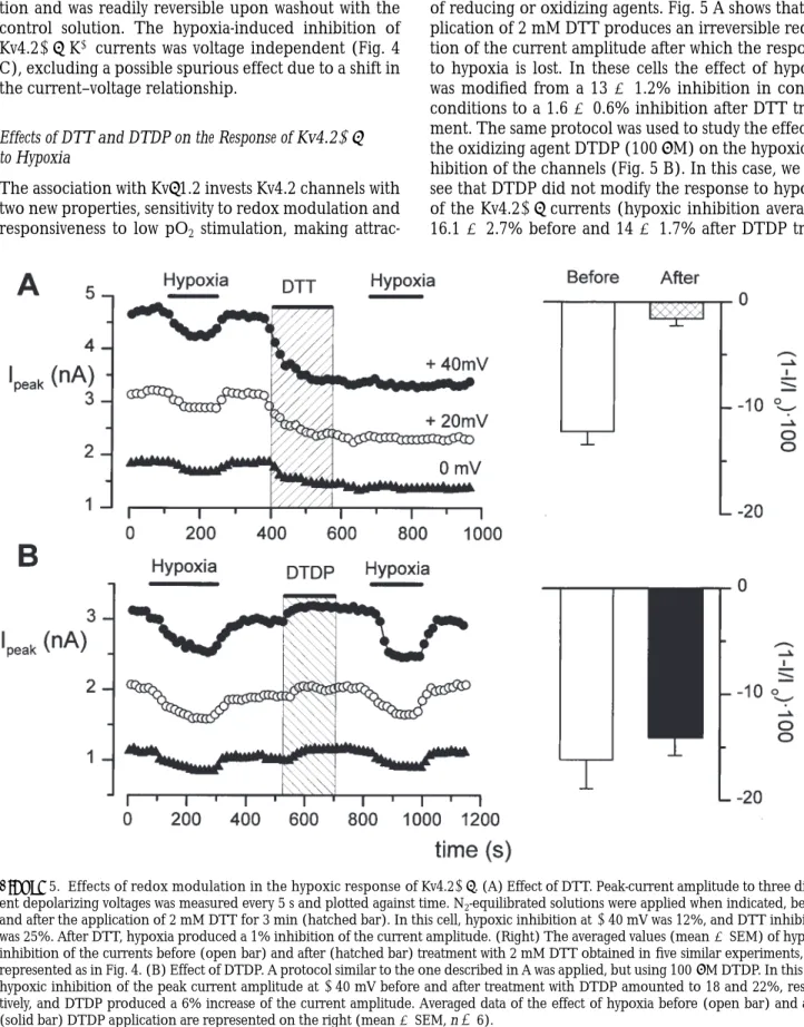
A shows typical records obtained in depolarizing pulses to 140 mV from a holding potential of 260 mV for each channel (endogenous, Shaker, and Kv4.2) alone or in combination with Kvb1.2. In each case, N2 -equili-brated solutions were applied for 2–5 min. Hypoxia did not modify in a significant way the amplitude or the time course of the endogenous currents in the mock-transfected (n 5 5) or b-transfected (n 5 6) cells. The same lack of effect was observed in all cells expressing Shaker channels alone (n 5 8). In cells transfected with Shaker1b, hypoxia produced a 10% reduction in the current amplitude only in one of eight cells studied (12.5%) and, in Kv4.2-transfected cells, there was a 9% reduction in the current amplitude in 2 of 13 cells (15%). However, when the effect of hypoxia was stud-ied in Kv4.21b-cotransfected cells, 38 of 41 cells stud-ied (93%) showed a reduction in the peak current am-plitude that ranged from 10 to 40% and averaged 15.48 6 0.93% at 140 mV (mean 6 SEM, n 5 38). Even though in some of the cells low pO2 application seemed to slow down the time course of inactivation, this effect was not found to be statistically significant.
The time course of hypoxic inhibition of the Kv4.21b
Figure 4. Effect of hypoxia on the endogenous and heterologous currents. (A) Current records elicited by pulses to 140 mV for each of the subunit combinations studied. The traces were obtained in control conditions (C), during the application of a N2-equilibrated solution
for 3–5 min (H), and after returning to the control solution (R). (B) Peak amplitudes of Kv4.2 1b currents obtained by depolarizing steps to three potentials applied every 5 s plotted against time. During the indicated periods, the cell was exposed to a N2-equilibrated solution.
1
cotransfected cells is shown in Fig. 4 B, where the peak current amplitude at three different voltages is repre-sented against time. The effect of hypoxia was fully achieved within 1 min after the exchange of the solu-tion and was readily reversible upon washout with the control solution. The hypoxia-induced inhibition of Kv4.21b K1 currents was voltage independent (Fig. 4 C), excluding a possible spurious effect due to a shift in the current–voltage relationship.
Effects of DTT and DTDP on the Response of Kv4.21b to Hypoxia
The association with Kvb1.2 invests Kv4.2 channels with two new properties, sensitivity to redox modulation and responsiveness to low pO2 stimulation, making
attrac-tive the hypothesis that the redox status of the Kv4.21b channels could be involved in the effect of hypoxia. This hypothesis was explored by studying the effect of hypoxic solutions on these channels after application of reducing or oxidizing agents. Fig. 5 A shows that ap-plication of 2 mM DTT produces an irreversible reduc-tion of the current amplitude after which the response to hypoxia is lost. In these cells the effect of hypoxia was modified from a 13 6 1.2% inhibition in control conditions to a 1.6 6 0.6% inhibition after DTT treat-ment. The same protocol was used to study the effect of the oxidizing agent DTDP (100 mM) on the hypoxic in-hibition of the channels (Fig. 5 B). In this case, we can see that DTDP did not modify the response to hypoxia of the Kv4.21b currents (hypoxic inhibition averaged 16.1 6 2.7% before and 14 6 1.7% after DTDP
treat-Figure 5. Effects of redox modulation in the hypoxic response of Kv4.21b. (A) Effect of DTT. Peak-current amplitude to three differ-ent depolarizing voltages was measured every 5 s and plotted against time. N2-equilibrated solutions were applied when indicated, before
ment, n 5 6), although it was able to recover the hy-poxic response of cells previously exposed to DTT (data not shown). These results indicate that the resi-dues of the Kv4.21b heteromultimers sensitive to DTT and DTDP treatment are involved in the response of the channel to acute hypoxia.
Effects of GSH on Kv4.21b Currents
Although the previous data suggest that the redox state could be one of the mechanisms involved in the hy-poxic modulation of Kv4.21b channels, we have ex-plored whether physiological redox modulators such as GSH have effects comparable to DTT on the currents and on their response to hypoxia. It is noteworthy that these two reducing agents modify in a similar way the time course of inactivation of cloned Shaker K1 chan-nels (Ruppersberg et al., 1991). Additionally, due to its lower membrane permeability, GSH could be helpful in indicating whether the effects are mainly due to modification of an intracellular or an extracellular site. We performed a series of experiments to explore the effect of pipette application of 5 mM GSH on the
am-plitude and the kinetics of Kv4.21b currents and on their response to low pO2 exposure. Parallel experi-ments in the same cultures with our normal pipette so-lution were used as controls. We found that inclusion of 5 mM GSH in the pipette solution did not change the amplitude of the currents (the peak current at 140 mV averaged 2.57 6 0.46 nA in control versus 2.37 6 0.49 nA in GSH-treated cells, n 5 9) nor the inactivation time course (the two time constants were 11.6 6 2.9 and 120 6 6 ms in control cells versus 15 6 4 and 135 6 10.2 ms in the presence of GSH, n 5 9). When the cells were bathed in a N2-equilibrated solution, all cells in the two groups showed a reduction of the peak current amplitude, averaging 16.25 6 1.6% in control and 16.86 6 1.83% in GSH-treated cells.
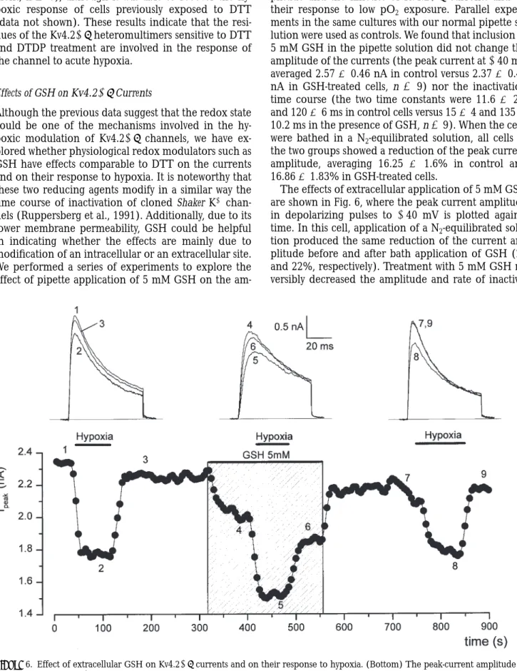
The effects of extracellular application of 5 mM GSH are shown in Fig. 6, where the peak current amplitude in depolarizing pulses to 140 mV is plotted against time. In this cell, application of a N2-equilibrated solu-tion produced the same reducsolu-tion of the current am-plitude before and after bath application of GSH (23 and 22%, respectively). Treatment with 5 mM GSH re-versibly decreased the amplitude and rate of
inactiva-Figure 6. Effect of extracellular GSH on Kv4.21b currents and on their response to hypoxia. (Bottom) The peak-current amplitude in depolarizing pulses to 140 mV was measured every 5 s and plotted against time. N2-equilibrated solutions were applied as indicated with
1
tion of Kv4.21b currents, but did not modify the mag-nitude of the effect of hypoxia. Similar results were ob-served in four more cells, in which the reduction produced by 5 mM GSH was somehow variable, averag-ing 33 6 7%.
Molecular Mechanism of Low pO2 Inhibition of Kv4.21b Currents
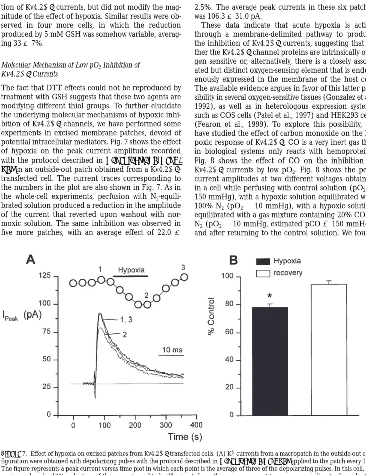
The fact that DTT effects could not be reproduced by treatment with GSH suggests that these two agents are modifying different thiol groups. To further elucidate the underlying molecular mechanisms of hypoxic inhi-bition of Kv4.21b channels, we have performed some experiments in excised membrane patches, devoid of potential intracellular mediators. Fig. 7 shows the effect of hypoxia on the peak current amplitude recorded with the protocol described in materials and meth-ods in an outside-out patch obtained from a Kv4.21b -transfected cell. The current traces corresponding to the numbers in the plot are also shown in Fig. 7. As in the whole-cell experiments, perfusion with N2 -equili-brated solution produced a reduction in the amplitude of the current that reverted upon washout with nor-moxic solution. The same inhibition was observed in five more patches, with an average effect of 22.0 6
2.5%. The average peak currents in these six patches was 106.3 6 31.0 pA.
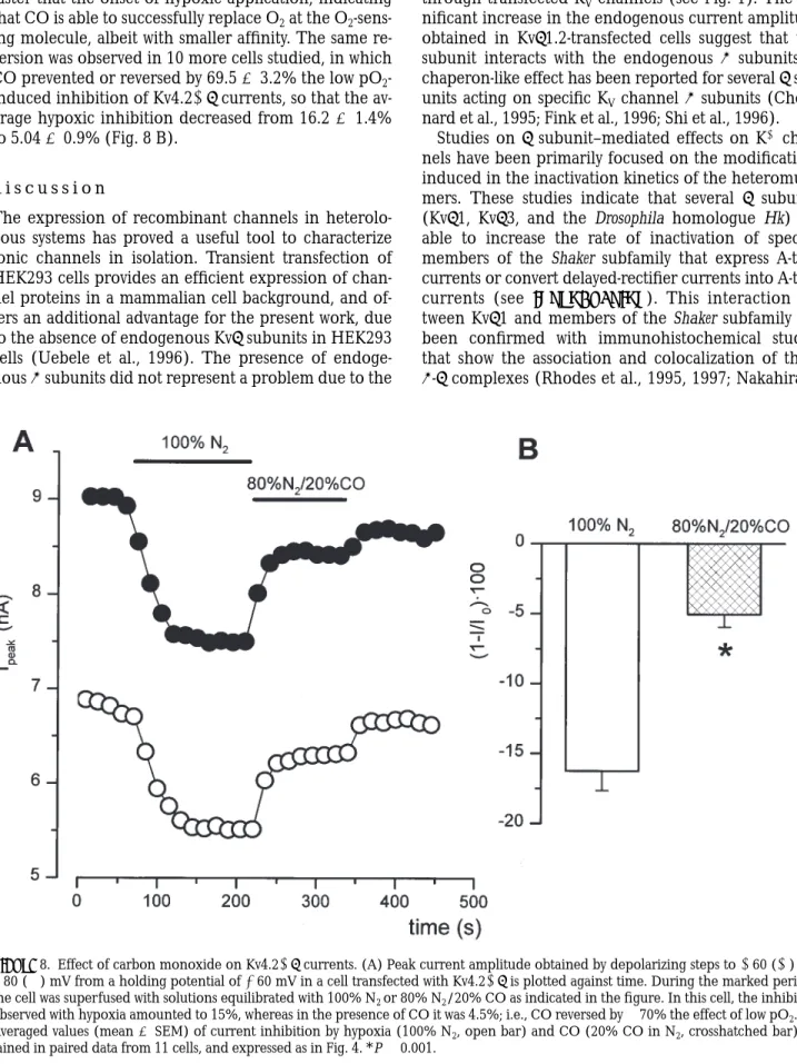
These data indicate that acute hypoxia is acting through a membrane-delimited pathway to produce the inhibition of Kv4.21b currents, suggesting that ei-ther the Kv4.21b channel proteins are intrinsically oxy-gen sensitive or, alternatively, there is a closely associ-ated but distinct oxygen-sensing element that is endog-enously expressed in the membrane of the host cell. The available evidence argues in favor of this latter pos-sibility in several oxygen-sensitive tissues (Gonzalez et al., 1992), as well as in heterologous expression systems such as COS cells (Patel et al., 1997) and HEK293 cells (Fearon et al., 1999). To explore this possibility, we have studied the effect of carbon monoxide on the hy-poxic response of Kv4.21b. CO is a very inert gas that in biological systems only reacts with hemoproteins. Fig. 8 shows the effect of CO on the inhibition of Kv4.21b currents by low pO2. Fig. 8 shows the peak current amplitudes at two different voltages obtained in a cell while perfusing with control solution (pO2 5 150 mmHg), with a hypoxic solution equilibrated with 100% N2 (pO2 , 10 mmHg), with a hypoxic solution equilibrated with a gas mixture containing 20% CO in N2 (pO2 , 10 mmHg, estimated pCO 5 150 mmHg), and after returning to the control solution. We found
that CO is reverting in a significant extent the inhibi-tion observed with hypoxia with a time course even faster that the onset of hypoxic application, indicating that CO is able to successfully replace O2 at the O2 -sens-ing molecule, albeit with smaller affinity. The same re-version was observed in 10 more cells studied, in which CO prevented or reversed by 69.5 6 3.2% the low pO2 -induced inhibition of Kv4.21b currents, so that the av-erage hypoxic inhibition decreased from 16.2 6 1.4% to 5.04 6 0.9% (Fig. 8 B).
d i s c u s s i o n
The expression of recombinant channels in heterolo-gous systems has proved a useful tool to characterize ionic channels in isolation. Transient transfection of HEK293 cells provides an efficient expression of chan-nel proteins in a mammalian cell background, and of-fers an additional advantage for the present work, due to the absence of endogenous Kvb subunits in HEK293 cells (Uebele et al., 1996). The presence of endoge-nous a subunits did not represent a problem due to the
clear differences, both in amplitude and kinetics, be-tween these endogenous currents and the currents through transfected KV channels (see Fig. 1). The sig-nificant increase in the endogenous current amplitude obtained in Kvb1.2-transfected cells suggest that this subunit interacts with the endogenous a subunits. A chaperon-like effect has been reported for several b sub-units acting on specific KV channel a subunits (Choui-nard et al., 1995; Fink et al., 1996; Shi et al., 1996).
Studies on b subunit–mediated effects on K1 chan-nels have been primarily focused on the modifications induced in the inactivation kinetics of the heteromulti-mers. These studies indicate that several b subunits (Kvb1, Kvb3, and the Drosophila homologue Hk) are able to increase the rate of inactivation of specific members of the Shaker subfamily that express A-type currents or convert delayed-rectifier currents into A-type currents (see introduction). This interaction be-tween Kvb1 and members of the Shaker subfamily has been confirmed with immunohistochemical studies that show the association and colocalization of these
a-b complexes (Rhodes et al., 1995, 1997; Nakahira et
Figure 8. Effect of carbon monoxide on Kv4.21b currents. (A) Peak current amplitude obtained by depolarizing steps to 160 (s) and
180 (d) mV from a holding potential of 260 mV in a cell transfected with Kv4.21b is plotted against time. During the marked periods, the cell was superfused with solutions equilibrated with 100% N2 or 80% N2/20% CO as indicated in the figure. In this cell, the inhibition
observed with hypoxia amounted to 15%, whereas in the presence of CO it was 4.5%; i.e., CO reversed by z70% the effect of low pO2. (B)
Averaged values (mean 6 SEM) of current inhibition by hypoxia (100% N2, open bar) and CO (20% CO in N2, crosshatched bar)
1
al., 1996; Yu et al., 1996). It has also been reported the existence of selective interaction between both Kvb1 and Kvb2 and the mammalian Shal homologue Kv4.2 (Nakahira et al., 1996), but functional analysis has failed to reveal a change in the inactivation properties of the members of the Shal subfamily when coexpressed with Kvb1 subunit (Heinemann et al., 1996; Yu et al., 1996). In agreement with these reports, we found that coex-pression with Kvb1.2 produces a significant change in the rate of inactivation of the Shaker channels due to a decrease in the fast time constant. Besides, the fact that the acceleration of the channel inactivation by Kvb1.2 does not reduce the peak current amplitude (Fig. 1 B) suggests that Kvb1.2 is also increasing the surface ex-pression of Shaker channels. Also in agreement with previous data, we found no changes in the inactivation rate of the Kv4.2 currents upon Kvb1.2 coexpression. However, the association is functionally assessed by the acquisition by the Kv4.21b currents of new property, namely the sensitivity to sulfhydryl group reagents (Fig. 3). Another proof of this functional association of Kv4.2 with Kvb1.2 is the capability of Kv4.21b currents to respond to low pO2. This response was only consis-tently observed in our expression system with this par-ticular a1b subunit combination, and consisted in a re-versible reduction of the current amplitude upon expo-sure to hypoxic solutions (Figs. 4–8). One important aspect to consider regarding this effect is whether we are dealing with a metabolic or an allosteric-type mech-anism. Given the speed of the effect of hypoxia, and the presence of 5 mM ATP in the intracellular solution, a direct action of hypoxia seems more likely than a re-sponse to altered cellular metabolism. Actually, hypoxic inhibition of native A-type K1 channels has been slow to occur in excised membrane patches (Ganfornina and Lopez-Barneo, 1991), and the data presented in Fig. 7, showing the same effect of hypoxia in excised patches in the absence of potential intracellular media-tors, strongly suggest that the response to hypoxia is a membrane-delimited mechanism. In addition, the per-sistence of the low pO2 inhibition in a cell-free prepara-tion confirms that Kv4.2 is able to coassemble with Kvb1.2.
The modifications in the hypoxic response after ap-plication of freely membrane-permeable oxidizing and reducing agents suggest that hypoxic sensitivity can be modulated by the redox status of the channel proteins and that the same cysteine residues modified by DTT and DTDP are involved in the low pO2 regulation of the Kv4.21b channels. However, the absence of effect of GSH when applied intracellularly argues against a role for redox modulation under physiological conditions, and also excludes the possibility that the effect of low pO2 on the Kv4.21b channels could be attributable to the redox status of the cytoplasmic b subunits. On the
other hand, extracellular GSH application does not in-terfere with the hypoxic response of the channel, sup-porting the idea that hypoxia and reducing agents can inhibit Kv4.21b currents through different mecha-nisms. The fact that Shaker and Shaker1b channels are also modified by these agents, but insensitive to hy-poxia, stresses out the fact that the effect of low pO2 as a physiological stimulus is not simply achieved by the reduction of a sulfhydryl group. Redox modulation of Shaker or Shaker1b currents was able to change their rate of inactivation, but none of these maneuvers ren-dered the channels sensitive to hypoxia (data not shown). Furthermore, in contrast with DTT effect, ap-plication of hypoxic solutions did not modify the rate of inactivation of the channels (see Fig. 4 A). These ob-servations indicate that O2 sensing must have some spe-cific structural requirements that seem to be achieved in our expression system by the combination of Kv4.2 a subunits with Kvb1.2 subunits. With respect to the mo-lecular nature of the O2-sensing mechanism, there are two possibilities: first, the Kv4.21b channels themselves are the O2-sensing devices and, second, there is some other O2-sensing molecule endogenously present in HEK293 cells (Wang and Semenza, 1993; Fearon et al., 1999) capable of interacting with Kv4.2 a subunits only when a b subunit is also present. Data on the literature showing that other structurally distinct channels are also O2 sensitive in this cell line (Fearon et al., 1997; McKenna et al., 1998) support the second possibility, and data on the present study locate this O2 sensor in the plasma membrane. Since the only known targets of CO in biological systems are reduced hemoproteins with accessible iron sites, our observation that CO is able to interact with this putative O2 sensor, replacing O2 and preventing the inhibition of K1 currents (Fig. 8), strongly suggests that the intrinsic O2 sensor of HEK293 cells is a hemoprotein.
We thank Drs. E. Marban and G.F. Tomaselli for kindly providing the plasmids used in this study, and M. de los Llanos Bravo for technical help.
This study was supported by Spanish Dirección General de Investigación Científica y Técnica grant PB97/0400 to C. González.
Original version received 18 December 1998 and accepted version received 31 March 1999.
r e f e r e n c e s
Chandy, K.G., and G.A. Gutman. 1995. Voltage-gated potassium channel genes. In Ligand- and Voltage-gated Ion Channels. R.A. North, editor. CRC Press, Inc., Boca Raton, FL. 1–71.
Chouinard, S.W., G.F. Wilson, A.K. Schlimgen, and B. Ganetzky. 1995. A potassium channel beta subunit related to the aldo-keto reductase superfamily is encoded by the Drosophila hyperkinetic locus. Proc. Natl. Acad. Sci. USA. 92:6763–6767.
Duprat, F., E. Guillemare, G. Romey, M. Fink, F. Lesage, M. Lazdun-ski, and E. Honore. 1995. Susceptibility of cloned K1 channels to reactive oxygen species. Proc. Natl. Acad. Sci. USA. 92:11796–11800. England, S.K., V.N. Uebele, J. Kodali, P.B. Bennett, and M.M. Tamkun. 1995. A novel K1 channel beta-subunit (hKv beta 1.3) is produced via alternative mRNA splicing. J. Biol. Chem. 270:28531–28534. Fearon, I.M., A.C.V. Palmer, A.J. Balmforth, S.G. Ball, G. Mikala, A.
Schwartz, and C. Peers. 1997. Hypoxia inhibits the recombinant
a1C subunit of the human cardiac L-type Ca21 channel. J. Physiol.
500:551–556.
Fearon, I.M., A.C.V. Palmer, A.J. Balmforth, S.G. Ball, G. Varadi, and C. Peers. 1999. Modulation of recombinant human cardiac L-type Ca21 channel a
1C subunits by redox agents and hypoxia. J.
Physiol. 514:629–637.
Fink, M., F. Duprat, F. Lesage, C. Heurteaux, G. Romey, J. Barha-nin, and M. Lazdunski. 1996. A new K1 channel beta subunit to specifically enhance Kv2. 2 (CDRK) expression. J. Biol. Chem. 271:26341–26348.
Ganfornina, M.D., and J. Lopez-Barneo. 1991. Single K1 channels in membrane patches of arterial chemoreceptor cells are modu-lated by O2 tension. Proc. Natl. Acad. Sci. USA. 88:2927–2930.
Gonzalez, C., L. Almaraz, A. Obeso, and R. Rigual. 1992. Oxygen and acid chemoreception in the carotid body chemoreceptors. Trends Neurosci. 15:146–153.
Heinemann, S.H., J. Rettig, F. Wunder, and O. Pongs. 1995. Molec-ular and functional characterization of a rat brain Kvb3 potas-sium channel subunit. FEBS Lett. 377:383–389.
Heinemann, S.H., J. Rettig, H.R. Graack, and O. Pongs. 1996. Functional characterization of Kv channel beta-subunits from rat brain. J. Physiol. 493:625–633.
Jan, L.Y., and Y.N. Jan. 1997. Cloned potassium channels from eu-karyotes and proeu-karyotes. Annu. Rev. Neurosci. 20:91–123. Lopez-Barneo, J. 1994. Oxygen-sensitive ion channels: how
ubiqui-tous are they? Trends Neurosci. 17:133–135.
Lopez-Barneo, J., J.R. Lopez-Lopez, J. Ureña, and C. Gonzalez. 1988. Chemotransduction in the carotid body: K1 current modu-lated by PO2 in type I chemoreceptor cells. Science. 241:580–582.
Majumder, K., M. De Biasi, Z. Wang, and B.A. Wible. 1995. Molecu-lar cloning and functional expression of a novel potassium chan-nel beta-subunit from human atrium. FEBS Lett. 361:13–16. Marshall, J., R. Molloy, G.W.J. Moss, J.R. Howe, and T.E. Hughes.
1995. The Jellyfish green fluorescent protein: a new tool for studying ion channel expression and function. Neuron. 14:211–215. McCormack, K., T. McCormack, M. Tanouye, B. Rudy, and W. Stüh-mer. 1995. Alternative splicing of the human Shaker K1 channel beta 1 gene and functional expression of the beta 2 gene prod-uct. FEBS Lett. 370:32–36.
McKenna, F., M.L.J. Ashford, and C. Peers. 1998. Hypoxia
revers-ibly inhibits the activity of cloned hyman brain BKCa channels stably expressed in HEK 293 cells. J. Physiol. 509P:188P. (Abstr.) Morales, M.J., R.C. Castellino, A.L. Crews, R.L. Rasmusson, and
H.C. Strauss. 1995. A novel beta subunit increases rate of inacti-vation of specific voltage-gated potassium channel alpha sub-units. J. Biol. Chem. 270:6272–6277.
Nakahira, K., G. Shi, K.J. Rhodes, and J.S. Trimmer. 1996. Selective interaction of voltage-gated K1 channel beta-subunits with alpha-subunits. J. Biol. Chem. 271:7084–7089.
Patel, A.J., M. Lazdunski, and E. Honore. 1997. Kv2.1/Kv9.3, a novel ATP-dependent delayed-rectifier K1 channel in oxygen-sensitive pulmonary artery myocytes. EMBO (Eur. Mol. Biol. Or-gan.) J. 16:6615–6625.
Pongs, O. 1995. Regulation of the activity of voltage-gated potas-sium channels by b subunits. Semin. Neurosci. 7:137–146. Post, J.M., J.R. Hume, S.L. Archer, and E.K. Weir. 1992. Direct role
for potassium channel inhibition in hypoxic pulmonary vasocon-striction. Am. J. Physiol. 262:C882–C890.
Rettig, J., S.H. Heinemann, F. Wunder, C. Lorra, D.N. Parcej, J.O. Dolly, and O. Pongs. 1994. Inactivation properties of voltage-gated K1 channels altered by presence of beta-subunit. Nature. 369:289–294.
Rhodes, K.J., S.A. Keilbaugh, N.X. Barrezueta, K.L. Lopez, and J.S. Trimmer. 1995. Association and colocalization of K1 channel al-pha- and beta-subunit polypeptides in rat brain. J. Neurosci. 15: 5360–5371.
Rhodes, K.J., B.W. Strassle, M.M. Monaghan, Z. Bekele-Arcuri, M.F. Matos, and J.S. Trimmer. 1997. Association and colocalization of the Kvb1- and Kvb2-subunits with Kv1 a-subunits in mammalian brain K1 channel complexes. J. Neurosci. 17:8246–8258.
Ruppersberg, J.P., M. Stocker, O. Pongs, S.H. Heinemann, R. Frank, and M. Koenen. 1991. Regulation of fast inactivation of cloned mammalian IK(A) channels by cysteine oxidation. Nature.
352:711–714.
Shi, G., K. Nakahira, S. Hammond, K.J. Rhodes, L.E. Schechter, and J.S. Trimmer. 1996. Beta subunits promote K1 channel surface ex-pression through effects early in biosynthesis. Neuron. 16:843–852. Uebele, V.N., S.K. England, A. Chaudhary, M.M. Tamkun, and D.J.
Snyders. 1996. Functional differences in Kv1.5 currents ex-pressed in mammalian cell lines are due to the presence of en-dogenous Kv beta 2.1 subunits. J. Biol. Chem. 271:2406–2412. Wang, G.L., and G.L. Semenza. 1993. General involvement of
hy-poxia-inducible factor 1 in transcriptional response to hypoxia. Proc. Natl. Acad. Sci. USA. 90:4304–4308.
Weir, E.K., and S.L. Archer. 1995. The mechanism of acute hypoxic pulmonary vasoconstriction: the tale of two channels. FASEB J. 9:183–189.
Wigler, M., A. Pellicer, S. Siverstein, and R. Axel. 1978. Biochemical transfer of single-copy eucaryotic genes using total cellular DNA as a donor. Cell. 14:725–731.
Youngson, C., C. Nurse, H. Yeger, and E. Cutz. 1993. Oxygen sens-ing in airway chemoreceptors. Nature. 365:153–155.