Role of nitric oxide in liver regeneration
Cristina E Carnovale,* Maria T Ronco** Instituto de Fisiología Experimental-CONICET. Facultad de Ciencias Bioquímicas y Farmacéuticas. Universidad Nacional de Rosario, Suipacha 570-2000 Rosario, Argentina.
ABSTRACT
The liver has a remarkable ability to regenerate in response to surgical removal or chemical insult. The me-chanisms regulating regenerative processes are complex, and incompletely understood. A large number ge-nes, which are not normally expressed in the quiescent liver, are activated. Immediately after partial hepatectomy (PH) (1-6 h), nitric oxide (NO) is synthesized by liver parenchymal and nonparenchymal cells from L-arginine, via induction of the inducible form of nitric oxide synthase (iNOS). NO is a highly reactive molecule, known to be involved in diverse biological processes in nearly all aspects of life. Liver regenera-tion is a major area within the field of NO research. Our review describes several processes that have been suggested to be modulated by the NO released following PH, including proliferation, apoptosis and angiogenesis in the remnant tissue. Because iNOS up regulation has such profound physiologic effects, its regulation is strictly controlled. The up regulation of iNOS after PH and the subsequent production of NO induce positive effects on the regulation of early stages of the regenerative process. However, overpro-duction (> 100%) can have detrimental effects, including apoptosis. Thus, the iNOS inoverpro-duction after PH is necessary, and enough to allow for the normal regenerative process.
Key words. Apoptosis. Oxidative stress. Nitric oxide. Liver regeneration. Vascular endothelial growth factor.
Correspondence and reprint request: Cristina Ester Carnovale, Ph.D. Instituto de Fisiología Experimental. CONICET, Facultad de Ciencias Bioquí-micas y Farmacéuticas. Universidad Nacional de Rosario
Suipacha 570-2000 Rosario, Argentina Fax: 54-341-4399473
E mail: [email protected]
Manuscript received: January 11, 2012. Manuscript accepted: April 16, 2012. LIVER REGENERATION
Liver regeneration after the loss of hepatic tis-sue is critical for the restoration of the homeostatic role of the organ. Loss of liver mass can be induced by administering hepatotoxic chemicals (e.g. car-bon tetrachloride). This is followed by an inflam-matory response, which removes tissue debris, followed by the regenerative response. Most com-monly, however, regeneration of the liver is studied by performing a surgical procedure which removes 2/3 of the liver mass in rodents (rats and mice), a technique known as 2/3 partial hepatectomy (PH).1,2 Due to the multi-lobe structure of the
ro-dent liver, three of the five liver lobes (representing 2/3 of the liver mass) can be removed by an easy
surgical procedure, without causing any tissue da-mage to the residual two lobes. The latter grow in size to restore an aggregate equivalent to the mass of the original five lobes. The process, in rats and mice, is complete within 5-7 days after surgery.3
The reproducibility of PH in terms of mass remo-ved and precision of timing of the sequence of en-suing events has made PH the preferred approach for experimental study of liver regeneration. In a clinical setting, this procedure is also done in hu-mans, in order to resect solitary liver metastases or repair trauma, etc.4
apoptosis of hepatocytes seen at the end of DNA synthesis suggests that this is a mechanism to correct an over-shooting of the regenerative res-ponse.3
One remarkable characteristic of the regenerative process after PH is the capacity of the liver to grow until the organ size and functionality are fully resto-red. Therefore, it is widely accepted the existence of a precise pattern of events (release and modulation of growth factors and cytokines) controlling the suc-cessive regenerative steps after PHx.1
PH induces rapid induction of more than 100 ge-nes not expressed in normal liver.5 These genes
rela-te directly or indirectly to preparative events for the entry of hepatocytes into the cell cycle. The precise role of the many genes expressed early in liver rege-neration is not always clear and the early changes in gene expression should be viewed as serving both the entry of hepatocytes into the cell cycle as well the orchestration of specific adjustments that hepa-tocytes have to make, so that they can carry all es-sential hepatic functions while going through cell proliferation.1,4,5
The events occurring in the early period of 0-1 h after PH have often been called “priming”.1 The
term is a useful one, in that it denotes not only events associated for preparation for entry into the cell cycle, but also events and strategies of he-patocytes aimed at modifying patterns of gene ex-pression so that they continue to deliver their homeostatic functions. During this phase, the ini-tial factors comprise interleukin-6 (IL-6) and tu-mor necrosis factor alpha (TNF-α). Following IL-6 binding to the gp130 receptor, activation of STAT 3 and C/EBP beta/nuclear factor -IL-6 takes place. Both cytokines TNF-α and IL-6 triggers the transition G0/G1 in the cell cycle. IL-6 and TNFR1 deficient animals fail to accomplish initia-tion and regenerative response.1,6,7 Another
chan-ge in the immediate hours following PH is the
in vivo induction of nitric oxide synthase and the release of nitric oxide (NO).2,8
Following to priming/initiation, several immedia-te early-phase genes relaimmedia-ted to hepatocyimmedia-te prolifera-tion are induced within 2 h. They comprise c-fos, c-jun and others. The progression of the priming/ competent hepatocytes through G1 and subsequent replication depends on the signaling mediated by he-patocyte growth factor (HGF), transforming growth factor alpha (TGFα) and epidermal growth factor (EGF) by another.4,7,9,10 Then, progression in the
cell cycle is regulated by the expression of cyclins and cyclin-dependent kinase.11
NITRIC OXIDE
Nitric oxide molecule
Nitric oxide (NO), a short-lived, highly reactive free radical, influences physiological processes in vir-tually every organ and tissue. It exhibits a remarka-bly broad spectrum of functions, including neurotransmission and memory formation, regula-tion of blood pressure, mediaregula-tion of the bactericidal and tumoricidal activity of macrophages, and liver regeneration.
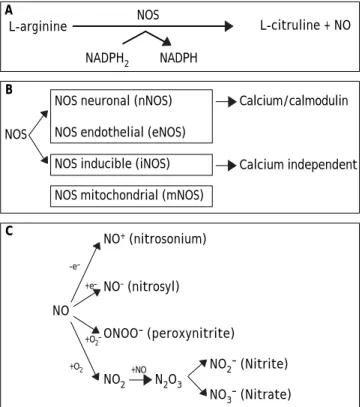
This small, unstable, gaseous, free radical perfor-ms its complex tasks by acting as an intra- or extra-cellular messenger molecule. Nitric oxide is produced from the amino acid L-arginine through a reaction catalysed by the enzymes nitric oxide syn-thases (NOS). These enzymes catalyse the oxidation by molecular oxygen of one of the guanidine nitro-gens in L-arginine to form NO and citrulline12
(Figure 1A).
NO synthesis
• NO synthase isoforms and subcellular loca-lization. Three main NOS isoforms have been identified and cloned so far: nNOS (neuronal or type I), iNOS (inducible or type II), and eNOS (endothelial or type III). In addition, new isofor-ms or mitochondrial variants of NOS (mtNOS) have been described recently in rat liver, thymus, and brain.13 eNOS and nNOS are constitutively
expressed, and produce relatively small amounts of NO. These isoforms depend on Ca2+
/calmodu-lin, and account for a rapid increase of NO in response to hormone receptor interaction.14,15
On the other hand, iNOS is up-regulated in liver under a number of conditions, including endo-toxemia, hemorrhagic shock, ischemia-reperfu-sion, hepatitis and liver regeneration. This isoform is Ca2+-independent, and synthesizes NO
for extended periods of time, at high concentra-tions, serving as an important regulator and effector during inflammation and infection.16 It is
expressed in different types of cells, including en-dothelial cells, hepatocytes, Kupffer cells and smooth-muscle cells.
NOS isoforms have differential localizations within the cells: nNOS and iNOS are mainly cytosolic, although recently, Stolz, et al. have es-tablished the peroxisome as a site of iNOS locali-zation in hepatocytes.17 On the other hand, eNOS
rough endoplasmic reticulum and in the nuclei of rat hepatocytes, and has been shown to be a membrane associated protein.18 Finally, mtNOS
activity is modulated by Ca2+, and it is located at
the inner mitochondrial membrane, where conti-nuously controls mitochondrial respiration.19
In liver, eNOS activity is normally detectable in Kupffer cells and at the plasma membrane of rat hepatocytes.7 In addition, functional eNOS has
been identified and characterized in sinusoidal endothelial cells with the liver and may contribu-te to local distribution of perfusion and portal pressure.20
In addition, hepatocytes, Kupffer and stellate cells are prompted to express an intense iNOS activity once exposed to effective stimuli, such as bacterial lipopolysaccharide (LPS) and cytokines1
(Figure 1B).
• NO and biological reaction. Reactive nitrogen intermediates (RNI) are now also recognized as important radicals. Under normal conditions, in-teraction of NO with oxygen results in the forma-tion of the relatively inactive end products nitrite (NO2-) and nitrate (NO
3-). NO can be also
conver-ted into different reactive chemical forms (NO_,
NO. and NO+), and this is the reason why it has a
wide range of chemical reactivity and regulatory functions in a variety of biological targets (Figure 1C). These functions include regulation of the car-diovascular system, smooth muscle relaxation, neurotransmission, coagulation and immune re-gulation. Despite these beneficial functions, the molecule has also a pivotal role in cell death, by having the ability to either induce or protect against apoptosis depending on its levels and ce-llular context. Furthermore, NO can turn an apoptotic response into a necrotic one. NO can also combine with O2- (superoxide anion) to form
peroxynitrite (ONOO–), which shares some
pro-perties with NO, such as its capability to freely di-ffuse via the intra- and the intercellular pathway, and also to act as a powerful oxidant.21
NO and apoptosis
Apoptosis, or programmed cell death, is essential to the normal development of multicellular organisms as well as to the physiologic cell turnover. NO is one of the most potent regulators of apoptosis since, as stated above, it has dual pro- and anti-apoptotic effects. The cellular threshold for apoptosis is highly regulated, especially by members of the Bcl-2 protein family. Members of this family are anti-apop-totic proteins (Bcl-2, Bcl-xL, Bcl-w) while others can promote programmed cell death (Bax, Bak, Bad, BNIP3).22 The oligomerization of apoptotic
pro-teins into the outer mitochondrial membrane leads to the formation of pores, and results in mitochon-drial release of cytocrhome c. The association of cytochrome c with an adapter molecule, Apaf-1, and caspase 9 in the cytoplasm activates the latter, which, in turn, activates downstream caspases.23
In addition, apoptosis can be induces by the TNF-induced death receptor pathway. They have been identified two families of receptor: the receptor of TNFα (TNFR1) and Fas receptor protein that in-duce apoptosis in a similar way. Binding of TNFα to TNFR1 results in receptor trimerization and the re-cruitment of a series of intracellular proteins24 that
ultimate bind caspase-8, leading to its activation.25
Activated caspase-8 initiates a proteolytic cascade L-arginine NOS
NADPH2 NADPH
L-citruline + NO
NOS
NOS neuronal (nNOS)
NOS endothelial (eNOS)
NOS inducible (iNOS)
NOS mitochondrial (mNOS)
Calcium/calmodulin
Calcium independent
NO
NO+ (nitrosonium)
NO– (nitrosyl)
ONOO–(peroxynitrite)
NO2–(Nitrite) NO2 N2O3
NO3–(Nitrate)
+NO -e–
+e–
+O2–
+O2 A
B
C
Figure 1. A. NO is synthesized endogenously by conversion of L-arginine to L-citruline, using NADPH as an electron do-nor. B. The reaction is catalyzed by a family of enzymes called nitric oxide synthases (NOS). Distinct isoforms of NOS are pre-sented. C. Under normal conditions, interaction of NO with oxygen results in the formation of the relatively inactive end products nitrite (NO2-) and nitrate (NO
3-). NO can be also
con-verted into different reactive chemical forms as nitrosonium (NO+) and nitroxyl (NO-) radicals, by gaining or losing
elec-trons, respectively. Finally, NO can combine with O2-
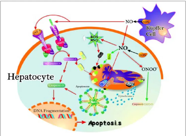
that results in release of lysosomal cathepsin B, cleavage of the pro-apoptotic Bcl-2 family member Bid, initiation of the mitochondrial death pathway with release of cytocrome c, and activation of do-wnstream effectors caspases that ultimately induces apoptosis26 (Figure 2).
NO induces apoptosis both in vivo27,28 and in
se-veral cell types in vitro, such as neuronal cells,23
macrophages,29 cardiac myocytes,30 endothelial
cells,31 lymphocytes, and thymocytes.32 The
mecha-nisms of NO-induced apoptosis are presently under intensive investigation and several mechanisms
un-derlying the effects of NO on apoptosis have now been elucidated. They include activation of the death receptor Fas through upregulation of Fas ligand expression, generation of the potent oxidant and cytotoxic mediator peroxynitrite, inhibition of mito-chondrial ATP synthesis, and inactivation of several antioxidant enzymes.33,34
Another site of action of NO on mitochondria is the mitochondrial permeability transition pore (MPTP). There is now increasing evidence that su-pport a redox regulation of cytochrome c release du-ring apoptosis.35 The MPTP plays an essential role
Figure 2. Schematic representation of the pro- and antiapoptotic effects exerted by NO. The NO production by up-regulation of iNOS in both hepatocytes and Kupffer cells leads to Fas stimulation. This produces caspase 8 activation, followed by increase of caspase 3 activity and, downstream, release of cytochrome c from mitochondria to cytosol, with further activation of the apop-tosome. Caspase 8 activation may promote Bid processing, leading eventually to apoptosis. Once peroxynitrite is increased, it produces increase of reactive nitrogen intermediaries (RNI) which may raise pro-apoptotic protein Bax levels, and downstream, cytochrome c release and activation of the apoptosome. Peroxynitrite can open membrane transition pores (MTP), and lead to cytochrome c release (red arrows).
in apoptosis; once opened, cytochrome c is released, resulting in the formation of the apoptosome, acti-vation of caspase-9 and execution of apoptosis. Recently, inhibition of MPTP opening was identified as a novel site of action for NO signaling in apopto-sis.36 Inhibition of MPTP opening would result in
less cytochrome c available to initiate apoptosis. This study suggests that a fine balance exists bet-ween the pro- and anti-apoptotic properties of NO at the level of the mitochondrion.
Apart from pro-apoptotic action, NO may protect apotosis death under certain conditions, depending on the cell type.
Several mechanisms have been proposed to elu-cidate the ability of NO to confer protection against apoptotic cell death. These can be divided into cGMP-dependent and cGMP-independent mechanisms:
• cGMP dependent mechanisms. It is known that NO mediates many of its physiological func-tions through the direct heme-dependent activa-tion of soluble guanilate cyclase, and the consequent increase in intracellular cGMP le-vels.37 NO-dependent generation of intracellular
cGMP has been shown to protect against apopto-sis in lymphocyte,38 eosinophils,39 embryonic
mo-tor neurons.40 Similarly, an increase of cGMP
suppresses apoptosis in hepatocytes through a marked activation of serine/threonine kinase Akt, suggesting a link between cGMP and the PI3K/Akt signaling pathway.37,41 Activation of
Akt can promote cell survival through phos-phorylation of the pro-apoptotic protein Bad and caspase 9, which favors Bad proteosomal degra-dation of Bad and caspase 9 inactivation, respec-tively, and by preventing cytochrome c release from mitochondria.42,43
Among the other genes regulated by NO is BNIP3, a protein known to promote apoptosis, which belongs to the Bcl-2 family. BNIP3 locali-zes at the mitochondria and other cytoplasmic membrane structures, and is found widely expre-ssed several mouse and human tissues. BNIP3 expression is markedly suppressed following iNOS up regulation and NO production in a cGMP-dependent manner, suggesting that this is another mechanism by which NO prevents apop-tosis mediated by cGMP-dependent pathways.44
Ray, et al. demonstrated recently that BNIP3 he-terodimerizes with Bcl-2/Bcl-xl and induces cell death independent of a BH3 domain at both mito-chondrial and non-mitomito-chondrial sites.45
• cGMP-independent mechanisms. NO protects apoptotic cell death in a cGMP-independent man-ner by preventing Bcl-2 cleavage, cytochrome c release, and the induction of protective proteins such as Hsp70 and Bcl-2.41 In addition, many of
the direct protective actions of NO are mediated by S-nitrosylation of proteins. S-nitrosylation in-volves the transfer of a nitric group to cysteine sulfhydryls, leading to the formation of a nitro-sothiol (RSNO). While the activity of NO is often limited due to its very short half-life, nitroso-thiols can be very stable compounds. Caspases contain a highly conserved cysteine residue within their active site, and therefore are a tar-get for inactivation via S-nitrosylation. In line with this, NO has been reported to protect against Fas-induced liver injury by inhibiting caspase activity. This caspase inhibition is rever-ted by DTT, suggesting that cysteine S- nitrosyla-tion is the underlying mechanism of caspase regulation by NO in this study.46
NO and liver
Under normal conditions, only the constitutive eNOS is present in the liver, and the low level of NO produced by eNOS regulates hepatic perfusion, among other functions. However, iNOS is readily up-regulated in the liver under a number of condi-tions, including endotoxemia, hemorrhagic shock, ischemia-reperfusion, sepsis, infection, hepatitis, ozone exposure, and liver regeneration.47 Once
iNOS is expressed, large amounts of NO are genera-ted in the liver in a sustained fashion, which func-tions as an important regulator and effector during inflammation and infection.48 Because the liver
plays a crucial role in m any metabolic and immune processes, physiological and pathophysiological functions of the NO generated in the liver have prompted numerous investigations in recent years. Both cytoprotective and cytotoxic effects of NO have been demonstrated in the liver.37,48,49
• Induction of hepatic iNOS after different stimulus. In response to endotoxin or proinfla-mmatory cytokines, such as tumor necrosis fac-tor-α (TNF-α), interleukin (IL)-1 and interferon-γ, as well as their combinations, iNOS is rapidly up-regulated within hours in hepato-cytes, and in resident hepatic macrophages (Kupffer cells) as well.50 These stimuli often act
iNOS in liver. The cytokine-mediated up-regula-tion of iNOS gene transcripup-regula-tion requires the transcriptional factor nuclear factor-κB (NF-κB) in both animals and humans.51 In addition,
hepa-tic endothelial cells and stellate cells can also produce NO through iNOS up regulation. There-fore, in liver inflammation, hepatocytes are si-tuated in an environment where NO is generated from surrounding cells, as well as from hepato-cytes themselves. In cell cultures has been repor-ted that the maximum production of NO, which is calculated as the release of nitrite, after stimu-lation with polysaccharides and/or cytokines in hepatocytes is between 45 and 1,200 nmol NO2-/106
cells per day, Kupffer cells between 50 and 100 nmol NO2-/106cells per day, and endothelial cells
between 10 and 16 nmol per day NO2-/106cells37,52
(Figure 2).
• NO and liver regeneration. One remarkable characteristic of the regenerative process after PH is the capacity of the liver to grow until the organ size and functionality are fully restored. Therefore, it is widely accepted the existence of a precise pattern of events (release and modulation of growth factors and cytokines) controlling the successive regenerative steps after PH.1
Howe-ver, the nature of the factors and early signals involved in the recruitment of cells to entry in the division cell cycle is far from been fully understood. The current view is based on the existence of a dynamic balance between positive and negative control. Equilibrium between sti-mulator and inhibitor gene of the cell cycle expre-ssed after PH, may explain why liver regeneration is tightly regulated growth process. Within 30 min after PH, several genes are indu-ced which contributes to regeneration. Among these genes, signal transducer and activation of transcription-3 (Stat-3), nuclear factor kB (NF-kB), CCAAT/enhancer binding protein b (C/ EBPb), and activating protein 1 (AP-1) are known to play a cooperative role in intracellular signaling cascades leading to DNA synthesis.53-55
These transcription factors regulate the expres-sion of many hepatocyte genes including iNOS,53,56 which is up-regulated within the
im-mediate hours after PH. NO begins to be released within 30 min after PH, reaching its maximal le-vel at 5 h (progression phase of cell cycle) and re-turns to basal levels at 18 h after surgery.1,33 In
this connection, our group has demonstrated a marked decrease in the peak of DNA synthesis in hepatectomised rats when they were pretreated
with two iNOS inhibitors (a specific inhibitor, aminoguanidine, and a non-specific one, NG
-mo-nomethyl-L-arginine, L-NAME).8 Likewise, Rai, et al. have shown impaired liver regeneration in iNOS-deficient mice.57 These results suggest a
positive effect of NO in the regulation of the rege-nerative process at early stages.
Interestingly, this NO seems to be delivered exclusively in the liver, and apparently, the molecule is completely consumed in the hepatic tissue. This conclusion is supported by the ob-servation of a total absence of NO in blood, as measured through the formation of a nitroxyl-he-moglobin complex, as well as by the absence of changes in the nitrite concentration in plasma, a NO-derived metabolite that is more stable than NO itself.2 After PH, both hepatic iNOS activity
and levels of iNOS messenger RNA have been de-tected exclusively in the liver, indicating that this is a local effect.2,8,12 The relative contribution
of each liver cell type (Kupffer cells, and hepato-cytes and, possibly, endothelial cells) to the total iNOS activity and NO synthesis seems to be di-fferent. However, because NO can easily diffuse through the cells, the origin of this molecule is not critical for the ability to promote intracellu-lar changes in neighboring cells.2
Evidence shows the importance of cytokines du-ring liver regeneration. When a large piece of li-ver is removed by PH, increased local expression of TNF-α triggers the production of another cytokine, IL-6, and both cytokines are required to initiate subsequent hepatocyte proliferation.58 In vivo analysis of murine iNOS genes indicate that neither TNF-α nor IL-6 alone are sufficient to activate iNOS transcription, but when these two cytokines are combined, up-regulation oc-curs.59,60
Ø Role of NO in apoptosis/proliferation ba-lance. The determinants of hepatocytes proli-feration during liver regeneration are highly complex, and different mechanisms operate between initiation of DNA synthesis and the termination of proliferative process.61 The
control of liver regeneration and the events involved in regulating the growth of the organ remain unknown. It has become increasingly apparent that apoptosis plays a key role in the cell cycle.62,63 Many of the proteins that can
induce cell death are components of the cell di-vision cycle.41 In previous studies by our
regu-lation of apoptosis/survival-related pro-teins.48,63
The relative prevalence of Bax and Bcl-xL pro-teins are critical factors influencing cell fate; they promote either cell survival or death, whose ultimate outcome largely depends on the Bcl-xL/Bax ratio.63 Large increases of the
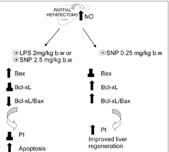
hepatic levels of NO (> 100%) (obtained ei-ther by the iNOS inducer LPS or by the direct NO donor sodium nitroprusiate (SNP, 2.5 mg/kg body wt, administered 5 h after PH) induces expression of the pro-apopoptotic protein Bax in the mitochondrial fraction, without chan-ges of the anti-apoptotic protein Bcl-xL as compared with PH-alone.48 On the contrary,
when the increase of NO is moderate, e.g. a 35%, obtained by low doses of SNP (0.25 mg/kg body wt), the pro-apoptotic protein Bax remains unchanged with respect to PH-alone, while the anti-apoptotic protein Bcl-xL is in-creased (Ronco, et al., unpublished data). The mechanism by which NO produces chan-ges of Bax/Bcl-xL gene expression remain unknown. Some investigators have focused on
p53 as a linkage between NO and Bax/Bcl-2 genes, since NO is known to induce p53 accu-mulation, which is a direct transcriptional ac-tivator of Bax gene and a transcriptional inhibitor of Bcl-2 gene.64 p53 acts as a
check-point control of the cell cycle, permitting the repair of damaged DNA. The blockage in G1/S transition that results from p53 activation has been suggested to cause apoptosis in the case of severe DNA damage. Expression of wild-type p53, a tumor suppressor gene, seems to be closely linked with apoptosis caused by most of DNA-damaging agents.65,66 Recent
data from our laboratory showed that a large augmentation of NO in the liver after PH (as those caused by LPS and SNP 2.5 mg/ kg body wt.), increases p53 protein levels with a correlation between cytosolic nitrate and p53 levels.48 On the other hand, a smaller increase
of NO (as that caused by SNP 0.25 mg/kg body wt) does not show differences in p53 ex-pression when compared with hepatectomized rats (Ronco, et al., unpublished data) (Figure 4). The apoptotic index revealed that a high
Figure 3. Schematic repre-sentation of the effects of the overproduction of NO after PH. The induction of iNOS with lipo-polisacaride (LPS, 2 mg/kg) and the treatment with NO do-nor SNP2 (2.50 mg/mL) produ-ce an over-increase of 100% of NO, leading to an increase of pro-apoptotic proteins Bax and p53, leading to apoptosis. Treatment with a NO donor, SNP1 (0.25 mg/mL), produces an increase lower than 35% of NO, thus leading to an increase of anti-apoptotic protein Bcl-xL,which improves liver rege-neration (⊥ not difference, ↑
Figure 4. Immnublottintg analysis of VEGF protein expres-sion in total liver lysates frac-tion at 72 h post-surgery. A. Line 1: Sh-Control, a midline la-parotomy with liver manipula-tion was carried out on surgical sham control rats. Line 2: PH-Control, two-thirds hepatec-tomy was performed to control rats. Line 3: PH-SNP1, two-thirds hepatectomy was perfor-med to rats that had received sodium nitropruside 0.25 mg/kg body weight, intravenously. B. Line 1: Sh-Control. Line 2: PH-Control. Line 3: PH-AG, two-thirds hepatectomy was performed to rats that had re-ceived aminoguanidine (AG) 100 mg/kg body weight, intraperito-neally. *p < 0.05 vs. Sh-Control.
#p < 0.05 vs. PH-Control (Ronco,
et al. 2007).
increase of NO induces cellular death by apop-tosis, while a mild increase does not modify this process. In agreement with other au-thors,64,65 our results suggest that high levels
of NO induce apoptosis by accumulation of p53, which induces Bax expression.48
What is the role of NO in the proliferative process? To answer this question, we analy-zed how modification of NO levels modulate the proliferative process that takes place after PH. The results obtained by immunohistoche-mistry detection of proliferation cellular nu-clear antigen (PCNA) indicate that no change occurs in the proliferation of the remaining li-ver under an increase of oli-ver 100% of NO. Nevertheless, an increase of NO lower than 35% enhances the proliferation index (Ronco,
et al., unpublished data), suggesting that a moderate increase of NO improve the prolife-rative process at early stages of liver regene-ration.
As we previously described, the specific role of NO in the balance of apoptosis/proliferation remains controversial considering the dual ac-tion of NO in either promoting or impairing programmed cell death. Analyzing our own sults focusing on the action of NO in the re-gulation of apoptosis/survival proteins in liver regeneration, we observed an expression of survival factors when NO is in “small concen-trations”, and an expression of pro-apoptotic proteins when NO is at “high concentrations”.
Figure 3 shows a schematic description of the regulation of apoptosis/proliferation balance after PH, mediated by the impact of increases in NO on anti-apoptotic proteins Bcl-xL and pro-apoptotic Bax.
Ø NO and Vascular Endothelial Growth Factor (VEGF). One of the well-characterized functions of NO is as a mediator of vascular dilatation and permeability, as well as in vascular remodeling.2,8 Angiogenesis, i.e. the
formation of new blood vessels, is a complex process that involves proliferation and migra-tion of endothelial cells. This phenomenon is required for remodeling liver architecture following liver resection.67-69 The initial wave
of hepatocyte proliferation is followed by endo-thelial cell proliferation and penetration of vascular hepatocellular islands leading to the formation of newly-formed sinusoids.70
After PH, both hepatocytes and nonparenchy-mal cells express VEGF mRNA, suggesting that VEGF plays a significant role in this pro-cess. Hepatocellular production of VEGF shows the maximal levels between 48 and 72 h after PH.71,73 An increase of VEGF production
by hepatocytes correlates with an increase in VEGF receptor expression on endothelial cells after PH. Moreover, recent studies have shown that inhibition of angiogenesis with angiostatin impairs liver regeneration. Fur-thermore, an increased expression of VEGF and its receptors induce the proliferation of endothelial cells.71,74
It is known that NO plays an important role in the processes of vascularization, angiogene-sis and permeabilization of tissue. Although there is a growing body of evidence that NO has angiogenic effects, partly mediated by VEGF, there is no unanimity of opinion on this regard.75,76 The effects of NO are greatly
dependent upon cell type, cellular redox sta-tus, and the amount and chemical nature of NO donors.21 In previous studies, we showed
that treatment of PH-animals with low doses of the direct NO donor SNP (0.25 mg/kg body wt) increases VEGF levels compared with PH-alone, and that the inhibition of NO synthesis (with the selective inhibitor of iNOS amino-guanidine) decreases VEGF protein levels, su-ggesting that NO is implicated in VEGF expression (Figure 4). Accordingly with ours results, other authors have reported that the exogenous addition of NO donors or increased levels of endogenous NO enhanced VEGF syn-thesis in rat vascular smooth muscle cells.58
Furthermore, in the rabbit cornea model of angiogenesis, VEGF-induced angiogenesis is blocked by L-NAME (non-selective NOS inhi-bitor), demonstrating that neovascularization is suppressed by the blockade of NO produc-tion.77 Besides, Taniguchi, et al. have shown
that VEGF expression in regenerating rat li-ver occurs predominantly in periportal hepato-cytes.73 They also demonstrated that VEGF is
involved in proliferation of hepatocytes asso-ciated with proliferation of sinusoidal endo-thelial cells after PH in rats. Histological studies revealed that NO donor treatment in-creases the number of vascular structures in portal areas in PH animals and that amino-guanidine treatment reduces this rate,78
sug-gesting that the augmentation of NO levels
increases periportal vascularization, probably via VEGF. In agreement with other authors, these studies provide further evidence that VEGF production is regulated by NO, and that this compound plays a central role in rat liver regeneration after PH.75,76 In our
stu-dies, we showed that the modification of NO levels at 5 h post-PH produces changes in VEGF protein levels at 72 h after PH.78 These
results suggest that the NO increase during early steps of liver regeneration initiates an adaptive response leading to activation of transcriptional factors that act as signal transducers between cytoplasm and nucleus, which results in the regulation of VEGF ex-pression.
CONCLUSION
Data revised and presented in this review lead to the assumption that NO activates, as a signaling mo-lecule, different cellular mechanisms that can promo-te either cell growth or cell death. NO plays an important and diverse role during liver regeneration, with the potential to be either anti- or pro-apoptotic. Experimental evidence shows that the expression of iNOS is induced at early stages of liver regeneration after PH, provided a high amount of NO is present. When conditions are right for peroxinitrite genera-tion, NO, via the formation of peroxynitrite, can da-mage cellular components such as mitochondria, leading to the opening of the membrane transition po-res (MTP) with the consequent release of cytochrome c into the cytoplasm. In addition, NO can increase the expression of the pro-apoptotic proteins Bax and p53, resulting in cell death.
At early stages of liver regeneration, the small amounts of NO produced appear to be necessary and perhaps sufficient to produce an increase in anti-apoptotic protein Bcl-xL, which results in protection from apoptotic cell death. Also, this level of NO en-sures a maximum increase of VEGF at 72 h post-he-patectomy, which is necessary to maintain sinusoidal perfusion and induce neovascularization.
the possibility of using these drugs, or even the de-signing new, hepatotropic drugs that regulate NO levels with the aim of improving the process of liver regeneration.
ACKNOWLEDGMENTS
This work was supported by research grants from ANPCyT (PICT Nº 32413, C.E. Carnovale PhD.) and from CONICET (PIP Nº 5531, C.E. Carnovale PhD.).
REFERENCES
1. Fausto N. Liver regeneration. J Hepatol 2000; 32: 19-31. 2. Hortelano S, Dewez B, Genaro AM, az-Guerra MJ, Bosca L.
Nitric oxide is released in regenerating liver after partial hepatectomy. Hepatology 1995; 21: 776-86.
3. Sakamoto T, Liu Z, Murase N, Ezure T, Yokomuro S, Poli V, Demetris AJ. Mitosis and apoptosis in the liver of interleu-kin-6-deficient mice after partial hepatectomy. Hepatolo-gy 1999; 29: 403-11.
4. Michalopoulos GK, De Frances MC. Liver regeneration. Science 1997; 276: 60-6.
5. Taub R. Liver regeneration: from myth to mechanism. Nat Rev Mol Cell Biol 2004; 5: 836-47.
6. Streetz KL, Luedde T, Manns MP, Trautwein C. Interleukin 6 and liver regeneration. Gut 2000; 47: 309-12.
7. Wuestefeld T, Klein C, Streetz KL, Betz U, Lauber J, Buer J, Manns MP, Muller W, Trautwein C. Interleukin-6/glyco-protein 130-dependent pathways are protective during li-ver regeneration. J Biol Chem 2003; 278: 11281-8. 8. Carnovale CE, Scapini C, Alvarez ML, Favre C, Monti J,
Ca-rrillo MC. Nitric oxide release and enhancement of lipid pe-roxidation in regenerating rat liver. J Hepatol 2000; 32: 798-804.
9. Kim TH, Mars WM, Stolz DB, Petersen BE, Michalopoulos GK. Extracellular matrix remodeling at the early stages of liver regeneration in the rat. Hepatology 1997; 26: 896-904. 10. Stolz DB, Michalopoulos GK. Synergistic enhancement of
EGF, but not HGF, stimulated hepatocyte motility by TGF-beta 1 in vitro. J Cell Physiol 1997; 170: 57-68.
11. Liu H, Di Cunto F, Imarisio S, Reid LM. Citron kinase is a cell cycle-dependent, nuclear protein required for G2/M transition of hepatocytes. J Biol Chem 2003; 278: 2541-8. 12. Muriel P. Regulation of nitric oxide synthesis in the liver. J
Appl Toxicol 2000; 20: 189-95.
13. Carreras MC, Poderoso JJ. Mitochondrial nitric oxide in the signaling of cell integrated responses. Am J Physiol Cell Physiol 2007; 292: C1569-C1580.
14. Moncada S, Palmer RM. Biosynthesis and actions of nitric oxide. Semin Perinatol 1991; 15: 16-9.
15. Moncada S, Higgs EA. Endogenous nitric oxide: physiology, pathology and clinical relevance. Eur J Clin Invest 1991; 21: 361-74.
16. Weidenbach H, Nussler AK, Shu Z, Adler G, Beckh K. Nitric oxide formation lowers norepinephrine-induced intrahepa-tic resistance without major effects on the metabolism in the perfused rat liver. Hepatology 1997; 26: 147-54. 17. Stolz DB, Zamora R, Vodovotz Y, Loughran PA, Billiar TR,
Kim YM, Simmons RL, Watkins SC. Peroxisomal localization of inducible nitric oxide synthase in hepatocytes. Hepato-logy 2002; 36: 81-93.
18. Gobeil F Jr, Zhu T, Brault S, Geha A, Vazquez-Tello A, For-tier A, Barbaz D, et al. Nitric oxide signaling via nucleari-zed endothelial nitric-oxide synthase modulates expression of the immediate early genes iNOS and mPGES-1. J Biol Chem 2006; 281: 16058-67.
19. Ghafourifar P, Richter C. Nitric oxide synthase activity in mitochondria. FEBS Lett 1997; 418: 291-6.
20. Liu S, Premont RT, Kontos CD, Zhu S, Rockey DC. A crucial role for GRK2 in regulation of endothelial cell nitric oxide synthase function in portal hypertension. Nat Med 2005; 11: 952-8.
21. Stamler JS, Singel DJ, Loscalzo J. Biochemistry of nitric oxide and its redox-activated forms. Science 1992; 258: 1898-902.
22. Tzung SP, Fausto N, Hockenbery DM. Expression of Bcl-2 family during liver regeneration and identification of Bcl-x as a delayed early response gene. Am J Pathol 1997; 150: 1985-95.
23. Wei T, Chen C, Hou J, Xin W, Mori A. Nitric oxide induces oxidative stress and apoptosis in neuronal cells. Biochim Biophys Acta 2000; 1498: 72-9.
24. Wajant H. Death receptors. Essays Biochem 2003; 39: 53-71. 25. Micheau O, Tschopp J. Induction of TNF receptor
I-media-ted apoptosis via two sequential signaling complexes. Cell 2003; 114: 181-90.
26. Wullaert A, van Loo G, Heyninck K, Beyaert R. Hepatic tu-mor necrosis factor signaling and nuclear factor-kappaB: effects on liver homeostasis and beyond. Endocr Rev 2007; 28: 365-86.
27. Donovan M, Carmody RJ, Cotter TG. Light-induced photo-receptor apoptosis in vivo requires neuronal nitric-oxide synthase and guanylate cyclase activity and is caspase-3-independent. J Biol Chem 2001; 276: 23000-8.
28. Nishikawa M, Sato EF, Kuroki T, Utsumi K, Inoue M. Macro-phage-derived nitric oxide induces apoptosis of rat hepa-toma cells in vivo. Hepatology 1998; 28: 1474-80.
29. D’Acquisto F de, Cristofaro F, Maiuri MC, Tajana G, Car-nuccio R. Protective role of nuclear factor kappa B against nitric oxide-induced apoptosis in J774 macropha-ges. Cell Death Differ 2001; 8: 144-51.
30. Andreka P, Zang J, Dougherty C, Slepak TI, Webster KA, Bishopric NH. Cytoprotection by Jun kinase during nitric oxide-induced cardiac myocyte apoptosis. Circ Res 2001; 88: 305-12.
31. Shen YH, Wang XL, Wilcken DE. Nitric oxide induces and inhibits apoptosis through different pathways. FEBS Lett 1998; 433: 125-31.
32. Okuda Y, Sakoda S, Shimaoka M, Yanagihara T. Nitric oxi-de induces apoptosis in mouse splenic T lymphocytes. Im-munol Lett 1996; 52: 135-8.
33. Almeida A, Bolanos JP. A transient inhibition of mitochon-drial ATP synthesis by nitric oxide synthase activation trig-gered apoptosis in primary cortical neurons. J Neurochem 2001; 77: 676-90.
34. Dobashi K, Pahan K, Chahal A, Singh I. Modulation of endo-genous antioxidant enzymes by nitric oxide in rat C6 glial cells. J Neurochem 1997; 68: 1896-903.
35. Kirkland RA, Franklin JL. Evidence for redox regulation of cytochrome C release during programmed neuronal death: antioxidant effects of protein synthesis and caspase inhi-bition. J Neurosci 2001; 21: 1949-63.
37. Li J, Billiar TR. Nitric Oxide. IV. Determinants of nitric oxi-de protection and toxicity in liver. Am J Physiol 1999; 276: G1069-G1073.
38. Mannick JB, Hausladen A, Liu L, Hess DT, Zeng M, Miao QX, Kane LS, et al. Fas-induced caspase denitrosylation. Scien-ce 1999; 284: 651-4.
39. Beauvais F, Michel L, Dubertret L. The nitric oxide do-nors, azide and hydroxylamine, inhibit the programmed cell death of cytokine-deprived human eosinophils. FEBS Lett 1995; 361: 229-32.
40. Estevez AG, Spear N, Thompson JA, Cornwell TL, Radi R, Barbeito L, et al. Nitric oxide-dependent production of cGMP supports the survival of rat embryonic motor neu-rons cultured with brain-derived neurotrophic factor. J Neurosci 1998; 18: 3708-14.
41. Kim YM, Kim TH, Seol DW, Talanian RV, Billiar TR. Nitric oxi-de suppression of apoptosis occurs in association with an inhibition of Bcl-2 cleavage and cytochrome c release. J Biol Chem 1998; 273: 31437-41.
42. Cardone MH, Roy N, Stennicke HR, Salvesen GS, Franke TF, Stanbridge E, Frisch S, Reed JC. Regulation of cell death protease caspase-9 by phosphorylation. Science 1998; 282: 1318-21.
43. Datta SR, Dudek H, Tao X, Masters S, Fu H, Gotoh Y, Greenberg ME. Akt phosphorylation of BAD couples survi-val signals to the cell-intrinsic death machinery. Cell 1997; 91: 231-41.
44. Zamora R, Alarcon L, Vodovotz Y, Betten B, Kim PK, Gibson KF, Billiar TR. Nitric oxide suppresses the expression of Bcl-2 binding protein BNIP3 in hepatocytes. J Biol Chem 2001; 276: 46887-95.
45. Ray R, Chen G, Vande Velde C, Cizeau J, Park JH, Reed JC, Gietz RD, et al. BNIP3 heterodimerizes with Bcl-2/Bcl-X(L) and induces cell death independent of a Bcl-2 homology 3 (BH3) domain at both mitochondrial and nonmitochondrial sites. J Biol Chem 2000; 275: 1439-48.
46. Kim YM, Kim TH, Chung HT, Talanian RV, Yin XM, Billiar TR. Nitric oxide prevents tumor necrosis factor alpha-induced rat hepatocyte apoptosis by the interruption of mito-chondrial apoptotic signaling through S-nitrosylation of caspase-8. Hepatology 2000; 32: 770-8.
47. Martin-Sanz P, Hortelano S, Callejas NA, Goren N, Casado M, Zeini M, Bosca L. Nitric oxide in liver inflammation and regeneration. Metab Brain Dis 2002; 17: 325-34.
48. Ronco MT, Alvarez M de L, Monti JA, Carrillo MC, Pisani GB, Lugano MC, Carnovale CE. Role of nitric oxide increase on induced programmed cell death during early stages of rat liver regeneration. Biochim Biophys Act 2004; 1690: 70-6. 49. Wiley JW. The many faces of nitric oxide: cytotoxic,
cyto-protective or both. Neurogastroenterol Motil 2007; 19: 541-4.
50. Ozaki T, Habara K, Matsui K, Kaibori M, Kwon AH, Ito S, Nishizawa M, et al. Dexamethasone inhibits the induction of iNOS gene expression through destabilization of its mRNA in proinflammatory cytokine-stimulated hepato-cytes. Shock 2010; 33: 64-9.
51. Taylor BS, Alarcon LH, Billiar TR. Inducible nitric oxide syn-thase in the liver: regulation and function. Biochemistry (Mosc) 1998; 63: 766-81.
52. Obolenskaya M, Schulze-Specking A, Plaumann B, Frenzer K, Freudenberg N, Decker K. Nitric oxide production by cells isolated from regenerating rat liver. Biochem Biophys Res Commun 1994; 204: 1305-11.
53. Cressman DE, Greenbaum LE, Haber BA, Taub R. Rapid ac-tivation of post-hepatectomy factor/nuclear factor kappa
B in hepatocytes, a primary response in the regenerating liver. J Biol Chem 1994; 269: 30429-35.
54. Cressman DE, Greenbaum LE, DeAngelis RA, Ciliberto G, Furth EE, Poli V, et al. Liver failure and defective hepato-cyte regeneration in interleukin-6-deficient mice. Science 1996; 274: 1379-83.
55. Diehl AM. Roles of CCAAT/enhancer-binding proteins in re-gulation of liver regenerative growth. J Biol Chem 1998; 273: 30843-6.
56. Fausto N, Campbell JS. The role of hepatocytes and oval cells in liver regeneration and repopulation. Mech Dev 2003; 120: 117-30.
57. Rai RM, Lee FY, Rosen A, Yang SQ, Lin HZ, Koteish A, Liew FY, Zaragoza C, et al. Impaired liver regeneration in indu-cible nitric oxide synthasedeficient mice. Proc Natl Acad Sci USA 1998; 95: 13829-34.
58. Knowles RG, Moncada S. Nitric oxide as a signal in blood vessels. Trends Biochem Sci 1992; 17: 399-402.
59. Akerman P, Cote P, Yang SQ, McClain C, Nelson S, Bagby GJ, Diehl AM. Antibodies to tumor necrosis factor-alpha in-hibit liver regeneration after partial hepatectomy. Am J Physiol 1992; 263: G579-G585.
60. Goldring CE, Reveneau S, Algarte M, Jeannin JF. In vivo foo-tprinting of the mouse inducible nitric oxide synthase gene: inducible protein occupation of numerous sites inclu-ding Oct and NF-IL6. Nucleic Acids Res 1996; 24: 1682-7. 61. Boulton R, Woodman A, Calnan D, Selden C, Tam F,
Hodg-son H. Nonparenchymal cells from regenerating rat liver generate interleukin-1alpha and -1beta: a mechanism of negative regulation of hepatocyte proliferation. Hepato-logy 1997; 26: 49-58.
62. Fan G, Kren BT, Steer CJ. Regulation of apoptosis-associa-ted genes in the regenerating liver. Semin Liver Dis 1998; 18: 123-40.
63. Ronco MT, de Alvarez ML, Monti J, Carrillo MC, Pisani G, Lugano MC, Carnovale CE. Modulation of balance between apoptosis and proliferation by lipid peroxidation (LPO) du-ring rat liver regeneration. Mol Med 2002; 8: 808-17. 64. Chae IH, Park KW, Kim HS, Oh BH. Nitric oxide-induced
apoptosis is mediated by Bax/Bcl-2 gene expression, tran-sition of cytochrome c, and activation of caspase-3 in rat vascular smooth muscle cells. Clin Chim Acta 2004; 341: 83-91.
65. Inoue Y, Tomiya T, Yanase M, Arai M, Ikeda H, Tejima K, Ogata I, Kimura S, Omata M, Fujiwara K. p53 May positi-vely regulate hepatocyte proliferation in rats. Hepatology 2002; 36: 336-44.
66. Messmer UK, Ankarcrona M, Nicotera P, Brune B. p53 ex-pression in nitric oxide-induced apoptosis. FEBS Lett 1994; 355: 23-6.
67. Assy N, Spira G, Paizi M, Shenkar L, Kraizer Y, Cohen T, Neufeld G, et al. Effect of vascular endothelial growth fac-tor on hepatic regenerative activity following partial he-patectomy in rats. J Hepatol 1999; 30: 911-5.
68. Kraizer Y, Mawasi N, Seagal J, Paizi M, Assy N, Spira G. Vascular endothelial growth factor and angiopoietin in li-ver regeneration.Biochem. Biophys Res Commun 2001; 287: 209-15.
69. Sturm J, Keese M, Zhang H, Bonninghoff R, Magdeburg R, Vajkoczy P, Dono R, et al. Liver regeneration in FGF-2-de-ficient mice: VEGF acts as potential functional substitute for FGF-2. Liver Int 2004; 24: 161-8.
71. Mochida S, Ishikawa K, Inao M, Shibuya M, Fujiwara K. In-creased expressions of vascular endothelial growth factor and its receptors, flt-1 and KDR/flk-1, in regenerating rat liver. Biochem Biophys Res Commun 1996; 226: 176-9. 72. Shimizu H, Miyazaki M, Wakabayashi Y, Mitsuhashi N, Kato
A, Ito H, Nakagawa K, et al. Vascular endothelial growth factor secreted by replicating hepatocytes induces sinus-oidal endothelial cell proliferation during regeneration af-ter partial hepatectomy in rats. J Hepatol 2001; 34: 683-9.
73. Taniguchi E, Sakisaka S, Matsuo K, Tanikawa K, Sata M. Ex-pression and role of vascular endothelial growth factor in liver regeneration after partial hepatectomy in rats. J Histochem Cytochem 2001; 49: 121-30.
74. Redaelli CA, Semela D, Carrick FE, Ledermann M, Candinas D, Sauter B, Dufour JF. Effect of vascular endothelial
growth factor on functional recovery after hepatectomy in lean and obese mice. J Hepatol 2004; 40: 305-12. 75. Dulak J, Jozkowicz A. Nitric oxide in vascular endothelial
growth factor synthesis and signaling. Circulation 2001; 104: E48-E49.
76. Frank S, Stallmeyer B, Kampfer H, Kolb N, Pfeilschifter J. Nitric oxide triggers enhanced induction of vascular endo-thelial growth factor expression in cultured keratinocytes (HaCaT) and during cutaneous wound repair. FASEB J 1999; 13: 2002-14.
77. Ziche M. Role of nitric oxide in the angiogenesis of avascular tissue. Osteoarthritis Cartilage 1999; 7: 403-5.