Otras secciones de este sitio:
☞ ☞ ☞ ☞
☞ Índice de este número ☞
☞ ☞ ☞
☞ Más revistas ☞
☞ ☞ ☞
☞ Búsqueda
Others sections in this web site:
☞ ☞ ☞ ☞
☞ Contents of this number
☞ ☞ ☞ ☞
☞ More journals
☞ ☞ ☞ ☞ ☞ Search
Artículo:
Current concepts in autoimmune hepatitis
Copyright © 2005: Mexican Association of Hepatology
Number 1 January-March 2 0 0 5
Volume 4
Annals of Hepatology 4(1) 2005: 6-24
6
edigraphic.com
Annals of Hepatology 2005; 4(1): January-March: 6-24Annals of Hepatology
1From the Division of Gastroenterology and Hepatology, Mayo
Clinic College of Medicine, Rochester, Minnesota.
Address for correspondence: Albert J. Czaja, M.D.
Mayo Clinic 200 First Street S.W. Rochester, Minnesota 55905 Telephone: 507-284-8118, Fax: 507-284-0538.
E-mail: [email protected]
Manuscript received and accepted:12 January, 2005.
Concise Review
Current concepts in autoimmune hepatitis
Albert J. Czaja1
Abstract
Autoimmune hepatitis has a global occurrence, di-verse clinical phenotype, and evolving treatment op-tions. The goals of this report are to review the codi-fied diagnostic criteria, spectrum of clinical presenta-tions, proposed pathogenic mechanisms, conventional treatment strategies, and promising interventions. The literature published in English from 1980-2005 was re-viewed and an updated current perspective provided. Autoimmune hepatitis affects all ages, may be asymp-tomatic, frequently has an acute onset, and can present as fulminant hepatitis. Perivenular (zone 3) necrosis is within the histological spectrum. Autoim-mune hepatitis can recur or develop de novo after liver transplantation. CD4+ T-helper cells and natural kill-er T cells have been implicated in the pathogenesis, and molecular mimicry may break self-tolerance. DRB1*0301 and DRB1*0401 are the susceptibility al-leles among white North Americans and northern Eu-ropeans, whereas diverse alleles of HLA DR4 have been associated with the disease in Japan, mainland China, and Mexico. DRB1*1301 is associated with au-toimmune hepatitis in South American children, and it may predispose to an indigenous etiologic agent. Anti-bodies to soluble liver antigen/liver pancreas may have prognostic importance, and cyclosporine and myco-phenolate mofetil must be assessed by clinical trial be-fore incorporation into management algorithms. Site-specific interventions are feasible, and they require a confident experimental animal model for evaluation. Variant syndromes lack diagnostic and therapeutic guidelines. In conclusion, autoimmune hepatitis must be considered in all patients with acute and chronic liver disease and those with allograft dysfunction after transplantation. New immunosuppressive agents and site-specific interventions promise to improve care.
Key words: Autoimmunity, hepatitis, genetic predisposi-tions, variants, treatment
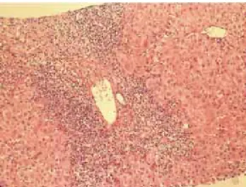
Autoimmune hepatitis is an unresolving inflammation of the liver of unknown cause.1 It is characterized by interface hepatitis on histological examination, hypergammaglobuline-mia, and autoantibodies. The sine qua non of the diagnosis is the presence of interface hepatitis on histological examina-tion. Lymphocytic, often lymphoplasmacytic, inflammatory infiltrates extend from portal tracts into parenchymal tissue where they are associated with hepatocyte injury (Figure 1). Parenchymal inflammation may be limited to periportal areas (interface hepatitis), or it may involve the entire acinus (panacinar or lobular hepatitis) (Figure 2). Plasma cells can be seen within the infiltrate (Figure 3), but they are not spe-cific or essential for the diagnosis. Plasma cells in groups or sheets in the portal tracts are present in 66% of patients, and this finding in conjunction with moderate-to-severe interface hepatitis and/or panacinar hepatitis has a diagnostic specific-ity of 81% and positive predictabilspecific-ity of 68%.2
Occurrence and ethnic variability
Autoimmune hepatitis has a global occurrence, and it has been described in African Americans, native Alaskans, Arabs, Asians, Europeans, Iranians, South Americans, and subcontinental Indians. Its incidence among white northern Europeans is 1.9 cases per 100,000 persons per year, and its point prevalence is 16.9 cases per 100,000 persons per year.3,4 In the United States, autoimmune hepatitis affects 100,000 to 200,000 persons, and it accounts for 5.9% of the liver transplantations in this country and 2.6% of the liver transplantations in Europe.5 The frequency of autoim-mune hepatitis among patients with chronic liver disease in North America is between 11% and 23%. Its prevalence among Alaskan natives is 43 per 100,000 population and higher than that reported elsewhere.6
M G
edigraphic.com
North American counterparts, and they have more severelaboratory abnormalities.9 African, Asian and Arab patients have early age onset disease, and they have a higher fre-quency of cholestatic laboratory findings, greater occur-rence of biliary changes on histological examination, and poorer initial response to standard therapy than other eth-nic groups.10 These findings suggest that geographical lo-cation and ancestry affect occurrence and behavior of the disease. Interwoven into the natural history of the disease in each racial group and geographical region are cultural and socioeconomic factors that remain unsorted. Differ-ences in the consequDiffer-ences of the liver disease must be cor-related with delays in diagnosis or difficulties in accessing medical care that are region-specific.11
Diagnostic clinical criteria
The clinical diagnosis of autoimmune hepatitis has been codified by an international panel, and these criteria must be applied to all patients12 (Table I). An acute, even fulminant, presentation is recognized and important to di-agnose quickly and treat promptly.13-16 The histological patterns that characterize acute onset autoimmune hepati-tis are a panacinar hepatihepati-tis that resembles an acute viral or drug-induced hepatitis16,17 (Figure 2) and a centrilobu-lar or perivenucentrilobu-lar (Rappaport zone 3) hepatitis that re-sembles an acute toxic injury16,18,19 (Figure 4).
Transitions from a perivenular (Rappaport zone 3) hepatitis to an interface hepatitis have been demonstrated
Figure 1. Interface hepatitis. Mononuclear inflammatory infiltrate dis-rupts limiting plate of portal tract and extends into parenchymal tis-sue. Hematoxylin and eosin. Original magnification, x100.
Figure 2. Pancinar hepatitis. Mononuclear inflammatory infiltrates line the sinusoidal spaces in association with liver cell degenerative and regenerative changes. Hematoxylin and eosin. Original magnifi-cation, x200.
Figure 3. Plasma cells. The hyperchromatic nuclei of plasma cells are attenuated by a cytoplasmic halo. Hematoxylin and eosin. Original magnification, x200.
8
edigraphic.com
in successive biopsy specimens from patients with acuteonset disease.19 These observations suggest that the periv-enular (Rappaport zone 3) pattern of injury may be an ear-ly histological manifestation of autoimmune hepatitis that is unrecognized in specimens obtained later in the course. An exacerbation of a previously unrecognized chronic liv-er disease may also have an acute presentation, and it should be suspected by the presence of hypoalbuminemia, thrombocytopenia, ascites, esophageal varices, and/or bridging (septal) fibrosis or cirrhosis on histological ex-amination.17,20
There are no disease-specific clinical or histological fea-tures. The diagnosis of autoimmune hepatitis requires the confident exclusion of other similar disorders including Wilson disease, genetic hemochromatosis, α-1 antitrypsin deficiency, chronic viral hepatitis, drug-related chronic liv-er disease, primary biliary cirrhosis (PBC), and primary sclerosing cholangitis (PSC)12 (Table I). Minocycline is the drug that has been most commonly implicated as a cause of the syndrome.21 A cholestatic form of autoimmune hepati-tis is not recognized, and the presence of pruritus or hyper-pigmentation compels another diagnosis.1,12
Diagnostic scoring criteria
An international scoring system for the objective diag-nosis of the disease has been developed and validated ret-rospectively12,22,23 (Table II). Multiple clinical, laboratory and histological features are graded, and a composite score is derived both before and after corticosteroid treat-ment. The scoring system was developed as a research tool to ensure comparability between patient populations in clinical trials. It is not a discriminative diagnostic in-dex, and it should not be used to distinguish between classical syndromes of chronic liver disease or to deduce that common non-disease-specific clinical and laboratory features connote a mixed or hybrid pathological state.
The virtues of the scoring system are that it quantifies the diagnosis, facilitates objective comparisons between patient populations, and accommodates individuals with atypical manifestations. Its drawbacks are its complexi-ty, and its failure to consistently distinguish cholestatic syndromes from autoimmune hepatitis. The sensitivity of the scoring system for autoimmune hepatitis ranges from 97%-100%, and its specificity for excluding au-toimmune hepatitis in patients with chronic hepatitis C ranges from 66%-92%. Its diagnostic specificity for ex-cluding autoimmune hepatitis in cholestatic syndromes ranges from 45%-65%.12
Types
Three types of autoimmune hepatitis have been pro-posed based on serological markers, but only two types have distinctive clinical phenotypes24 (Table III). None has been ascribed a unique cause, individual management strategy or special behavior, and they have not been en-dorsed as separate entities by the International Autoim-mune Hepatitis Group. The designations are used mainly as clinical descriptors.
Type 1 autoimmune hepatitis is the most common form worldwide, constituting 80% of all cases, and it is characterized by the presence of antinuclear antibodies (ANA) and/or smooth muscle antibodies (SMA) (Table III). Seventy-eight percent of patients are female, and the female:male ratio is 3.5. Earlier reports that autoimmune hepatitis had a bimodal age distribution between ages 10 years and 30 years and between 40 years and 50 years were probably affected by referral patterns to tertiary medical centers.25 Current experiences suggest that au-toimmune hepatitis occurs as commonly across all age ranges and that it may be under-diagnosed in the elder-ly.26,27 Forty-eight percent of patients are less than 40 years old, and the disease can affect infants.
Table I. International criteria for the diagnosis of autoimmune hepatitis.
Diagnostic Criteria
Definite AIH Probable AIH
Normal α-1 AT phenotype Partial α-1 AT deficiency
Normal ceruloplasmin level Nondiagnostic ceruloplasmin/copper levels Normal iron and ferritin levels Nondiagnostic iron and/or ferritin changes No active hepatitis A, B and/or C infection No active hepatitis A, B and/or C infection Daily alcohol <25 g/day Daily alcohol <50 g/day
No recent hepatotoxic drugs No recent hepatotoxic drugs
Predominant serum AST/ALT abnormality Predominant serum AST/ALT abnormality Globulin, γ-globulin or IgG level >1.5 times upper limit of normal Hypergammaglobulinemia of any degree ANA, SMA, or anti-LKM1 >1:80 in adults and >1:20 in children; ANA, SMA or anti-LKM1>1:40 in adults;
no AMA other autoantibodies
Interface hepatitis, moderate to severe Interface hepatitis, moderate to severe
No biliary lesions, granulomas or prominent changes suggestive No biliary lesions, granulomas or prominent changes suggestive of
of another disease another disease
M G
edigraphic.com
An abrupt onset of symptoms occurs in 40%, and afulminant presentation is possible.16 Thirty-eight percent of individuals have concurrent immune diseases, espe-cially autoimmune thyroiditis, synovitis, or ulcerative colitis, and 25% have cirrhosis already established at the time of presentation.28 Cholangiography is warranted in patients with ulcerative colitis to exclude PSC. Forty-one percent will have abnormal cholangiograms that support the diagnosis of PSC, and this finding may explain a re-fractory response to corticosteroid therapy.29 The high frequency of cirrhosis at presentation indicates that type 1 autoimmune hepatitis has an indolent, aggressive stage.
Thirty-four percent of patients with type 1 autoim-mune hepatitis may be asymptomatic at initial consulta-tion, and they are most commonly men with lower serum aminotransferase and immunoglobulin levels than symp-tomatic patients.31 Histological features are similar be-tween asymptomatic and symptomatic patients, and both groups respond as well to corticosteroids. Most asymp-tomatic patients become sympasymp-tomatic during follow-up, and differences between the asymptomatic and symptom-atic state may reflect variations in disease activity and pa-tient tolerance.
Type 2 autoimmune hepatitis is characterized by the presence of antibodies to liver/kidney microsome type 1 (anti-LKM1)31 (Table III). This disease occurs mainly in children, but 20% of patients with type 2 disease in Eu-rope are adults. Concurrent immune diseases are also common, especially insulin-dependent diabetes mellitus, vitiligo, and autoimmune thyroiditis. Organ specific
au-toantibodies are frequent, including antibodies to parietal cells, islets of Langerhans, and thyroid. As in type 1 dis-ease, a fulminant presentation is possible and important to recognize early.14
Type 2 autoimmune hepatitis is the only form in which the target autoantigen has been identified. It is the cyto-chrome mono-oxygenase, CYP2D6, which is an impor-tant drug-metabolizing enzyme within the cytosol of the hepatocyte.32-34 The antigen has been sequenced, cloned, and mapped, and five antigenic sites located between peptides 193-212, 257-269, 321-351, 373-389, and 410-429 are recognized by anti-LKM1.35 The amino acid se-quence spanning 193-212 of the CYP2D6 molecule is the target of anti-LKM1 in 93% of patients.
Homologies have been recognized between epitopes on the CYP2D6 molecule and the genome of the hepatitis C virus (HCV).34-37 The detection of anti-LKM1 in occa-sional patients with chronic hepatitis C in Europe (<10%) may reflect this molecular mimicry and antibody cross-reactivity. The hexameric amino acid sequence spanning 193-212 of the CYP2D6 molecule is homologous to the sequence spanning region 2985-2990 of the HCV ge-nome and identical to the sequence spanning region 130-135 of the cytomegalovirus (CMV) genome.35 These ho-mologies suggest that multiple exposures to viruses mim-icking self may be a mechanism by which to break self-tolerance and induce type 2 autoimmune hepatitis. Antibodies to LKM1 are extremely rare in North Ameri-can patients with chronic hepatitis C, and this rarity may reflect differences in the indigenous virus or the genetic
Table II. International scoring system for diagnosis of autoimmune hepatitis.
Gender Female +2 HLA DR3 or DR4 +1
AP:AST (or ALT) ratio >3 -2 Immune disease Thyroiditis, colitis, +2
<1.5 +2 others
γ-globulin >2.0 +3 Other markers Anti-SLA/LP, +2
or IgG level 1.5-2.0 +2 actin, LC1, pANCA
above normal 1.0-1.5 +1
<1.0 0
ANA, SMA, >1:80 +3 Histological features Interface hepatitis +3
or anti-LKM1 1:80 +2 Plasmacytic +1
titers 1:40 +1 Rosettes +1
<1:40 0 None of above -5
Biliary changes -3 Other features -3
AMA Positive -4 Treatment response Complete +2
Relapse +3
Viral markers Positive -3
Negative +3
Drugs Yes -4 Pretreatment score
No +1 Definite diagnosis >15
Probable diagnosis 10-15
Alcohol <25 g/day +2 Post-treatment score
>60 g/day -2 Definite diagnosis >17 Probable diagnosis 12-17
10
edigraphic.com
susceptibility of the host.38,39 Studies in Germany40 andIt-aly41 have not found an association between structural changes within the viral genome and the presence of anti-LKM1. A host factor for anti-LKM1 expression has been implicated.40
Type 2 autoimmune hepatitis occurs in 15% of patients with autoimmune polyendocrinopathy-candidiasis-ecto-dermal dystrophy (APECED).42 This syndrome consists of multiple endocrine organ failure, mucocutaneous candidia-sis, and ectodermal dystrophy in various syndromic combi-nations that may include autoimmune hepatitis. APECED is caused by a single-gene mutation located on chromo-some 21q22.3 which affects the generation of the autoim-mune regulator (AIRE).43 AIRE is a transcription factor that is expressed in epithelial and dendritic cells within the thymus, and it regulates clonal deletion of autoreactive T cells (negative selection). APECED has an autosomal re-cessive pattern of inheritance, and it lacks HLA-DR associ-ations and female predilection. The autoantigens associat-ed with APECED are CYP1A2 and CYP2A6.
Type 3 autoimmune hepatitis is the least established form, and it is characterized by the presence of antibodies to soluble liver antigen/liver-pancreas (anti-SLA/LP).44,45 These antibodies are directed against a 50 kDa cytosolic protein46 which is a transfer RNA complex (tRNP(ser)sec) involved in the incorporation of selenocysteine into polypeptide chains.47 Patients with type 3 disease are mostly women (91%) with a mean age of 37 years (range, 17 to 67 years).44 Other autoantibodies, including ANA, SMA and anti-LKM1, can co-exist with anti-SLA/LP, and only 26% of patients have anti-SLA/LP as their sole
serological finding.44 Patients with anti-SLA/LP are indis-tinguishable from patients with type 1 autoimmune hepa-titis by clinical or laboratory features, HLA phenotype, or response to corticosteroids.48,49 The designation of a type 3 autoimmune hepatitis has been largely abandoned.
Pathogenic mechanisms
The pathogenic mechanisms of autoimmune hepatitis are unknown. The most popular hypotheses evoke a con-stellation of interactive factors that include a triggering agent, a genetic predisposition, and various determinants of autoantigen display, immunocyte activation, and effec-tor cell expansion.50,51 Multiple triggering factors have been proposed, and they include infectious agents, drugs, and toxins. The multiplicity of etiologic agents that have been implicated in the pathogenesis of the disease sug-gests that the triggering epitope is a short amino acid se-quence that is common in many antigens. There can be a long lag time between exposure to the trigger and onset of the disease, and the triggering factor is not needed for perpetuation of the disorder.
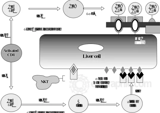
The CD4+ T-helper cell is the principal effector cell, and its activation and differentiation are the initial steps in the pathogenic pathway50,51 (Figure 5). Natural killer T (NKT) cells are abundant in the liver, and they have also been im-plicated in the pathogenesis of the disease. NKT cells are produced in the bone marrow rather than the thymus, lack antigen-specific receptors, and produce interferon (IFN)-γ and tumor necrosis factor (TNF)-α. They are inhibited by cells with normal expression of the major histocompatibility
Table III. Types of autoimmune hepatitis
Clinical features Type 1 Type 2
Signature markers SMA, ANA Anti-LKM1
Autoantigen Unknown CYD2D6
Age (years) Infancy to old age Pediatric (2-14)
Female (%) 78 89
Acute or fulminant onset Yes Yes
Immune diseases (%) 38 34
Typical concurrent immune diseases Thyroiditis Thyroiditis
Graves’ disease Vitiligo
Ulcerative colitis Type 1 diabetes APECED
HLA associations B8, DR3, DR4 B14, DR3, C4A-Q0, DR7
Allelic risk factors DRB1*0301, DRB1*0401 DRB1*07
DRB1*0404, DRB1*0405 DRB1*1301 DRB1*1501 (protective)
Autoimmune promoter genes (putative) Tumor necrosis factor-α Unknown
Cytotoxic T lymphocyte antigen 4 (CTLA-4) Vitamin D receptor (VDR) Tyrosine phosphatase CD45
Fas
MHC class I chain-related A (MICA)
Steroid responsive +++ ++
M G
edigraphic.com
complex (MHC) and by inhibitory receptors activated byglycolipid. Conversely, they target cells with aberrant MHC expression, defend against cells altered by viruses or cancer, and seem to promote hepatic regeneration.
The immunoregulatory cytokines orchestrate immuno-cyte differentiation through cross-regulatory actions and result in cellular and humoral mechanisms of liver cell in-jury50-52 (Figure 5). Interleukin (IL)-2, IFN-γ, and TNF-α constitute the type 1 (Th1) cytokine response which regu-lates cellular immune mechanisms by facilitating clonal expansion of cytotoxic T lymphocytes. Interleukin-4, IL-5, IL-6, IL-8, IL-10 and IL-13 constitute the type 2 (Th2) cytokine response which influences the humoral immune response by activating B cells and stimulating autoanti-body production. The type 1 cytokine response favors liv-er injury by expanding sensitized tissue-infiltrating cyto-toxic T cells (cellular cytocyto-toxicity), and the type 2 cytok-ine response favors liver cell injury by generating immunoglobulin complexes on the hepatocyte surface that are targeted by NKT cells (antibody-dependent cell-mediated cytotoxicity). The type 2 cytokine response also has anti-inflammatory effects that counter the type 1 cy-tokine actions.
Molecular mimicry of a foreign antigen and a an-tigen is the most common explanation for the loss of self-tolerance, but this mechanism has not been established in human disease.51 Cross-reacting autoantibodies between foreign and self antigens have been described, but cross-reacting immunocytes have been more difficult to dem-onstrate. Recently, a murine model of type 2 autoimmune hepatitis based on DNA immunization against self-anti-gens has supported this possibility.53 Most immunized
mice developed peak serum alanine aminotransferase ab-normalities 4 and 7 months after the last of three plasmid injections that contained the antigenic regions of human CYP2D6 and human formiminotransferase cyclodeami-nase, the target antigen of antibodies to liver cytosol type 1 (anti-LC1). Affected mice expressed anti-LKM1 and anti-LC1, but they also had cytotoxic T lymphocytes within the liver that were sensitized against the antigens in the plasmid constructs. This murine model indicated that DNA immunization against human autoantigens could break self-tolerance and cause liver injury by mo-lecular mimicry between foreign and self-antigens in-volving cross-reacting humoral and cellular responses.
HLA Associations
Genetic factors affect the occurrence, clinical expres-sion and treatment outcome of type 1 autoimmune hepati-tis. HLA DR3 is the main susceptibility factor in white northern European and North American patients, and HLA DR4 is a secondary but independent risk factor54 (Table III). Eighty-five percent of white patients from these regions have HLA DR3, DR4 or DR3-DR4. Differ-ent geographical regions and ethnic groups have differDiffer-ent susceptibility factors. HLA DR3 occurs rarely in the Jap-anese population, and HLA DR4 is the principal risk fac-tor for autoimmune hepatitis in this ethnic group.55 HLA DR4 is also the principal susceptibility factor for autoim-mune hepatitis in mainland China.56 In contrast, HLA DR3, but not HLA DR4, is the susceptibility factor in Ita-ly,57 and HLA DR13 is associated with childhood autoim-mune hepatitis in South America.9,58-60
Figure 5. Cytokine pathways of im-munocyte differentiation. The prin-cipal effector is the CD4+ T helper cell, and its differentiation depends on the counter-regulatory effects of the interleukins (IL) and tumor ne-crosis factor-α (TNF-α). The type 1 cytokine response promotes clonal expansion of liver infiltrating cyto-toxic T lymphocytes (CD8 CTL). The type 2 cytokine response pro-motes the expansion of plasma cells and immunoglobulin G (IgG) pro-duction. The immunoglobulin com-plexes with normal membrane pro-teins on the hepatocyte surface, and natural killer T (NKT) cells target these complexes and cause cytoly-sis by an antibody-dependent cell-mediated form of cyotoxicity. Type 1 and type 2 cytokine responses are counter-regulatory.
CD4 Th1
IL-2
CD8
TNF-a
CD8
CTL CD8CTL CD8CTL
Normal membrane
proteins
IL-10
IgG
Type 2 cytokine response IL-10
IL-4 IL-12
Type 1 cytokine response
B cellB
cell PlasmaPlasmacellcell
Activated CD4
Activated CD4
CD4 Th2
MHC class IMHC class I
Liver cell
Liver cell
12
edigraphic.com
Patients with HLA DR4 in North America are olderand more commonly female than patients with HLA DR3.61 They also have higher serum levels of γ-globulin and immunoglobulin G (IgG), higher titers of ANA, and a greater frequency of concurrent immune diseases. Pa-tients with HLA DR4 respond more readily to corticoster-oid therapy than those with HLA DR3 by entering remis-sion more commonly and failing treatment less often. The bases for these effects on clinical phenotype and outcome are unknown, but they may relate to the diversity of alle-les associated with each susceptibility factor.
HLA DR3 is associated with only 2 alleles that might affect autoreactivity, and DRB1*0301 is the only allele common in the United States.62 In contrast, HLA DR4 is associated with 26 alleles that may affect autoreactivity, of which 10 are common in the United States. HLA DR4-positive patients have a greater diversity of susceptibility alleles, and they can present a wider spectrum of antigens to immunocytes than patients with HLA DR3. This diver-sity of antigenic presentation may in turn enhance the spectrum of clinical manifestations and the frequency of concurrent immune diseases.
The genetic associations of type 2 autoimmune hepati-tis are not well defined because the disease is infrequent in some geographical regions and type 1 autoimmune hepatitis is the predominant form in all experiences.
Pa-tients with type 2 autoimmune hepatitis from Germany have DRB1*03 and DRB1*04 less commonly and DRB1*07 more frequently than white North American patients with type 1 autoimmune hepatitis and normal control subjects63 (Table III). DRB1*07 has also been as-sociated with the disease in Brazil,64 whereas HLA B14, HLA DR3 and C4A-QO have been incriminated as genet-ic risk factors in northern Europe.65 Recent studies have suggested that the expression of anti-LKM1 may be asso-ciated with DRB1*07 regardless of the autoimmune or vi-ral basis of the liver disease.66 Host-specific genetic fac-tors in addition to disease-specific etiologic agents proba-bly contribute to the production of anti-LKM1 and influence its occurrence in different geographical areas and racial groups. Unlike type 1 autoimmune hepatitis, the HLA phenotype of type 2 autoimmune hepatitis has not yet been ascribed a clinical relevance.
Allelic associations
High resolution DNA-based techniques have indicated that the alleles associated with susceptibility, clinical ex-pression and outcome in white northern European and North American patients with type 1 autoimmune hepati-tis are DRB1*0301 and DRB1*0401.67 These findings im-plicate the DRB1 locus as the principal susceptibility
re-Figure 6. Key susceptibility sequence within the antigen binding groove of the class II MHC molecule. The six amino acid sequence between posi-tions 67 and 72 on the DRβ chain of the class II MHC molecule (insert) is the critical sequence encoded by the susceptibility alleles. DRB1*0301 and
DRB1*0401 encode the identical sequence of LLEQKR (insert) where lysine (K) is the critical determinant at position DRβ71. DRB1*0404 and
DRB1*0405, which are the susceptibility alleles in Mexican, Japanese, mainland Chinese and Argentine adults, encode a similar sequence except for
an arginine (R) at the DRβ71 position. DRB1*1501, which protects against type 1 autoimmune hepatitis in white North Americans and northern Eu-ropeans, encodes an isoleucine (I) for leucine (L) at position DRβ67 and an alanine (A) for lysine (K) at position DRβ71. DRB1*1301, which is asso-ciated with type 1 autoimmune hepatitis in Argentine children and Brazilian patients, encodes ILEDER at positions DRβ67-72 where glutamic acid (E), aspartic acid (D), and glutamic acid (E) are at positions DRβ69, 70 and 71, respectively. The structural and electrostatic properties of the antigen binding groove determine the antigens that can be presented.
DR
Antigen binding groove
DRb
CHO CHO
DRB1*0301 DRB1*0401
L
L E E
Q Q K
K R
DR 67-DR- 72b b
DRa DRB1* 0404, *0405DRB1*1501
DRB1*1301
M G
edigraphic.com
gion of the MHC. Patients with DRB1*0301 are youngerthan patients with DRB1*0401, and they fail corticoster-oid therapy more often, die of liver failure or require liv-er transplantation more commonly, and have a signifi-cantly greater frequency of an adverse treatment outcome than patients with DRB1*0401.68
The risk of type 1 autoimmune hepatitis may relate to amino acid sequences in the antigen binding groove of the class II MHC molecule, and multiple alleles may en-code the same or similar sequence69,70 (Figure 6). The critical shared motif in white North Americans and north-ern Europeans with type 1 autoimmune hepatitis is a six-amino-acid sequence represented by the code, LLEQKR.67,71 This sequence is located between positions 67 and 72 of the DRβ polypeptide chain of the class II MHC molecule, and lysine (K) in position 71 is the critical determinant of susceptibility.
DRB1*0301 and DRB1*0401 encode identical amino acid sequences in the DRβ 67-72 region, and they affect susceptibility similarly.67 DRB1*0404 and DRB1*0405 are the susceptibility alleles in Mexican,72 Japanese,55 mainland Chinese56 and Argentine adults,58,59 and they en-code a similar sequence except for an arginine (R) for lysine (K) at the DRβ 71 position. Arginine is a positive-ly charged amino acid that is structuralpositive-ly similar to positive-lysine, and its substitution for lysine would not greatly alter the antigen binding properties of the class II MHC molecule. In contrast, DRB1*1501 protects against type 1 autoim-mune hepatitis in white North Americans and northern Eu-ropeans, and this allele encodes an isoleucine (I) for leu-cine (L) at position DRβ 67 and an alanine (A) for lysine (K) at position DRβ 7167,71 (Figure 6). Alanine is a neutral, nonpolar amino acid whose substitution for lysine would greatly affect antigen presentation and immunocyte activa-tion. As in other autoimmune diseases such as type 1 dia-betes mellitus, the substitution of a single amino acid at a critical location in the antigen binding groove of the class II MHC molecule may affect disease occurrence. By un-derstanding the requirements for optimal autoantigen pre-sentation, it is possible to predict the ideal antigenic pep-tide. This ideal peptide must have a negatively charged res-idue, either aspartic acid or glutamic acid, at position P4 from the N terminus to complement the positively charged residue at position DRβ 71 (either lysine or arginine).70
DRB1*1301 is associated with type 1 autoimmune hepatitis in Argentine children and Brazilian patients, and it encodes ILEDER at positions DRβ 67-72.70,73 Glutamic acid (E), aspartic acid (D), and glutamic acid (E) are at positions DRβ 69, 70 and 71, respectively, in the class II MHC molecule. These critically located but negatively charged amino acid residues create a different antigen-presenting milieu within the class II MHC molecule than that encoded by the DRB1*0301 and DRB1*04 alleles. These findings have generated a “molecular footprint hy-pothesis” of pathogenesis which holds that susceptibility to type 1 autoimmune hepatitis in different regions and
racial groups relates to indigenous factors or agents fa-vored by certain genetic phenotypes.73
In South America, DRB1*1301 is associated with pro-tracted hepatitis A virus infection,74 and individuals with this allele may be selected from their environment to have prolonged exposure to viral and hepatic antigens that fa-vor the development of autoimmune hepatitis.75 The indi-vidual susceptibility allele in a geographic region may be a “footprint” by which to track the cause of the disease.
Autoimmune promoters
Genetic autoimmune promoters inside and outside the MHC may also affect the occurrence of autoimmune hep-atitis either in synergy with the principal susceptibility al-leles (epistasis) or in lieu of them73 (Table III). Polymor-phisms of the tumor necrosis factor-α gene (TNFA*2)76,77 and the cytotoxic T lymphocyte antigen 4 gene (CTLA-4)78,79 have been associated with increased immune reac-tivity and disease severity in type 1 autoimmune hepatitis in white North American and northern European patients. These promoters are host-related and not disease-specific. Constellations of them in varying combinations may af-fect the occurrence, clinical phenotype, and outcome of autoimmune hepatitis. Their occurrence and impact may also vary by geographical region and ethnic group.80
Other genetic promoters that have been implicated in the pathogenesis of autoimmune hepatitis include poly-morphisms of the vitamin D receptor (VDR) gene,81 point mutation of the tyrosine phosphatase CD45 gene,82 poly-morphisms of the Fas gene (tumor necrosis factor receptor super family-6 or TNFRSF6),83 and polymorphisms of the MHC class I chain-related A gene (MICA)84 (Table III). Cytokine imbalances perhaps related to genetic polymor-phisms that control cytokine production and receptor func-tion are undoubtedly important in affecting the cascade of immune-mediated interactions resulting in hepatocyte inju-ry. In this context, transforming growth factor-β (TGF-β) has recently been implicated as an important protective mechanism against autoimmune hepatitis by suppressing infiltration of the liver with autoreactive T cells.85
Gender effects
The female predisposition for autoimmune hepatitis and autoimmune disease in general is unexplained.86 HLA DR4-positive women with type 1 autoimmune hepatitis have a greater variety of HLA DR4 alleles associated with their disease than HLA DR4-positive men.62 Women may thereby have a greater facility to be sensitized to self or foreign antigens than men. They may also be exposed to unique antigens, and/or they may respond to common antigens against which men do not react.
14
edigraphic.com
exposures to fetal cells or antigens in the maternalcircula-tion may stimulate autoreactivity through microchimerism. The diversity and strength of the antigenic stimulation in women may in turn be modulated by the repertoire of HLA DR4 alleles and various hormonal factors.
Estrogen levels do modulate immune reactivity, and they may be contributory to the autoimmune propensity in women.87 High estrogen levels favor a type 2 cytokine response which drives activated immunocytes towards antibody production and an anti-inflammatory effect (Figure 5). Normal or low estrogen levels promote a type 1 cytokine response which drives the clonal expansion of tissue infiltrating cytotoxic T cells and causes liver dam-age (Figure 5).
The female propensity for autoimmune hepatitis is ap-parent among children and among pre- and post-meno-pausal adults.26,88 The presence or absence of estrogen, therefore, is an insufficient explanation for the risk of dis-ease. Interactions between growth hormone, prolactin, testosterone and estrogen may constitute a changing hor-monal milieu that affects immune responsiveness differ-ently at various ages and favors certain antigens during different stages of maturation. The female gender may be the critical determinant affecting the hormonal blend of the interactive network at each age.
The importance of hormonal effects on the pathogenic mechanisms of autoimmune hepatitis is evident during pregnancy. Autoimmune hepatitis commonly improves during pregnancy, possibly because high estrogen levels promote a switch from type 1 cytokine actions which are
cytotoxic to type 2 cytokine actions which are anti-inflam-matory. Autoimmune hepatitis may then worsen after de-livery, possibly because low estrogen levels promote a switch back to the type 1 cytokine actions which are cyto-toxic.89 The role of other hormonal interactions during and after pregnancy in modulating this effect is unknown.
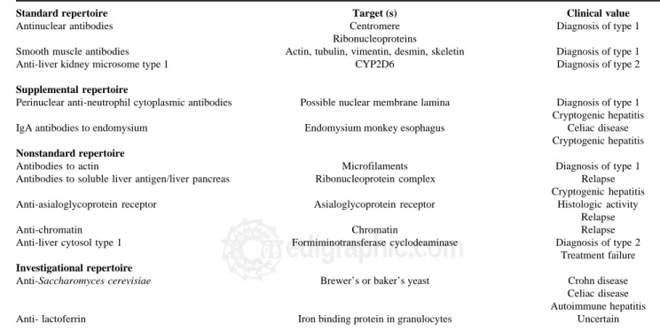
Autoantibodies
ANA, SMA, and anti-LKM1 constitute the standard repertoire of autoantibodies that are assessed in autoim-mune hepatitis1,12,90-92 (Table IV). Antibodies to soluble liver antigen/liver pancreas (anti-SLA/LP), neutrophil cy-toplasm (pANCA), endomysium, and tissue transglutam-inase are ancillary markers that are useful in evaluating patients who are seronegative for the standard battery.92 Other autoantibodies that have been described in autoim-mune hepatitis which are either not generally available, investigational in nature, or of limited clinical value are antibodies to asialoglycoprotein receptor (anti-ASGPR),93 actin,94 chromatin,95 liver cytosol type 1,96 double-strand-ed DNA,97 histones,98 Saccharomyces cerevisiae,99,100 and lactoferrin101 (Table IV).
New autoantibodies continue to be characterized in the hope that they will reveal critical pathogenic mechanisms or have prognostic value. Antibodies to SLA/LP are present in 26% of patients with autoimmune hepatitis who are otherwise seronegative,44,45 and they are useful in re-classifying cryptogenic chronic hepatitis as autoimmune hepatitis.102 These autoantibodies also identify individuals
Table IV. Autoantibodies associated with autoimmune hepatitis
Standard repertoire Target (s) Clinical value
Antinuclear antibodies Centromere Diagnosis of type 1
Ribonucleoproteins
Smooth muscle antibodies Actin, tubulin, vimentin, desmin, skeletin Diagnosis of type 1
Anti-liver kidney microsome type 1 CYP2D6 Diagnosis of type 2
Supplemental repertoire
Perinuclear anti-neutrophil cytoplasmic antibodies Possible nuclear membrane lamina Diagnosis of type 1 Cryptogenic hepatitis IgA antibodies to endomysium Endomysium monkey esophagus Celiac disease
Cryptogenic hepatitis Nonstandard repertoire
Antibodies to actin Microfilaments Diagnosis of type 1
Antibodies to soluble liver antigen/liver pancreas Ribonucleoprotein complex Relapse Cryptogenic hepatitis Anti-asialoglycoprotein receptor Asialoglycoprotein receptor Histologic activity
Relapse
Anti-chromatin Chromatin Relapse
Anti-liver cytosol type 1 Formiminotransferase cyclodeaminase Diagnosis of type 2 Treatment failure Investigational repertoire
M G
edigraphic.com
who have more severe disease than seronegativecounter-parts and who invariably relapse after corticosteroid with-drawal.103-105 Since anti-SLA/LP are closely associated with HLA DR3, they may be surrogate markers of a genetic pro-pensity for relapse or refractory disease.103,105 Analysis of the immunoprecipitated RNAs extracted from HeLa cell extracts is the most powerful, sensitive and specific meth-od to detect anti-tRNP(ser)sec/SLA/LP autoantibodies, but an enzyme-linked immunosorbent assay (ELISA) is available as a commercial kit and performance parameters between the methods are comparable.106
Perinuclear anti-neutrophil cytoplasmic antibodies (pANCA) and immunoglobulin A (IgA) antibodies to en-domysium (EMA) and tissue transglutaminase (tTG) are ancillary markers of autoimmune hepatitis that are avail-able in the clinical laboratory91,92 (Table IV). pANCA are found with great frequency (50%-92%) and in high titer in type 1 autoimmune hepatitis, and they can be the sole serological markers of this disease.107,108 IgA EMA have a sensitivity of 94% and specificity of 99% for celiac dis-ease,109,110 and they are less likely to be falsely positive in chronic hepatitis than IgA antibodies to tTG.100,111-113 Se-rological screening for celiac disease is important in pa-tients with autoimmune hepatitis and in papa-tients with chronic undifferentiated liver disorders since celiac ease can occur coincidentally with autoimmune liver dis-ease114,115 or cause liver dysfunction that may improve with gluten restriction.116-119
The autoantibodies that are still investigational and that have promise as prognostic indices include GPR and anti-actin (Table IV). The presence of anti-AS-GPR correlates with histological activity and the pro-pensity to relapse after corticosteroid withdrawal.93,120 Continuation of treatment until disappearance of anti-ASGPR has been associated with a sustained remission. Antibodies to actin identify patients with a higher fre-quency of treatment failure and death from liver failure or requirement for liver transplantation than seronega-tive patients, but they are restricted to those individuals with SMA.94
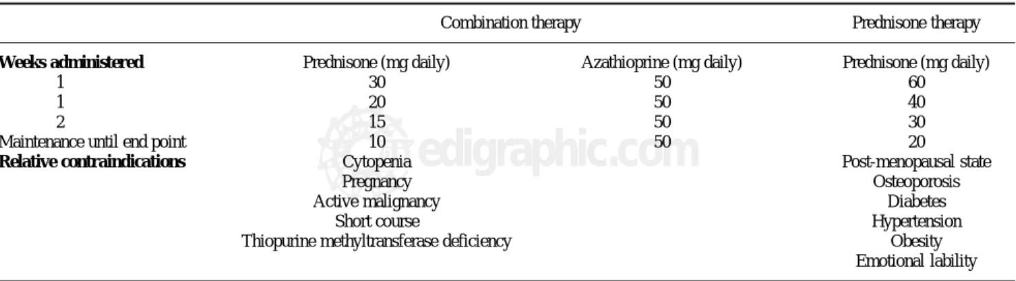
Conventional treatment schedules
The preferred treatment schedule for all forms of au-toimmune hepatitis in adults is prednisone in combination with azathioprine121-123 (Table V). Prednisone alone in higher dose is as effective as the combination regimen, but it is associated with a higher frequency of drug-relat-ed side effects (44% versus 10%). The preferrdrug-relat-ed treat-ment schedule in children is prednisone alone in a dose of 2 mg/kg daily (up to 60 mg daily).1 Azathioprine or 6-mercaptopurine can also be introduced as a corticoster-oid-sparing measure. Tapering schedules or alternate day corticosteroid regimens are commonly used in children to reduce deleterious effects on linear growth, bone devel-opment, and physical appearance.
Therapy is continued until remission, treatment failure, incomplete response, or drug toxicity.122,123 Remission im-plies the absence of symptoms, resolution of all laboratory indices of liver inflammation, and histological improve-ment to normal or minimal inflammatory activity. A serum aspartate aminotransferase level of less than twice the up-per limit of normal is an acceptable laboratory end point if the histological examination confirms the absence of inter-face hepatitis. Treatment failure connotes deterioration during therapy, and it is characterized by worsening of the serum aspartate aminotransferase or bilirubin level by at least 67% of previous values, progressive histological ac-tivity, or onset of ascites or encephalopathy. Incomplete re-sponse connotes improvement that is insufficient to satisfy remission criteria after 3 years of continuous treatment, and drug toxicity implies severe intolerance of the medica-tion. Histological improvement lags behind clinical and laboratory improvement by 3-8 months, and liver tissue examination prior to drug withdrawal is the only means of ensuring inactive disease.124,125
Treatment outcomes
Sixty-five percent of treated patients satisfy remission criteria within 18 months, and 80% achieve this result
Table V. Conventional treatment regimens.
Combination therapy Prednisone therapy
Weeks administered Prednisone (mg daily) Azathioprine (mg daily) Prednisone (mg daily)
1 30 50 60
1 20 50 40
2 15 50 30
Maintenance until end point 10 50 20
Relative contraindications Cytopenia Post-menopausal state
Pregnancy Osteoporosis
Active malignancy Diabetes
Short course Hypertension
16
edigraphic.com
within 3 years.122,123 The average duration of treatment untilremission is 22 months,28 and the 10-year life expectancies of treated patients with and without cirrhosis at accession are 89% and 90%, respectively.126 These survivals are com-parable to those of an age- and sex-matched normal popu-lation from the same geographical region. Patients with rhosis respond as well to treatment as patients without cir-rhosis, and they should be treated similarly with the same expectation of success.126,127 Twenty-one percent of indi-viduals who enter remission sustain this result long term after drug withdrawal (median 7interval of follow-up, 76 months), and an effort should be made to discontinue ini-tial therapy in all patients with inactive disease.128 Thirteen percent develop side effects that justify premature discon-tinuation of medication (drug toxicity); 9% deteriorate de-spite compliance with therapy (treatment failure); and 13% improve but not to a degree to satisfy criteria for remission (incomplete response).122,123
Corticosteroid therapy may also reduce hepatic fibro-sis.129-132 Fibrosis scores improved in 56% of patients fol-lowed for 55+9 months, and they did not progress in 33% of patients followed for 62+14 months.132 Histological ac-tivity indices decreased concurrently, and patients in whom the histological activity indices improved had a higher frequency of improvement in the fibrosis scores (80% versus 25%, p=0.002).132,133 These findings suggest that improvement in hepatic fibrosis occurs in conjunc-tion with reducconjunc-tions in liver inflammaconjunc-tion and that corti-costeroid therapy facilitates the disappearance of fibrosis by suppressing inflammatory activity. Small case studies have also suggested that cirrhosis can disappear during
treatment, but this possibility must await confirmation by assays more reliably reflective of cirrhosis than conven-tional needle biopsy of the liver.129,132
Treatment of suboptimal responses
Treatment failure is managed by administering high dose prednisone alone (60 mg daily) or prednisone (30 mg daily) in conjunction with azathioprine (150 mg dai-ly).122,123 Doses of prednisone and azathioprine are then reduced by 10 mg and 50 mg, respectively, for each month of laboratory improvement until conventional maintenance levels of drug are achieved. Seventy-five percent of patients treated in this fashion enter clinical and laboratory remission, but only 20% have histological resolution. These patients remain at risk for progressive liver disease and drug toxicity.
Drug toxicity requires premature dose reduction or dis-continuation of the offending drug and continued use of the other tolerated medication in adjusted dose.122,123 Cor-ticosteroid-related side effects are the most common causes for drug withdrawal, and they include intolerable cosmetic changes or obesity (47%), osteoporosis with vertebral compression (27%), brittle diabetes (20%), and peptic ulceration (6%).134 Azathioprine can be adminis-tered as a corticosteroid-sparing agent with doses in-creased to 2 mg/kg daily. The emergence of a cholestatic hepatitis, pancreatitis, rash, progressive cytopenia, or gas-trointestinal upset indicates azathioprine toxicity and the need for its withdrawal. The dose of prednisone is then adjusted to suppress disease activity.
Table VI. Evolving Drug Therapies and Site-specific Interventions.
New drug regimens Site-specific Interventions
Agent Possible Uses Intervention Mechanism(s)
Cyclosporine158-165 Treatment failure Blocking synthetic peptides180 Blocks 1st co-stimulatory signal
(5-6 mg/kg daily) Steroid intolerance First-line therapy
Mycophenolate mofetil167,168 Azathioprine intolerance Soluble cytotoxic T Blocks 2nd co-stimulatory signal
(2 g daily) Steroid withdrawal lymphocyte antigen-4182
6-mercaptopurine173 Azathioprine intolerance Oral tolerance regimens183 Dose dependent
(1.5 mg/kg daily) Treatment failure suppression immune response
Tacrolimus170,171 Treatment failure T cell vaccination184 Clonal depletion of
(4 mg twice daily) Steroid intolerance activated cytotoxic T lymphocytes
Budesonide172 Front-line therapy Cytokine manipulations186,187 Recombinant cytokines
(3 mg twice daily) Osteopenia Monoclonal antibodies
Ursodeoxycholic acid175 Mild disease in Japanese Gene therapy188 Limit fibrosis
(13-15 mg/kg daily) Promote repair
Counter cytokines
Methotrexate176 Treatment failure
Cyclophosphamide177 Steroid intolerance
Deflazacort174 Steroid sparing
M G
edigraphic.com
An incomplete response is declared after 3 years ofconventional therapy without remission.122,123 Patients im-prove but not to a degree to satisfy remission criteria, and they are at risk for drug-related side effects associated with standard doses of prednisone. Low dose prednisone or long-term maintenance therapy with azathioprine (2 mg/kg daily) is a treatment option.
Relapse after remission and drug withdrawal
Relapse occurs in 20%-86% of patients depending on the criteria for remission prior to drug withdrawal.135-137 Diminished stamina, arthralgias, and increase in the se-rum aspartate aminotransferase level to more than three-fold normal characterize this occurrence. Re-treatment with the original regimen typically induces another remis-sion, but relapse recurs in 79% within 6 months after drug withdrawal.135 With each re-treatment and relapse, the frequency of drug-related side effects increases, and this risk outweighs the low probability of a sustained remis-sion with repeated conventional treatments.138 Alternative therapies with either low dose prednisone or azathioprine are warranted after the second relapse.
The low dose prednisone regimen requires induction of clinical and laboratory remission on standard therapy and then reduction in the dose of prednisone by 2.5 mg each month of clinical and laboratory stability.139 The lowest dose that prevents symptoms and keeps serum aspartate aminotransferase levels below three-fold normal is main-tained. Eighty-seven percent of patients can be managed on prednisone, 10 mg daily or less (median dose, 7.5 mg dai-ly). Side effects associated with earlier conventional treat-ments improve or disappear in 85%; new side effects do not develop; and survival is unaffected.
Maintenance therapy with azathioprine also requires induction of clinical and laboratory remission by conven-tional treatments.140,141 The corticosteroid component is then withdrawn, and the dose of azathioprine is increased to 2 mg per kg daily and maintained indefinitely. Eighty-seven percent of adult patients managed in this fashion remain in remission during a median observation interval of 67 months. Follow-up liver biopsy assessments dis-close inactive or minimal histological disease in 94%; corticosteroid-related side effects improve or disappear in most patients; and the drug is generally well-tolerated. The most common side effects are withdrawal arthralgias (63%), lymphopenia (57%), and myelosuppression (7%). Neoplasms involving diverse cell types occur in 8%.
Relapse does not preclude permanent discontinuation of medication late in the course of the disease.128,138 Twen-ty-eight percent of patients who relapse and are re-treated develop inactive disease and can be withdrawn from medication. The probability of a sustained remission after initial or subsequent therapy is 47% during 10 years of follow-up. Conventional re-treatment schedules are able to induce a sustained remission more commonly than
long-term maintenance schedules (59% versus 12%, p=0.00002), but all management schedules should be withdrawn periodically to assess this outcome.
Liver transplantation
Liver transplantation is an effective treatment for the decompensated patient. Patient and graft survival after liv-er transplantation ranges from 83% to 92%, and the actuar-ial 10-year survival after transplantation is 75%.5,142 Recur-rence is recognized in at least 17% of patients after 5+1 years, especially in individuals receiving inadequate im-munosuppression.143 Adjustments in the immunosuppres-sive regimen are usually able to suppress recurrent disease, but rarely cirrhosis or graft failure occurs.144 Patients trans-planted for autoimmune hepatitis may also have a greater frequency of acute and chronic rejection than patients transplanted for non-autoimmune conditions.145,146 These potential consequences have tempered efforts to rapidly withdraw corticosteroids after the procedure.
Autoimmune hepatitis can develop de novo in children and adult recipients who undergo transplantation for non-autoimmune liver disease.147-152 Children seem to have a predilection for the syndrome; immunosuppression with cyclosporine is a common feature; and treatment with prednisone and azathioprine is typically effective. De novo autoimmune hepatitis is rare, occurring in 3%-5% of allografts, and it can result in graft loss if not treated with corticosteroids.153 In children, de novo disease may reflect defective negative selection of autoreactive cells by the thymus, generation of promiscuous lymphocytes by ex-cessive antigenic exposure, and/or impaired apoptosis of autoreactive cells by cyclosporine or tacrolimus. In adult patients, these same mechanisms are pertinent except for thymic dysfunction.154,155
Emerging drug therapies
There is no shortage of new drugs that have been pro-posed for autoimmune hepatitis156,157 (Table VI). Many have emerged from the transplantation arena, but none has been rigorously evaluated or incorporated into a con-ventional management algorithm. Of the drugs that prom-ise greater blanket immunosuppression than prednisone or azathioprine, cyclosporine and mycophenolate mofetil have shown the most promise. Controlled clinical trials are sorely needed to establish their efficacy.
Cyclosporine has been used successfully as salvage ther-apy in patients who have failed conventional corticosteroid treatment or been intolerant of the medication.158-165 It has also been used as first-line therapy in children and adults. 163-165 The medication binds cyclophilin and inhibits the
18
edigraphic.com
:rop odarobale FDPVC ed AS, cidemihparG
arap
acidémoiB arutaretiL :cihpargideM sustraídode-m.e.d.i.g.r.a.p.h.i.c
sustraídode-m.e.d.i.g.r.a.p.h.i.c cihpargidemedodabor
Mycophenolate mofetil is an ester prodrug hydrolyzed by liver esterases to produce the active metabolite, myco-phenolic acid, which in turn acts as a non-competitive, re-versible inhibitor of inosine monophosphate dehydrogena-se.166 Inosine monophosphate dehydrogenase is the rate-limiting enzyme for de novo synthesis of purines, and by inhibiting its action, mycophenolate mofetil selectively prevents the proliferative responses of T and B cells to mi-togens or antigens. Mycophenolate mofetil is a purine an-tagonist like azathioprine, and it has a low frequency of side effects (mainly leukopenia) and independence from the thiopurine methyltransferase pathway of catabolism.
Two small uncontrolled clinical experiences have shown improvement in the laboratory indices of liver in-flammation after the administration of mycophenolate mofetil, 1 gram twice daily, to patients unsuccessfully treated with azathioprine or intolerant of this drug.167,168 The prospect of replacing corticosteroids with mycophe-nolate mofetil has also stimulated its empiric use. Other preliminary experiences have failed to demonstrate a po-tent salvage effect or consispo-tent corticosteroid-sparing ac-tion, and the proper role and target population for this drug remain uncertain.169
Tacrolimus (4 mg twice daily),170,171 budesonide (3 mg thrice daily),172 6-mercaptopurine (1.5 mg/kg daily),173 deflazacort (7.5 mg for each 5 mg prednisone dose dai-ly),174 ursodeoxycholic acid (13-15 mg/kg daily),175 meth-otrexate,176 and cyclophospamide177 have all been used with anecdotal success in treating corticosteroid resistant or intolerant patients. Only ursodeoxycholic acid has been evaluated by randomized controlled clinical trial as a salvage therapy, and it is the one negative experience.178 Budesonide is currently undergoing clinical trial as front-line therapy after earlier studies had demonstrated its lim-itations as a salvage treatment.179
Emerging site-specific interventions
Site-specific interventions are designed to target key steps in the pathogenic pathway and thereby control the disease without inducing blanket immune suppression.157 These therapies are at theoretical or preliminary stages of development, and they await full clarification of the mo-lecular mechanisms of the disease and the availability of suitable animal models to assess their feasibility. The var-ious intracellular signaling pathways affecting immuno-cyte activation and proliferation are prime targets for these strategies.
Competitive inhibition of autoantigen presentation has already been applied in rheumatoid arthritis, and it can be considered in autoimmune hepatitis after full character-ization of its autoantigens.180 Synthetic peptides that com-pete with the autoantigen for presentation by class II MHC molecules can be used to block the first co-stimula-tory signal in immunocyte activation. Soluble cytotoxic T lymphocyte antigen-4 (CTLA-4) interferes with the
sec-ond co-stimulatory signal and dampens immunocyte acti-vation.181 It has already been used to blunt immune reac-tivity in mismatched blood marrow recipients, and it is a powerful tool by which to manipulate the immune re-sponse.182 Oral tolerance regimens induce systemic non-responsiveness to an autoantigen by oral feedings, and this intervention has already been used in the treatment of multiple sclerosis, rheumatoid arthritis, insulin dependent diabetes mellitus, myasthenia gravis and thyroiditis.183 Low dose regimens stimulate cytokine production and suppression of the immune response, whereas high dose regimens cause clonal deletion of immunocytes and aner-gy. The ingested antigen is delivered directly to the liver by the portal circulation, and the treatment may be espe-cially effective in autoimmune hepatitis once the critical epitope has been defined.184 T cell vaccination can deplete clones of activated cytotoxic T lymphocytes, and it is the only site-specific intervention that has been assessed in experimental murine autoimmune hepatitis.185 Identifica-tion and precise targeting of the critical T cell clones re-main challenges for this modality. Cytokine manipula-tions are feasible by administering drugs that disrupt in-tracellular signaling pathways involved in cytokine transcription or by using recombinant cytokines (such as IL-10) or monoclonal antibodies (such as anti-TNF) to al-ter the type of cytokine response. Similar inal-terventions are already being evaluated in inflammatory bowel dis-ease186 and chronic hepatitis C.187 Gene therapy also has promise in the treatment of a polygenic disorder such as autoimmune hepatitis if genes can be delivered that coun-terbalance the over-production of certain regulatory cy-tokines, limit fibrosis, or promote regeneration.188
Variant syndromes
Codification of the clinical criteria for the diagnosis of autoimmune hepatitis has facilitated recognition of vari-ant syndromes.189-192 These syndromes include patients with autoimmune hepatitis and another type of chronic liver disease (overlap syndrome) or findings suggestive but non-diagnostic of autoimmune hepatitis (outlier syn-drome). Overlap syndromes include patients with mixed features of autoimmune hepatitis and PBC or PSC, and outlier syndromes include patients with autoimmune cho-langitis (or AMA-negative PBC) and cryptogenic chronic hepatitis. These variant conditions currently lack an es-tablished identity, official designation, and treatment strategy. Their occurrences, however, must be recog-nized, and they should not be assimilated into diagnoses that hide their individuality or imperil the homogeneity of the classical diseases.
M G
edigraphic.com
cholestasis at presentation, and this is most easilyas-sessed by determining the magnitude of the serum alka-line phosphatase level above the upper limit of normal. Patients with variant syndromes and serum alkaline phos-phatase levels greater than twofold normal are unlikely to respond to this treatment.193-196
Management of the variant syndromes is empiric and based on the predominant manifestations of the disease. Patients with autoimmune hepatitis and features of PBC who have serum alkaline phosphatase levels less than twofold normal can be treated with corticosteroids.193-195 Patients with higher serum alkaline phosphatase levels and those with florid duct lesions on histological exami-nation are candidates for treatment with corticosteroids and ursodeoxycholic acid.196 Patients with autoimmune hepatitis and PSC lack an effective treatment, but they are candidates for a trial of therapy with prednisone and high dose ursodeoxycholic acid (15-20 mg/kg daily).193 Pa-tients with autoimmune cholangitis can be treated with prednisone, ursodeoxycholic acid, or both depending on the serum alkaline phosphatase level.193,197 Multicenter, collaborative studies are needed to codify diagnostic cri-teria and establish treatment algorithms.11
Summary
Autoimmune hepatitis should be considered in all pa-tients with acute or chronic hepatitis of undetermined cause, including patients with fulminant presentations and those who have been transplanted. Recurrent autoimmune hepatitis is possible after liver transplantation, and the dis-ease can develop de novo in children and adults transplant-ed for non-autoimmune diseases. The histological spec-trum of autoimmune hepatitis includes centrilobular or perivenular (Rappaport zone 3) necrosis, and background histological features of bile duct injury do not dissuade the diagnosis or alter therapy. Prednisone in combination with azathioprine is the preferred treatment in adults, and it may reduce fibrosis by suppressing inflammatory activity. New autoantibodies have promise as diagnostic and prognostic tools, and several are now available as commercial kits. Genetic factors influence disease expression and behavior, and they may be clues to region-specific etiologic agents. New treatments are evolving that promise better blanket immunosuppression and site-specific intervention. Variant syndromes are common, and they should be sought in all patients, especially in those who are refractory to corticos-teroid therapy.
References
1. Czaja AJ, Freese DK. Diagnosis and treatment of autoimmune hepa-titis. Hepatology 2002; 36: 479-497.
2. Czaja AJ, Carpenter HA. Sensitivity, specificity and predictability of biopsy interpretations in chronic hepatitis. Gastroenterology 1993; 105: 1824-1832.
3. Boberg KM, Aadland E, Jahnsen J, Raknerud N, Stiris M, Bell H. Incidence and prevalence of primary biliary cirrhosis, primary scle-rosing cholangitis, and autoimmune hepatitis in a Norwegian popula-tion. Scand J Gastroenterol 1998; 33: 99-103.
4. Boberg KM. Prevalence and epidemiology of autoimmune hepatitis.
Clin Liver Dis 2002; 6: 635-647.
5. Seaberg EC, Belle SH, Beringer KC, Schivins JL, Detre KM. Liver transplantation in the United States from 1987-1998: updated re-sults from the Pitt-UNOS liver transplant registry. In: Cecka JM, Terasaki PI, eds. Clinical Transplants 1998. Los Angeles: UCLA Tis-sue Typing Laboratories 1999: 17-37.
6. Hurlburt KJ, McMahon BJ, Deubner H, Hsu-Trawinski B, Williams JL, Kowdley KV. Prevalence of autoimmune hepatitis in Alaska na-tives. Am J Gastroenterol 2002; 97: 2402-2407.
7. Lim KN, Casanova RL, Boyer TD, Bruno CJ. Autoimmune hepatitis in African Americans: presenting features and responses to therapy.
Am J Gastroenterol 2001; 96: 3390-3394.
8. Nakamura K, Yoneda M, Yokohama S, Tamori K, Sato Y, Aso K, Aoshima M, Hasegawa T, Makino I. Efficacy of ursodeoxycholic acid in Japanese patients with type 1 autoimmune hepatitis. J
Gastroenterol Hepatol 1998; 13: 490-495.
9. Czaja AJ, Souto EO, Bittencourt PL, Cancado ELR, Porto G, Goldberg AC, Donaldson PT. Clinical distinctions and pathogenic implications of type 1 autoimmune hepatitis in Brazil and the United States. J Hepatol 2002; 37: 302-308.
10. Zolfino T, Heneghan MA, Norris S, Harrison PM, Portmann BC, McFarlane IG. Characteristics of autoimmune hepatitis in patients who are not of European Caucasoid ethnic origin. Gut 2002; 50: 713-717. 11. Czaja AJ, Bianchi FB, Carpenter HA, Krawitt EL, Lohse AW, Manns MP, McFarlane IG, Mieli-Vergani G, Toda G, Vergani D, Vierling J, Zeniya M. Treatment challenges and investigational opportunities in autoimmune hepatitis. Hepatology 2005; 41: 207-215.
12. Alvarez F, Berg PA, Bianchi FB, Bianchi L, Burroughs AK, Cancado EL, Chapman RW, Cooksley WGE, Czaja AJ, Desmet VJ, Donaldson PT, Eddleston ALWF, Fainboim L, Heathcote J, Homberg J-C, Hoofnagle JH, Kakumu S, Krawitt EL, Mackay IR, MacSween RNM, Maddrey WC, Manns MP, McFarlane IG, Meyer zum Buschenfelde K-H, Mieli-Vergani G, Nakanuma Y, Nishioka M, Penner E, Porta G, Portmann BC, Reed WD, Rodes J, Schalm SW, Scheuer PJ, Schrumpf E, Seki T, Toda G, Tsuji T, Tygstrup N, Vergani D, Zeniya M. Inter-national Autoimmune Hepatitis Group report: review of criteria for diagnosis of autoimmune hepatitis. J Hepatol 1999; 31: 929-938. 13. Amontree JS, Stuart TD, Bredfeldt JE. Autoimmune chronic active
hepatitis masquerading as acute hepatitis. J Clin Gastroenterol 1989; 11: 303-307.
14. Porta G, Da Costa Gayotto LC, Alvarez F. Anti-liver-kidney microsome antibody-positive autoimmune hepatitis presenting as fulminant liver failure. J Pediatric Gastroenterol Nutr 1990; 11: 138-140.
15. Nikias GA, Batts KP, Czaja AJ. The nature and prognostic implica-tions of autoimmune hepatitis with an acute presentation. J Hepatol 1994; 21: 866-871.
16. Kessler WR, Cummings OW, Eckert G, Chalasani N, Lumeng L, Kwo PY. Fulminant hepatic liver failure as the initial presentation of acute autoimmune hepatitis. Clin Gastroenterol Hepatol 2004; 2: 625-631. 17. Burgart LJ, Batts KP, Ludwig J, Czaja AJ. Recent onset autoimmune hepatitis: biopsy findings and clinical correlations. Am J Surg Pathol 1995; 19: 699-708.
18. Singh R, Nair S, Farr G, Mason A, Perrillo R. Acute autoimmune hepatitis presenting with centrizonal liver disease: case report and review of the literature. Am J Gastroenterol 2002; 97: 2670-2673. 19. Okano N, Yamamotos K, Sakaguchi K, Miyake Y, Shimada N, Hakoda
T, Terada R, Baba S, Suzuki T, Tsuji T. Clinicopathological features of acute-onset autoimmune hepatitis. Hepatol Res 2003; 25: 263-270. 20. Davis GL, Czaja AJ, Baggenstoss AH, Taswell HF. Prognostic and