Capillary electrophoresis determination of non protein amino acids as quality markers in foods
56
0
0
Texto completo
(2) CAPILLARY ELECTROPHORESIS DETERMINATION OF NON PROTEIN AMINO ACIDS AS QUALITY MARKERS IN FOODS. Raquel Pérez-Míguez, María Luisa Marina, María Castro-Puyana*. Department of Analytical Chemistry, Physical Chemistry and Chemical Engineering. Faculty of Biology, Environmental Sciences and Chemistry. University of Alcalá, Ctra. Madrid-Barcelona Km. 33.600, 28871 Alcalá de Henares (Madrid), Spain.. Keywords: capillary electrophoresis, non-protein amino acids, foods, quality markers, microchip electrophoresis.. Correspondence: Dr. María Castro-Puyana, Department of Analytical Chemistry, Physical Chemistry and Chemical Engineering. Faculty of Biology, Environmental Sciences and Chemistry. University of Alcalá, Ctra. Madrid-Barcelona Km. 33.600, 28871 Alcalá de Henares (Madrid), Spain. E-mail: [email protected] Tfn/Fax: 34-918856430/34-918854971. 1.
(3) Abstract Non-protein amino acids mainly exist in food as products formed during food processing, as metabolic intermediates or as additives to increase nutritional and functional properties of food. This fact makes their analysis and determination an attractive field in food science since they can give interesting information on the quality and safety of foods. This article presents a comprehensive review devoted to describe the latest advances in the development of (achiral and chiral) analytical methodologies by capillary electrophoresis and microchip capillary electrophoresis for the analysis of nonprotein amino acids in a variety of food samples. Most relevant information related to sample treatment, experimental separation and detection conditions, preconcentration strategies and limits of detection will be provided.. 2.
(4) 1. Introduction The determination of protein amino acids has been exploited for years in the food field because they can provide relevant information on the quality and safety of food samples [1,2,3]. In addition to these compounds, it is also possible to find other amino acids of non-protein origin in foods which exist as metabolic intermediates, as products formed during food processing or as additives in food to increase some nutritional and functional properties [4,5]. Non-protein amino acids have been defined as those amino acids that are not found in protein main chain either for lack of a specific transfer RNA and codon triplet or because they do not arise from protein amino acids by posttranslational modification [6]. Even though non-protein amino acids have been studied to a lesser extent, they have also shown to play an important role in the quality and safety of foods. Thus, different research works have demonstrated the relevance of determining non-protein amino acids to detect food adulterations, to investigate fermentation, storage and thermal treatments, to evaluate the nutritional quality of foods or to determine their toxic effects [7,8,9,10,11,12]. Non-protein amino acids can be chiral having one or more chiral centers providing, therefore, at least one pair of enantiomers. Obviously, each enantiomer can originate different effects when interacting with chiral environments as enzymes, proteins and receptors [3]. Although the L-enantiomers are the natural form of amino acids, their exposure to certain processing conditions may originate their racemization to Denantiomers. Processing-induced amino acid racemization includes from heat treatments, fermentation or storage to microbiological processes [13,14]. Moreover, D-enantiomers can be found in foods as a consequence of the fraudulent addition of racemic mixtures of non protein amino acids in supplemented foodstuffs where regulations establish the use of pure L-enantiomers. The use of racemic mixtures originates economical benefits due 3.
(5) to the minor cost that these mixtures have with respect to the use of pure enantiomers. In general terms, enantioselective separations may be relevant in different food areas in order to propose quality markers to assess food authenticity and to detect adulterations, to evaluate the effects of processing, fermentation, microbiological activity and storage, to analyze chiral metabolites and to investigate health-promoting compounds or evaluate flavor and fragrance aromas [2,3,15,16]. In the specific case of chiral non-protein amino acids, the analysis of their enantiomers in foods is a very useful tool not only to assess food quality and authenticity [17], but also to evaluate processing, manufacturing of food supplements and detect microbiological contaminations [12,18]. Undoubtedly, these research activities are of great interest in the area of Food Analysis due to the increasing concern of consumers about the quality of food. Therefore, taking into account that assuring product food quality, authenticity, and safety is the main demand in the food field, there is an increasing need of analytical methodologies enabling the determination of non-protein amino acids in foodstuffs. Among the advanced analytical techniques that can be used to solve some of the challenges that Food Science has to face, Capillary Electrophoresis (CE) has already demonstrated its high potential for the (achiral and chiral) determination of many compounds in foods, including amino acids, to ensure compliance with food and trade laws [1,19,20,21]. Among the CE modes used to determine non protein amino acids in food samples, CZE (based on the free mobility of analytes in the aqueous solution under an applied electric field) and MEKC (based on the combination of electrokinetic migration and the partitioning mechanism between the bulk solution and micelles) are the most employed, whereas EKC and CEC (based on the interaction of each enantiomer with a chiral selector present in the mobile phase or with a chiral stationary phase, respectively) are the most employed CE modes to achieve an enantiomeric separation. 4.
(6) The present article reviews the most recent advances in the analysis of non protein amino acids in foods using capillary electrophoretic methodologies (CZE, MEKC, EKC, and CEC) and microchip electrophoresis under achiral as well as chiral conditions covering the research articles published during the period February 2007 to February 2015, following the previous article published by our research group on this topic [1]. To make easier the discussion of the literature data and demonstrate the usefulness of CE to face different challenges related to food quality, this review has been divided in two different sections focused on the achiral and chiral determination of non-protein amino acids in foods. These sections gather the non-protein amino acids according to their structure and describe the different CE approaches (including experimental conditions, preconcentration strategies and sample treatments) developed to analyze these compounds in a great variety of food samples.. 2. Determination of non-protein amino acids in foods by CE under achiral conditions With the aim of providing an updated view on the achiral CE methodologies developed for the analysis of non-protein amino acids in the period of time reviewed in this article, Table 1 summarizes the main characteristics of the developed methodologies. It can be observed that, as expected, most of the research articles published employed CZE as separation mode, although the use of other modes such as electrokinetic chromatography using micelles as pseudostationary phase (MEKC mode), CEC and microchip capillary electrophoresis (MCE) has also been reported. With respect to the detection systems, UV and fluorescence are the most popular despite the need to include an additional analytical step (derivatization) to add a chromophore or fluorophore group into the molecule. o-phthaldialdehyde (OPA), fluorescein isothiocyanate (FITC), 45.
(7) chloro-7-nitrobenzofurazan (NDB-Cl) or 9-fluoroenylmethylchloroformate (FMOC), among others, have mainly been used as reagents for the derivatization of non-protein amino acids. To a lesser extent, mass spectrometry (MS), inductively coupled plasmaMS (ICP-MS), electrochemical and capacitively coupled contactless conductivity (C4D) have also been reported as detection systems coupled to CE for determining non-protein amino acids. It should be highlighted that a broad range of food samples, from beverages (such as tea, soy based beverages, cow milk, energy drinks, etc), to flour products, shellfish, vegetables (tomato, brassica or allium species, etc), rice, meat products, or vegetable and olive oils have been analyzed by the developed CE approaches as shown in Table 1. The achiral determination of non-protein amino acids in these food matrices has been carried out mainly with quality control purposes. Thus, research works focused on the detection of adulterations, the study of the effects of fermentation, storage or thermal treatments, or the evaluation of the nutritional quality of different foods samples have enabled to point out these compounds as potential markers of food quality. A more detailed description of the different achiral CE methodologies developed for the determination of non protein amino acids and their applications in the area of Food Analysis will be provided next.. 6.
(8) Table 1. Characteristics of the analytical methodologies developed for the achiral determination of non protein amino acids in foods by CE. Classification. Name (Abbr.). Aliphatic monoamiomonocarboxyl aminoacids. γ- aminobutyric acid (GABA). CE-mode/detection Separation conditions CZE-Fluorescence (λex 200-400 nm; λem 495 nm). Separation from: Ala. Sample treatment. Application. LOD*. Ref. Boil, filtration, and dilution with water before in-capillary derivatization with OPA/2-ME. Determination of GABA and Ala in tea samples. 0.004 µM. [22]. Ala. Boil, filtration, and dilution with ACN:0.1 mM NaCl (2:1 v/v) before in-capillary derivatization with OPA/2-ME. Determination of GABA and Ala in tea samples. 0.7 nM. [23]. 16 protein amino acids. Extraction with 75 % ethanol (v/v), ultrasonication, centrifugation, addition of 1 M HCl (0.9 mL supernatant: 0.1 mL HCl) prior to CE analysis. Simultaneous determination of free amino acids in several types of royal jelly products and honey. 0.84 µg/g. [24]. 7 Catechins, 3 xanthines, gallic acid, vitamin C, and theaflavin. Different treatment for making non-fermented, partiallyfermented and fully-fermented tea, drying, water extraction (under orbital shaking), and filtration before CE injection. Determination of tea fermentation through simultaneous analysis of catechins, xanthines, gallic acid, vitamin C, thea and theaflavins. -. [25]. 13 protein amino acids + GABA. Extraction with hot water, centrifugation, filtration and precapillary derivatization with NDB-Cl. Simultaneous determination of free amino acids in different tea leaves. 0.1 ng/L. [26]. 30 mM sodium borate (pH 10.0); capillary, 50 µm x 65cm; 21 kV; 23ºC CZE-Fluorescence (λex 200-400 nm; λem 495 nm) 50 mM sodium borate (pH 10.0); capillary, 50 µm x 65cm; 21 kV; 23ºC CZE-MS2 1 mM formic acid (pH 1.8); capillary, 50 µm x 100 cm; 30 kV, 20ºC. Aliphatic amino acids with nitrogen in the side chain. Theanine (Thea). MEKC-UV 10 mM sodium dihydrogenphosphate + 4 mM sodium borate + 45 mM SDS + 0.5% ethanol (pH 7.0); capillary, 50 µm x 38.5 cm; 20 kV; 30 ºC MEKC-LIF (λex488 nm; λem520 nm) 20 mM Brij 35 + 10% ACN (v/v) in 20 mM sodium borate (pH 8.5);capillary, 75 µm x 40cm; 20 kV; 25 ºC. 7.
(9) Table 1. Continued Classification. Name (Abbr.) Homoarginine (Har). CE-mode/detection Separation conditions CZE-UV (200 nm). Separation from: -. Sample treatment. Application. LOD*. Extraction with ethanol/water (6:4 v/v) and centrifugation prior to CE analysis. Analysis of Har accumulation in Grass Pea dry Seeds. -. [27]. -. Sequential extraction using TCA and microwave-digested with HCL, drying, and solution in deuterated water. Determination of BMAA in 18 strains of estuarine cyanobacteria. 0.5 mg/L. [28]. 5 nonprotein amino acids (β-ala, GABA, allo, cit, pyro) -. Extraction with methanol/chloroform (2:1 v/v), centrifugation, drying, butanol derivatization, evaporation and dilution in ACN: water (40:60 v/v). Acid hydrolysis, filtration, cleanup (C18 cartridge), drying and formic acid (0.2 % (v/v)) dilution prior to CE injection. Potential of non-protein amino acids as novel markers of adulterations of olive oils with seeds oils. 0.04 ng/g (Orn, βala, GABA, allo) 0.19 ng/g (cit, pyro). [7]. Evaluation of protein damage throughout determination of furosine in commercial soybased beverages (soy milk and cow's milk supplemented with soy isoflavones). 5.30 mg/100 g protein a. [29]. -. Acid hydrolysis, filtration, drying, dissolution with the running buffer and re-filtration before CE analysis. Qualitative and quantitative analysis of furosine in food products (flours, pasta, milk, and tigelle bread). 0.07 mg/L. [30]. -. Acid hydrolysis, filtration, drying, dissolution with the running buffer and re-filtration before CE analysis. Quantification of furosine as marker for the assessment of thermal treatment of a cerealbased model food (tigelle bread). -. [9]. 18.5 mM sodium borate (pH 9.2) + 10 mM sodium sulfate; capillary, 50 µm x 42 cm; 25 kV, 30 ºC β-N-methylamino-Lalanine (BMAA). CZE-UV (192 nm). Ornithine (Orn). CZE-MS2. 5 mM sodium borate (pH 9.0); 75 µm x 50 cm; 25 kV, 25 ºC. 0.1 M formic acid (pH 2.0); capillary, 50 µm x 60 cm; 25 kV, 25 ºC Heterocycle amino acids. Furosine. CZE-UV (280 nm) 50 mM sodium phosphate (pH 7.0); bubble capillary, 50 µm x 40 and 56 cm; 25 kV, 25ºC CZE-MS2 50 mM ammonium formate (pH 2.7); capillary, 50 µm x 60 cm; 25 kV, 30ºC CZE-MS2 50 mM ammonium formate (pH 2.7); capillary, 50 µm x 60 cm; 25 KV, 30ºC. Ref.. 8.
(10) Table 1. Continued Classification. Name (Abbr.) Domoic acid (DA). CE-mode/detection Separation conditions pCEC-UV (242 nm). Separation from: -. Sample treatment. Application. LOD*. Ref. Extraction with methanol/water (1:1 v/v), centrifugation, and filtration prior to CEC analysis.. Determination of DA in shellfish tissues. 0.5 µg/mL (equivalent to 2 µg DA/g, ww of mussel tissue). [11]. -. Extraction with methanol/water (1:1 v/v), centrifugation, filtration. Then non competitive inmunoreaction between free domoic acid antigen and (HRP)-labeled antidomoic acid antibody, incubation before CE analysis. Quantitative analysis of DA in contaminated shellfish samples(mussels, oysters, clams and scallops). 0.02 ng/mL. [31]. -. Derivatization with FITC (Energy drink was diluted 500fold before CE analysis). Quantification of Tau in an energy drink and a cow´s milk. -. [32]. Caffeine. Sonication (to remove dissolved gases) and dilution (10-fold) with the BGE. Simultaneous determination of taurine and caffeine in energy drinks. 24 mg/L. [33]. -. Buffer dissolution, derivatization with NBD-Cl, and dilution (10-fold) in separation buffer prior to CE analysis. Quantitative determination of Tau energy and sports drinks. 1 x 10⁻⁶M. [34]. ACN: 5 mM Tris (pH 8.0) (60:40 v/v) at a flow rate of 0.05 mL/min; packed capillary column, 100 µm x 55 cm (total length of which 20 cm was packed with ODS particles); -13kV; supplementary pressure 7.2 MPa CZE-EIA-EC (Ed: -0.35 V) 1 % PVP + 1 mM H2O2 in 10 mM BR buffer (pH 5.0); separation capillary, 50 µm x 20 cm; reaction capillary, 50 µm x 5 cm; 15 kV. Sulfur amino acids. Taurine (Tau). CZE-LIF (λex and λem unspecified) 20 mM sodium phosphate (pH 11.8); capillary, 75µ x 40 cm; 22 kV, 23 ºC MEKC-C4D (450 kHz; 17 Vpp) 40 mM CHES + 15 mM NaOH + 50 mM SDS ( pH 9.36); capillary, 50 µm x 8 cm; 5 kV MCE-Fluorescence (λem 545 and 605 nm). 9.
(11) Table 1. Continued Classification. 50 mM borate, (pH 9.3); glass microchip with an orthogonal channel design (10 x 40 mm) and a channel cross section of 20 x 50 µm. Name (Abbr.). CE-mode/detection Separation conditions MCE-LIF (λex635 nm; λem 495 nm). Separation from: Lys, vitamin B3. Sample treatment. Application. Two-fold dilutions with 40 mM sodium borate, pH adjustment (pH 8.6), derivatization with Cy5 and dilution with sample buffer (10 mM sodium borate, pH 9.88). Simultaneous analysis of amino acids (Tau and Lys) and vitamin B3in functional drinks.. 0.50 nM. [35]. GSH, Cys, γ-GluCys, CysGly, and NAC. Extraction with PBS, trichloroacetic acid, and EDTA (added in order) centrifugation, neutralization with sodium hydroxide, and derivatization with DMDSPAB-I before CE analysis. Determination of Hcy and other thiols in cucumber and tomato samples. 0.15 nM. [36]. Separation between methiin and alliin.. Boiling and water extraction (microwave), water dilution, and filtration before CE analysis.. Analyze of S-alk(en)ylcysteine S-oxides in allium and brassica vegetables. -. [37]. Separation between Methiin, Alliin, Ethiin Isoalliin Propiin. Extraction with methanol/water (90:10 v/v) + 10 mM HCl, drying until 10-15 ml and dilution with 20 mM sodium borate (pH 9.2), filtration, and derivatization with FMOC. Simultaneous determination of S-alk(en)ylcysteine-Soxidesin alliaceous and cruciferous vegetables (e.g. garlic, onion, leek, chive, cabbage, radish, cauliflower and broccoli). 0.2 pmol. [38]. 100 mM sodium borate (pH 9.88); glass microchip with a simple cross channel design; separation channel, (60 mm x 25 μmx 70 μm (length x depth x width)) 45 mm from injection to the detector Homocysteine (Hcy). CZE-LIF (635 nm) 16 mM sodium citrate (pH 7.0) + 60% v/v ACN; capillary, 75 µm x 50 cm; 22,5 kV, 25 ºC. S-alk(en)ylcysteine-Soxides. CZE-UV (350 nm) 20 mM sodium benzoate + 0.5 mM TTAB (pH 12.0); capillary, 50µm x 91.5 cm; - 30 kV, 25 ºC MECK-indirect UV (265 nm) 20 mM sodium borate + 20mM SDS + 10% (v/v) MeOH (pH 9.2); capillary, 75µm x 67 cm; 20 kV, 25 ºC. LOD*. Ref. 10.
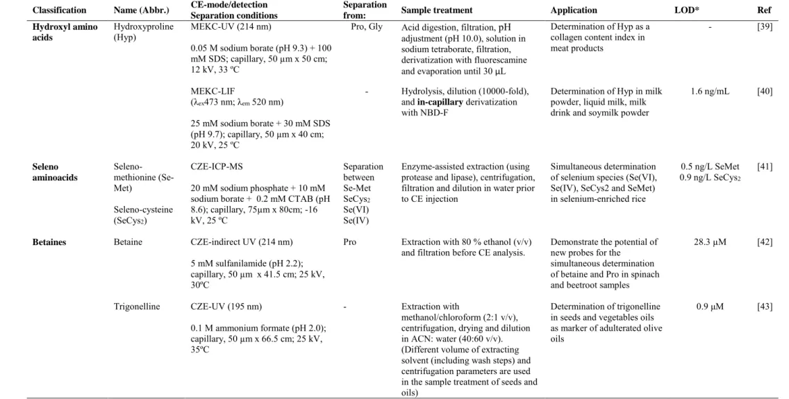
(12) Table 1. Continued Classification. Name (Abbr.). Hydroxyl amino acids. Hydroxyproline (Hyp). CE-mode/detection Separation conditions MEKC-UV (214 nm). Separation from: Pro, Gly. Sample treatment. Application. Acid digestion, filtration, pH adjustment (pH 10.0), solution in sodium tetraborate, filtration, derivatization with fluorescamine and evaporation until 30 µL. Determination of Hyp as a collagen content index in meat products. Hydrolysis, dilution (10000-fold), and in-capillary derivatization with NBD-F. Seleno-cysteine (SeCys2). 20 mM sodium phosphate + 10 mM sodium borate + 0.2 mM CTAB (pH 8.6); capillary, 75µm x 80cm; -16 kV, 25 ºC. Separation between Se-Met SeCys2 Se(VI) Se(IV). Betaine. CZE-indirect UV (214 nm). [39]. Determination of Hyp in milk powder, liquid milk, milk drink and soymilk powder. 1.6 ng/mL. [40]. Enzyme-assisted extraction (using protease and lipase), centrifugation, filtration and dilution in water prior to CE injection. Simultaneous determination of selenium species (Se(VI), Se(IV), SeCys2 and SeMet) in selenium-enriched rice. 0.5 ng/L SeMet 0.9 ng/L SeCys2. [41]. Pro. Extraction with 80 % ethanol (v/v) and filtration before CE analysis.. Demonstrate the potential of new probes for the simultaneous determination of betaine and Pro in spinach and beetroot samples. 28.3 µM. [42]. -. Extraction with methanol/chloroform (2:1 v/v), centrifugation, drying and dilution in ACN: water (40:60 v/v). (Different volume of extracting solvent (including wash steps) and centrifugation parameters are used in the sample treatment of seeds and oils). Determination of trigonelline in seeds and vegetables oils as marker of adulterated olive oils. 0.9 μM. [43]. 0.05 M sodium borate (pH 9.3) + 100 mM SDS; capillary, 50 µm x 50 cm; 12 kV, 33 ºC -. 25 mM sodium borate + 30 mM SDS (pH 9.7); capillary, 50 µm x 40 cm; 20 kV, 25 ºC. Betaines. Selenomethionine (SeMet). CZE-ICP-MS. 5 mM sulfanilamide (pH 2.2); capillary, 50 µm x 41.5 cm; 25 kV, 30ºC Trigonelline. CZE-UV (195 nm) 0.1 M ammonium formate (pH 2.0); capillary, 50 µm x 66.5 cm; 25 kV, 35ºC. Ref -. MEKC-LIF (λex473 nm; λem 520 nm). Seleno aminoacids. LOD*. 11.
(13) Table 1. Continued Classification. Name (Abbr.) trigonelline glycine betaine proline betaine carnitines. Carnitine (Carn). CE-mode/detection Separation conditions CZE-MS2. Separation from: -. 0.1 M ammonium formate (pH 2.0); capillary, 50 µm x 60 cm; 25 kV, 25 ºC. CZE-C4D (320 kHz; 280 Vpp). -. 500 mM acetic acid + 0.05 % (v/v) tween-20 (pH 2.6); capillary, 50 µm x 32 cm; 20 kV Others. N-phenylpropenoylL-amino acids (NPA). CZE-UV (325 nm) CE system 1: 50 mM sodium borate (pH 8.8), capillary, 70 µm x 70 cm; 30 kV, 27 ºC CE system 2: 50 mM sodium borate (pH 8.8), capillary, 50 µm x 50 cm; 20 kV, 27 ºC. Separation between Caff-Asp pC-Asp Caff-DOPA. Sample treatment. Application. LOD*. Ref. Extraction with methanol/chloroform (2:1 v/v), centrifugation, drying, butanol derivatization, evaporation and dilution in ACN: water (40:60 v/v).. Potential of betaines as novel markers of adulterations of olive oils with seeds oils. 0.050 ng/g (Carnitines, trigonelline) 0.075 ng/g (glycine and proline betaine). [8]. Dilution with 500 mM acetic acid, centrifugation and injection in the CE system. Quantification of carnitine in foodstuffs (fruit, juices, milk, yogurt, cheese, red meat and chicken meat) and food supplements. 2.6 µM. [44]. Cocoa beans and shells: deffating (soxhlet), extraction with acetone/water (7:3 v/v), centrifugation, rotatory evaporation, and centrifugation prior to CE analysis Flowers, leaves, husk, pulp, and shells: same extraction protocol without deffating. Analysis of main NPA in cocoa and cocoa products. -. [45]. *LODs units expressed as in the original work. These LODs are referred to the injected solutions of standard samples except for a) in which LODs is referred to the injected solutions of food samples. **Capillary dimension expressed as internal diameter x effective length (cm to the detector). ACN, acetonitrile; Ala, alanine; Allo, allo-isoleucine; BR, Britton-Robinson buffer; Caff-Asp, N-[3',4'-dihydroxy-(E)-cinnamoyl]-L-aspartic acid; Caff-DOPA, N-[3',4'-dihydroxy-(E)cinnamoyl]-3-hydroxy-L-tyrosine; C4D, capacitively coupled contactless conductivity; CE-EIA, capillary electrophoresis based enzyme immunoassay; CHES, 2-(N-cyclohexylamino)ethane sulfonic acid; Cit, citruline; CTAB, cetyltrimethylammonium bromide; Cy5, Sulfoindocyanine succinimidyl ester; Cys, cysteine; CysGly, cysteinylglycine; CZE, capillary zone electrophoresis; DMDSPAB-I, 1,7-dimethyl-3,5-distyryl-8-phenyl-(4'-iodoacetamido) difluoroboradiaza-s-indacene; EC, electrochemical; EDTA, ethylene diamine tetra acetic acid; EMMA, electrophoretically. 12.
(14) mediated microanalysis; FICT, fluorescein isothiocyanate; FMOC, 9-fluorenyl-methyloxycarbonyl chloride; GSH, glutathione; γ-GluCys, γ-glutamylcysteine; Gly, glycine; HRP, horseradish peroxidase; LIF, laser-induced fluorescence; Lys, lysine; 2- MCE, microchip capillary electrophoresis; ME, 2-mercaptoethanol; MEKC, micellar electrokinetic chromatography; MS2, tandem mass spectrometry; NDB-Cl, 4-chloro-7-nitrobenzofurazan; ODS, octadecyl silica; OPA, o-phthaldialdehyde; PBS, phosphate buffer saline; pCEC, pressurized capillary electrochromatography; pCTyr, N-[4'-hydroxy-(E)-cinnamoyl]-L-tyrosine; pC-Asp, N-[4'-hydroxy-(E)-cinnamoyl]-L-aspartic acid; PDA, diode array detector; Pro, proline; PVP, polyvinylpyrolidone; Pyro, pyroglutamic acid; SDS, sodium dodecyl sulfate; Se, selenium; TCA, trichloroacetic acid; TTAB, tetradecyltrimethyl-ammonium bromide.. 13.
(15) 2.1 Aliphatic monoamino-monocarboxyl amino acids γ-Aminobutyric acid (GABA) is a non-protein amino acid whose structure contains one carboxylic group and one primary amino group attached to the gamma carbon atom. It is distributed throughout the nervous system and it is the main inhibitory neurotransmitter in the mammalian brain which helps to regulate neuron activity and keep nerve cells firing normally [46,47]. In addition, GABA has demonstrated to have potential to maintain the balance of blood pressure in individuals with hypertensive cardiovascular disease [48]. In the period of time covered by this review, different CE methodologies coupled to fluorescence o MS detection systems were developed for the determination of GABA in tea, commercial dietary supplements containing royal jelly and honey (see Table 1). The lack of a chromophore group in GABA makes essential to carry out a derivatization step to enhance fluorescence detection. Thus, an in-capillary labeled derivatization reaction was investigated with the aim of determining the content of GABA and alanine (Ala) in different tea samples. The in-capillary derivatization was based on the use of ophthalaldehyde/2-mercaptoethanol (OPA/2-Me) as labeling reagent. OPA reacts rapidly with primary amines in presence of a thiol co-reactant under alkaline conditions, and 2ME (neutral co-reactant) minimized the instability of the resulting fluorophores. Under the optimized derivatization and CZE conditions (30 mM sodium tetraborate (pH 10.0)), GABA and Ala were analyzed in only 14 min (including in-capillary derivatization and CZE separation) with LODs of 0.004 and 0.02 M, respectively. Due to the existence of matrix interferences, a 1000-fold dilution of GABA-rich tea was needed before applying the developed methodology to the analysis of both amino acids in tea samples [22]. However, those tea samples with a low content of GABA could not be diluted. Then, to make possible the detection after dilution, an in-capillary sample stacking 14.
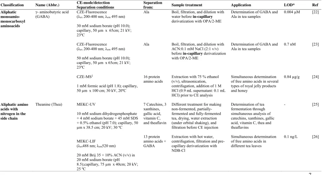
(16) preconcentration was subsequently included in the CZE-fluorescence methodology (see Figure 1A) [23]. By using this strategy, both GABA and Ala were detected in jasmine green tea sample (which has a low content of GABA) as it is shown in Figure 1B. The use of sample stacking preconcentration allowed achieving LODs of 0.7 and 0.8 nM for GABA and Ala, respectively. This implies a relevant enhancement of the LODs, from M up to nM level, which clearly shows that the dilution of sample solution followed by a preconcentration strategy is an interesting option to carry out the sensitive CE detection of analytes in complex matrix samples. Moreover, both methodologies could be applied with quality control purposes since they enabled to monitor the GABA content in the GABA-rich tea manufacturing process. From an analytical point of view, these methodologies are of great interest since they include, in just one analysis, from an easy sample treatment (dilution to avoid interferences and derivatization to enhance fluorescence) to an in-capillary preconcentration strategy to improve sensitivity. Undoubtedly, both methodologies provide an easy, fast, and sensitive option for determining GABA in routine analysis. The simultaneous determination of GABA and sixteen protein amino acids could also be performed without derivatization using a CZE-MS2 method which did not require a concentration step for sample preparation [24]. Using 1M formic acid (pH 1.8) as BGE and 50 % (v/v) methanol as sheath liquid, it was possible to carry out the identification and simultaneous determination of all the amino acids studied (see Table 1) in different dietary supplements containing royal jelly (tablets, capsules, powder, liquid drinks and raw materials) and honey samples. LOD for GABA was 0.84 µg/g. Since the product analyzed contained specific proportions of amino acids, the developed methodology was useful to distinguish among different royal jelly products. Moreover, taking into account that royal jelly raw material has a different composition from honey, this CE-MS2 strategy 15.
(17) along with the analysis of the content of trans-10-hydroxy-2-decenoic acid (the main fatty acid in royal jelly) could be employed to detect the intentionally use of honey instead of royal jelly. Figure 1.. Fig. 1. A) Diagram of the in-capillary labeled derivatization and stacking in ACN/salt solution: (a) working sample solution and labeling agents were introduced successively to the anodic end of the capillary column; (b) labeled derivatization occurred in the stacking process of analytes; (c) stacking of the labeled derivatives; (d) CZE began once the stacked ions left the sample zone solution. B) Electropherograms of GABA and alanine in jasmine green tea sample: (a) undiluted; (b) via stacking. CZE conditions: BGE, 50 mM sodium borate (pH 10); uncoated fused-silica capillary, 50 µm x 65 cm; voltage; 21 kV; temperature, 23 ºC. Peaks: 1, GABA adduct; 2, Ala adduct. Reprinted with permission from [23]. Copyright (2010) American Chemical Society.. 16.
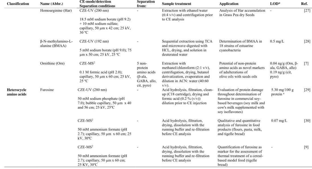
(18) 2.2 Aliphatic amino acids with nitrogen in the side chain Theanine (Thea), L-homoarginine (Har), β-N-methylamino-L-alanine (BMAA), and ornithine (Orn) are non protein amino acids containing nitrogen in their side chains. Theanine, the chief non-protein amino acid in tea (representing up to 50 % of the total amino acid content), is the main responsible of the taste of tea. It also has demonstrated to have an important role in different biological activities such as promoting relaxation, decreasing levels of serotonine and norepinephrine in brain, reducing blood pressure and enhancing anti-tumor activity [49]. A MEKC method using a BGE (pH 7.0) containing 10 mM phosphate, 4 mM sodium tetraborate, 45 mM sodium dodecyl sulfate (SDS), and 0.5% ethanol, and UV detection (205 and 266 nm) was developed by Hsiao et al. [25] for the simultaneous determination of theanine, seven catechins, three xanthines, gallic acid, vitamin C and theaflavins in non-fermented, partially-fermented and fully-fermented tea samples. The successful separation of the studied compounds within 8 min enabled the application of the proposed method for the quality control of tea fermentation (in particular, the theanine content did not change significantly). Lately, Yan et al. [26] developed for the first time a high sensitive MEKC approach with LIF detection for the simultaneous analysis of 13 protein amino acids, theanine and GABA in five different tea leaves as shown in Figure 2. Brij 35 was used as surfactant and 4-chloro-7nitrobenzofurazan (NBD-Cl) was chosen as labeling reagent to avoid time-consuming sample cleanup procedure because it does not react with other water soluble extracts in tea. This MEKC-LIF methodology enabled the detection up to 0.5 ng/mL and 0.1 ng/mL of GABA and theanine respectively, and has proved to be an efficient method for determining amino acids in tea. The two MEKC methodologies developed during the time covered by this review have a great potential to carry out an exhaustive characterization 17.
(19) of tea samples since both enable the determination of not only theanine but also of a broad variety of compounds that can be of high interest to evaluate manufacturing processes. Figure 2.. Fig. 2. MEKC electropherograms of amino acids in different tea varieties. (A) black, (B) jasmine, (C) green, (D) maofeng, and (E) biluochun. Derivatization conditions: 40 mM NBD-Cl, 30 mM sodium borate (pH 8.5), reaction time of 30 min at 60 ºC. Separation conditions: BGE, 20 mM sodium borate (pH 8.5) with 20 mM Brij 35 and 10 % acetonitrile (v/v); voltage, 20 kV; temperature, 25 ºC. Peak 1, Lys; 2, Phe; 3, Leu; 4, Met; 5, Val; 6, Thea; 7, His; 8, GABA; 9, Thr; 10, Ala; 11, Ser; 12, Gly; 13, Cys; 14, Glu; 15, Asp. Reprinted from [26], copyright (2014) with permission from Elsevier.. 18.
(20) L-Homoarginine (Har) and β-diaminopropionic acid (β-ODAP) are the main non protein amino acids in grass pea seeds. The impact of Har in humans and animal diets has given rise to contrasting opinions. A positive effect is considered due to its conversion into lysine and its potential to modulate β-ODAP toxicity [50,51]. However, other studies suggested that Har modulates the biosynthesis of NO decreasing the excitation of neuronal receptors, and its presence in gene activator-repressor histones could be a cause of different cancer types [52,53]. The variation of the Har amount among grass pea genotypes cultivated in different Italian regions (with different soil properties and climatic conditions) in two consecutive years was investigated by using a CZE method [27]. The Har analysis was accomplished with UV detection at 200 nm using a 18.5 mM sodium borate (pH 9.2) containing 10 mM sodium sulfate as running buffer. This methodology was successfully applied to obtain information about the variation of Har in different grass pea genotypes observing that in all cases there was a trend towards increasing Har content in the second season being Har contents always higher in one of the regions investigated (Guardia Perticara). Moreover, the variation of grain yield did not affect the Har storage. β-N-methylamino-L-alanine (BMAA) is a neurotoxic non-protein amino acid involved in the Amyotrophic Lateral Sclerosis which has been found in strains of cyanobacteria in fresh water and marine environment [54,55]. Due to its toxicological effect its quantitation in cyanobacteria is of great interest. A CZE method based on the use of 5 mM sodium borate (pH 9.0) as BGE and UV detection at 192 nm was developed by Baptista et al. [28] enabling the separation of BMMA from its isomer 2,4diaminobutyric acid (DBA). Eighteen strains of lyophilized estuarine cyanobacteria were analyzed employing the proposed method after a sequential BMMA extraction using trichloroacetic acid (TCA) and hydrochloric acid. Hydrochloric acid extraction was more 19.
(21) effective than TCA extraction (except for one type of cyanobacteria which is called Nostoc sp.). An interesting CE-MS2 methodology was developed enabling the determination of non-protein amino acids in vegetable oils [7]. The simultaneous separation of ornithine, β-alanine, GABA, alloisoleucine, citrulline, and pyroglutamic acid, previously derivatized with butanol, was achieved in only 15 min with a 0.1 M formic acid buffer (pH 2.0). LODs between 0.04 and 0.19 ng/g were achieved when a normal stacking was used as preconcentration technique. Under these conditions, different vegetable oils (soybean, sunflower, corn and extra virgin olive oils) were analyzed in order to identify the selected non-protein amino acids. β-alanine, GABA, and pyroglutamic acid were detected in all the vegetable oils analyzed, whereas ornithine and alloisoleucine were only detected in soybean, corn and sunflower oils, and citrulline was not detected in any sample. Bearing these results in mind, and corroborating the presence of ornithine and alloisoleucine in seed oils but not in olive oils by MS2 experiments, these two non-protein amino acids were proposed as novel markers for the detection of olive oil adulterations with sunflower, soybean and corn seed oils.. 2.3 Heterocycle amino acids Furosine and domoic acid are non protein amino acids characterized by the presence of a heterocycle group in the side chain and were analyzed by CE in the period of time covered by this review as it can be seen in Table 1. Furosine is originated in the reaction between lysine and reducing carbohydrates and is one of the Maillard reaction products most widely used as markers of the nutritional quality of foods. Its presence and amount in foods is related to the Maillard reaction so that it has demonstrated to be a reliable indicator of thermal treatment in many food 20.
(22) products. In the last eight years furosine was analyzed by CE with the aim of evaluating protein damage in commercial soy-based beverages [29], to study the effect of milling, drying or thermal processes in different food products (flours, pasta, milk and tigelle bread) [30] or to investigate the cooking effect of different ovens on a cereal-based model food (tigelle bread) [9]. All the strategies developed with these purposes are based on simple sample treatments and fast CE analysis (around 4 min), both conditions of high interest for setting up an analytical methodology in food control laboratories. To evaluate the protein quality of soy beverages, furosine was determined by CZE in different types of soy milk or cow's milk supplemented with soy isoflavones. A 50 mM sodium phosphate buffer (pH 7.0) was employed as BGE together with UV detection. The results were comparable with those obtained by an HPLC method. Even though HPLC proved to be slightly more sensitive than CZE (LODs of 1.30 and 5.30 mg/100 g of protein for HPLC and CZE, respectively), CZE was less time consuming (4 min versus 20 min) and caused less contamination, which showed the feasibility of the proposed CZE method [29]. The influence of different thermal processes on the quality of several food products was also investigated through the determination of furosine by CZE coupled to MS detection. Bignardi et al. [30] optimized different experimental conditions, such as capillary length, BGE concentration and pH, and applied voltage to establish a fast and reliable CZE-MS2 for determining the furosine content in flours, pasta, milk and tigelle bread (treated under different milling, drying or thermal processes). Under the optimized conditions (see Table 1), furosine was analyzed in 4 min achieving a LOD of 0.07 mg/L. Afterwards, these authors employed the same CE-MS2 method to evaluate the effect of different cooking treatments (two different ovens, modifying time and temperature) on tigelle bread through the determination of the furosine amount (along with the measure 21.
(23) of maltose: maltulose ratio or colour index) [9]. The negative correlation between the furosine amount and the different cooking processes, suggested that under the cooking parameters tested furosine was transformed in other molecules. Domoic acid (DA) is a crystalline water soluble kainoid amino acid which has neurotoxic effects. The consumption of shellfish containing high concentration of DA could be responsible of the amnesic shellfish poisoning (ASP) syndrome that produces abdominal cramps, vomits, disorientation and memory loss [56]. Despite the great evolution of CEC as separation technique, its potential in the analysis of non-protein amino acids in foods has not yet been achieved. This fact can be clearly observed in Table 1 since the only CEC methodology proposed in the period of time covered by this review to carry out the achiral determination of non-protein amino acids was applied to the rapid quantitation of DA in shellfish tissue extracts [11].To avoid practical problems related with bubble formation and column drying, an additional pressure was applied to the column inlet. Thus, a pressurized CEC-UV (pCEC-UV) method based on the use of a packed capillary column with octadecyl silica (ODS) particles, an isocratic separation (flow rate of 0.05 mL/min), and a supplementary pressure of 7.2 MPa, was developed to separate DA in shellfish matrices within 6 min achieving a LOD of 0.5 µg/mL. A second CE methodology based on enzyme immunoassay and electrochemical detection was also proposed to carry out the quantitative analysis of DA in shellfish samples [31]. This method, based on noncompetitive immunoreaction between DA antigen (Ag) and horseradish peroxidase (HRP)-labeled antidomoic acid antibody tracer (Ab*) in liquid phase, was able to separate the immmunocomplex (Ab*-Ag) and unbound (Ag*) in 4 min. The electrochemical detection was accomplished measuring amperometrically the enzymatic product obtained from the oxidation of o-aminophenol (OAP) with hydrogen peroxide. An LOD of 0.02 ng/ml was obtained enabling this approach to improve around 22.
(24) 16 times the sensitivity reached by a commercial ELISA check kit. Although the proposed methodology provides a sensitive approach for the trace determination of DA in shellfish samples, the need to carry out a noncompetitive immune reaction between DA antigen and (HRP)-labeled antidomoic acid antibody as well as the use of an electrochemical detector can difficult its implementation in routine food laboratories.. 2.4 Sulfur amino acids. Taurine (Tau), Homocysteine (Hcy) and a group of S-alk(en)ylcysteine-S-oxides (namely, methiin, ethiin, isoallin, propiin and butiin) were the sulfur-containing non protein amino acids analyzed by CE in the reviewed period. Taurine is a semi-essential amino acid that plays an important role in a variety of physiological. functions (antioxidation, neuromodulation, etc), pharmacological. properties (liver protection, low blood pressure, etc), and pathological effects (change of taurine´s levels in tissues and physiological fluids has a close relationship with different diseases such as Alzheimer, cardiovascular diseases or epilepsy, among others) [32]. Nowadays, the high consumption of energy and sport drinks makes that the normal daily uptake of taurine (one of the main components of these drinks) could be exceeded which can have undesirable effects. Therefore, reliable analytical methodologies are needed to determine taurine in foods from a quality control viewpoint. Bearing in mind this purpose, different strategies based on CZE, MEKC and microchip capillary electrophoresis (MCE) have been proposed. For instance, a CZE method with LIF detection was developed by Zinello et al. [32] to determine taurine in food and clinical samples (energy drinks and cow´s milk). The use of a high incubation temperature (100 ºC) enabled to reduce the reaction time between fluorescein isothiocyanate (FITC) and taurine from 6-14 h to 20 min. After optimization of different electrophoretic variables (buffer concentration, pH, 23.
(25) and temperature), the use of 20 mM sodium phosphate at pH 11.8, a temperature of 23 ºC, and a separation voltage of 22 kV allowed the analysis of taurine in less than 12 min. Different energy drinks were analyzed observing taurine contents in good agreement with the labeled ones. MEKC was also employed to determine taurine in energy drinks. In this case, the aim was to determine the caffeine and taurine contents (the major components) simultaneously in energy drinks, using a dual detection system in a short capillary (10.5 cm) employing a laboratory-home-made instrument [33]. Caffeine was detected by UV detection, whereas contactless conductivity detection (C4D) was employed for determining taurine since it does not absorb in the UV/Vis region. Using a simple sample treatment and a 40 mM CHES buffer containing 15 mM sodium hydroxide and 50 mM SDS, taurine and caffeine analysis was accomplished in only 1 min. This method has many outstanding features including the elimination of derivatization step and the possibility to carry out a fast separation of the two main compounds in energy drinks. Regarding MCE, it is one of the most relevant applications of micro-fluidics which offers some advantages such as miniaturization, short analysis times, and low solvent and sample consumption. From its introduction, MCE has experienced a substantial growth so that it is being considered as a new trend capable of solving a variety of problems in food analysis. In the period of time covered by this review, two different MCE methodologies were developed to determine taurine in beverages with fluorescence and LIF detection. On the one hand, using a simple sample pretreatment (just two dilution steps) and 4-chloro-7-nitro-1,2,3-benzofurazan (NBD-Cl) as labeling reagent, it was possible to quantify taurine in energy and sports drinks in only 12 s reaching a LOD of 1 x 10⁻⁶ M [34]. On the other hand, MCE with LIF detection was recently applied to the sensitive analysis of taurine, lysine, and B3 vitamin derivatized with sulfoindocyanine succinimidyl ester (Cy5) in functional drinks [35]. Even though LIF has shown to be a 24.
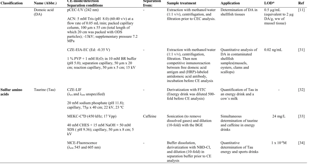
(26) potent detection method in sample analysis, the MCE-LIF sensitivity is sometimes not enough in sample analysis because of the low sample volume and short optical pathlength of the microchannels. For this reason, the development of on-line preconcentration methods able to enhance the sensitivity of MCE is relevant. With this aim, an on-line preconcentration approach combining field-amplified stacking (FASS) and reversed-field stacking was developed for the first time to achieve a sensitive analysis. The schematic mechanism of this strategy is shown in Figure 3A. The electrophoretic profiles obtained for the different concentration steps (see Figure 3B) demonstrated that both steps were crucial to achieve enhancement factors of 165-, 285- and 236-fold compared to the signal intensity without concentration. Under optimal conditions, taurine, lysine and B3 vitamin were focused and separated within 4 min achieving LODs at the nM level. The MCE-LIF methodology was successfully applied to the analysis of these ingredients in eight different functional drinks samples with a satisfactory recovery (see Figure 3C) and results were in good agreement with those listed in the label of the products. Figure 3. 1. 2. 3. 4. B 3 2 1. C 1. 2. 3. 4. 5. 6. 7. 8. 25.
(27) Fig. 3. A) Diagram of the MCE sample loading, on line preconcentration, and separation of nutritious compounds: (1) preloading, (2) FASS, (3) reversed-field stacking, and (4) separation. The dark blue zone represents the concentrated sample by FASS before using reversed-field stacking technique, the black zone represents the area of the concentrated sample after reversed-field stacking, the light blue zone represents the sample matrix, and the clear zone represents the running buffer. B) Signal enhancement of the multiple concentration: (1) signal intensity without concentration (buffer, 100 mM borate (pH 9.88), sample diluted with the buffer, sample injection time, 2 s), (2) signal intensity with FASS (sample prepared in a 10-fold-diluted buffer, sample injection time, 2 s), (3) signal intensity with a combination of FASS and reversed-field stacking (sample injection time, 10 s; reversed-polarity time, 8 s). The concentrations of Lys, Tau, and NA in (1) were 0.6, 0.9, and 0.6 μM, respectively. The sample concentrations in (2) and (3) were 1/10 of that in (1). Peak 1, excess of Cy5; 2, Lys; 3, Tau; and 4, NA. C) MCE electropherograms of eight different Cy5-labeled functional drink samples after dilution. Dilution fold: (1), 32000; (2), 1600; (3), 2000; (4), 2000; (5), 32000; (6), 64000; (7), 320000; and F8, 2000. Reprinted from [35], copyright (2015) with permission from Elsevier. Homocysteine (Hcy) is a non-protein thiol amino acid (contains a sulfhydryl group in its structure) formed during methionine´s metabolism. Thiols play an important role in abiotic and biotic stress resistance involved in the detoxification of xenobiotics in many organisms and they have nutritional value for humans when they are present in fruits and vegetables [57,58]. To achieve thiol determination by CZE with LIF detection, a fluorescent labeling reagent is always required because these compounds do not have detectable fluorescence. Taking this into account, Zhang et al. [36] developed a CZE-LIF methodology to determine Hcy along with other thiol compounds (cysteine, cysteinylglycine, γ-glutamylcysteine, glutathione, and N-acetylcysteine) in cucumber and 26.
(28) tomato samples using a new near-infrared fluorescent probe (namely 1,7-dimethyl-3,5distyryl-8-phenyl-(4'-iodoacetamide)difluoroboradiaza-s-indacene (DMDSPAB-I)) as labeling reagent. By using the appropriate derivatization protocol (45ºC for 25 min), and the best separation conditions (16 mM sodium citrate at pH 7.0 containing 60% (v/v) ACN), the studied thiols were analyzed within 14 min. The LOD obtained for Hcy with the proposed methodology was 0.15 nM. The authors proposed the method as a good alternative to investigate the biological function of low molecular weight thiols at trace levels. S-Alk(en)ylcysteine-S-oxides are non protein amino acids which appear as secondary metabolites in plants, fungi and algae and are precursors of a great variety of sensory-active and healthy compounds of Allium (onion, garlic, leek, etc) and Brassica vegetables (broccoli, cabbage, cauliflower, etc) [38]. Hideki et al. [37] developed a simple and rapid CZE method to analyze methiin and alliin in Allium and Brassica vegetables using indirect UV detection (at 350 nm) and 20 mM sodium benzoate containing 0.5 mM tetradecyltrimethyl-ammonium bromide (TTAB) as BGE. The use of a sample treatment based on boiling, extraction, dilution and filtration, (without derivatization), gave rise to a total analysis time for methiin and alliin in vegetables less than 25 min per sample. An interesting advantage of this CZE methodology was the possibility to detect pyruvate which is a useful marker of unsuccessful sample preparations (the peak for pyruvate appeared instead of methiin and alliin peaks when the samples were extracted without boiling (blanching)). Determination of the whole range of S-Alk(en)ylcysteine-S-oxides in alliaceous and cruciferous vegetables (fresh vegetables and garlic made products) was also carried out by MEKC with UV detection (previous derivatization with FMOC) [38]. Among the compounds investigated, isoalliin determination generated a special interest because it is 27.
(29) the responsible of the pungency of onion and the undesirable decoloration of garlic [59]. The developed MEKC methodology, based on the use of 20 mM sodium borate containing 20 mM SDS and 10 % methanol as running buffer, enabled the simultaneous separation of S-Alk(en)ylcysteine-S-oxides (methiin, alliin, isoalliin, propiin, and ethiin) within 20 min and a LOD at the pmol level. Alliin was found only in garlic whereas isoalliin was the main compound in other Allium species (such as onion, leek, chive and shallot). On the other hand, methiin was the only compound contained in plants from the Cruciferae family (occasionally along with traces of ethiin) [38].. 2.5 Hydroxyl amino acids. Hydroxyproline (Hyp) is the only hydroxyl amino acid analyzed in food samples by CE in the period of time covered by this review. It is formed by hydroxylation of proline and it is the most abundant component in collagen so that it can be used as marker of collagen content index (collagen is often added in its natural form or as hydrolysates in some products as a protein source being its levels regulated due to collagen effects on food quality). Two MEKC methods were developed to determine Hyp content in different matrix (meat and milk products) employing UV or LIF detection. To carry out the determination of Hyp (as collagen content index) with UV detection, the MEKC method was based on the use of fluorescamine as labeling reagent and 0.05 M sodium borate containing 100 mM SDS as running buffer [39]. Under these conditions, Hyp was clearly separated not only from other amino acids (Pro and Gly) present in collagen but also from other compounds present in meat samples. Since the results obtained using the developed MEKC-UV method were in agreement with the AOAC official colorimetric method, it can be considered as an alternative for Hyp analysis in meat products. Regarding the MEKC method with LIF detection, it was developed with the aim of providing a rapid 28.
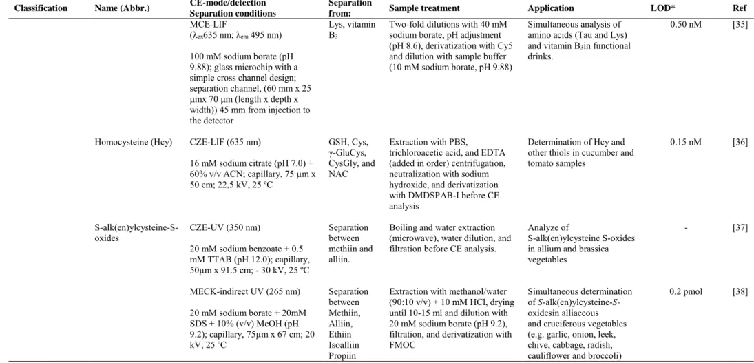
(30) and sensitive determination of Hyp in different dairy products for food quality assurance [40]. An in-capillary derivatization with 4-fluoro-7-nitro-2,1,3-benzoxadiazole (NBD-F) and a 25 mM sodium borate buffer containing 30 mM SDS as running buffer were employed. Derivatization and separation was completed in only 7 min and the LOD for Hyp was 1.6 ng/mL, both remarkable features enabling to consider the proposed methodology as an alternative for the quality control of dairy products. In addition, these analytical features are relevant advantages compared with other chromatographic alternatives (HPLC, GC or CE), either with LIF or other types of detector, described in the literature to perform the determination of Hyp. In fact this methodology provided comparable or higher sensitivity and decreased the analysis time thanks to the use of an in-capillary derivatization which also allows a full automatization.. 2.6 Seleno amino acids. Selenium is an essential trace element for human health whose deficiency causes serious nutritional and health problems. An effective way for providing selenium is through the selenium-enriched foods intake. To carry out the nutritional and toxic evaluation of selenium compounds (Se (IV), Se (VI) and the non-protein amino acids selenocysteine (SeCys2) and selenomethionine (SeMet)) in nutritional food supplements, Zhao et al. [41] developed a CZE-inductively coupled plasma MS (CZE-ICP-MS) methodology. As it can be seen in Table 1, sample treatment included an enzyme-assisted extraction approach to extract all selenium species and the CZE separation was accomplished using 20 mM sodium phosphate, 10 mM sodium borate and 0.2 mM cetyltrimethylammonium bromide (CTBA) as separation buffer (pH 8.6). LODs of 0.5 ng/L and 0.9 ng/L for SeMet and SeCys2 respectively, and recoveries in the range 90-103 % were achieved. Figure 4 shows the electropherograms obtained for a selenium29.
(31) enriched rice and the same sample spiked with the selenium species studied. As Figure 4 shows, only the non-protein amino acid SeMet was detected in selenium-enriched rice in the range of 0.136-0.143 mg Se/g dried weight. Figure 4.. Fig. 4. The electropherograms of Se(VI), Se(IV), SeCys2 and SeMet under the optimal CZE-ICP-MS conditions: BGE, 20 mM sodium phosphate-10 mM sodium borate-0.2 mM CTAB (pH 8.6); uncoated fused-silica capillary, 75 µm x 80 cm; voltage; -16 kV; temperature, 25 ºC. (A) selenium-enriched rice; (B) selenium enriched rice spiked with 1.0 µg/g of Se(VI), Se(IV), SeCys2 and SeMet. Reprinted from [41], copyright (2011) with permission from Elsevier. 30.
(32) 2.7 Betaines. Betaines are zwitterionic non protein amino acids which possess a quaternary ammonium group and a carboxylic group in their structure. They play a key role in many plants as osmoregulating compounds that help to tolerate environmental stress [60]. Betaine, trigonelline, glycine betaine, proline betaine, and carnitine have been the betaines analyzed by CE in different food samples during the time covered by this review. The simultaneous determination of betaine and the protein amino acid proline was accomplished using a CZE methodology with indirect UV detection [42]. Among a variety of different probes (imidazole, creatinine, 4-aminopyridine, 4-aminobenzoic acid, sulfanilamide, etc), sulfanilamide demonstrated to be the most appropriate for the indirect detection of the studied compounds due to its slow mobility and good molar absorptivity. Under the optimal experimental conditions (see Table 1) and using 5 mM sulfanilamide, it was possible to achieve a LOD of 28.3 µM for betaine. The applicability of this methodology was demonstrated with the identification and quantification of betaine in extracts obtained from spinach and beetroot samples. Trigonelline belongs to pyridine betaines group. It has demonstrated to have different health-promoting effects such as hypocholesterolemic, antitumor, or antimigraine, among others [61]. The determination of trigonelline in seeds and vegetables oils by CZE with UV detection (195 nm) enabled to propose this compound as a novel marker for the detection of adulterations in olive oils [43]. Under the experimental conditions detailed in Table 1 and using an in-capillary normal stacking as sample preconcentration strategy, trigonelline was detected in both soy and sunflower seeds (and their oils) but not in olives or olives oils at least above the LOD of the developed method (LOD up to 0.9 µM) [43]. Taking into account the limitation of this 31.
(33) CE-UV methodology to detect trigonelline in olives or olive oils, a CZE-MS2 strategy was further carried out with the aim of detecting olive oil adulterations with seed oils through the simultaneous analysis of trigonelline and other betaines, such as proline betaine, glycine betaine (see Figure 5A) [8]. Following the same sample treatment described for the determination of ornithine in olive and seed oils [7] and using a derivatization with butanol to improve not only the analytical sensitivity but also the selectivity (by improving mass differentiation among analytes) the separation was achieved within 10 min using a 0.1 M formic acid (pH 2.0) as running buffer. The LOD obtained using MS detection (1 ng/g) was 20 times lower than that obtained previously with UV detection. Figure 5B shows the extracted ion electropherogram obtained for glycine betaine, trigonelline and total carnitines in soybean oil sample, extra virgin olive oil sample, and oil mixture of extra virgin olive oil sample with a 5% w/w of soybean oil sample, as well as their MS2 spectra in the oil mixture. The improved sensitivity enabled to detect low quantities of trigonelline in olive oils, so that at this low level it cannot be used as adulteration marker. By contrast, carnitines were not detected or not quantifiable in extra virgin olive oils; however, they were present in seed oils. This fact made possible to propose them as a feasible novel marker for the detection of adulteration of olive oils with seed oils [8]. From the results obtained using the developed methodology for the determination of non-protein amino acids [7] and betaines [8], both included among the variety of compounds constituting the unsaponifiable fraction of oils, the potential of ornithine, alloisoleucine and carnitines as novel markers for the detection of olive oil adulteration was demonstrated .. 32.
(34) Figure 5. A. B. a). b). Fig. 5.. a. a. a. b. b. b. c. c. c. d. d. d. A) CZE-MS base-peak electropherogram (a) and simultaneous CZE-MS2. extracted-ion electropherogram (EIE) (b) for standard betaines mixture (5 µg/mL each one injected at 50 mbar x 15 s (a) or 50 s (b)). B) CZE-MS2 EIE for glycine betaine, trigonelline and total content of carnitines in (a) soybean oil sample, (b) extra virgin olive oil sample, (c) oil mixture of extra virgin olive oil sample with a 5 % w/w of soybean oil sample, and (d) MS2 spectra for the peaks obtained in (c) of glycine betaine, trigonelline or carnitines in the oil mixtures of extra virgin olive oil sample with a 5 % w/w of soybean oil sample. CZE conditions: BGE, 0.1M formic buffer (pH 2.0); uncoated fused-silica capillary, 50 µm id x 60 cm; voltage, 25 kV; temperature, 25 ºC. ESI conditions: positive ion mode (4.5 kV); sheath liquid, isopropanol/water (50/50, v/v) with 0.1 % formic acid at 3.3 µL/min; drying gas flow, 3 L/min; drying temperature, 300 ºC; nebulizer pressure, 2 psi. Ion trap conditions: maximum accumulation time, 300 ms; averages, 1; scan, 50280 m/z. MS2 transitions in MRM mode with width, 4 m/z; fragmentation amplitude 1.00 V and fragmentation time, 40 ms. Reprinted from [8], copyright (2011) with permission from John Wiley and Sons. 33.
(35) Carnitine (Carn) is produced from lysine and methionine in low levels in human, so it is mostly introduced by diet. Due to the important role that carnitine plays in the fatty acids metabolism, its deficiency could give rise to different health problems such as hypoglycemia, hyperammonemia and hypoketosis [62,63]. A CZE method with C4D detection was developed for the quantitative determination of carnitine in a great variety of foodstuffs (fruit, juices, milk, yogurt, cheese, chicken meat and red meat) [44]. This method consisted in the use of a 500 mM acetic acid buffer containing 0.05 % tween-20 (to prevent the wall interaction of the larger species) and a simple sample treatment. A LOD of 2.6 µM was obtained. The main advantage of the developed CZE-C4D methodology is, undoubtedly, the elimination of the derivatization step or indirect approaches to carry out the determination of carnitine.. 2.8 Other amino acids. N-Phenylpropenoyl-L-amino acids (NPAs) are a group of non protein amino acids which are among the main contributors for the astringent taste of cocoa. For this reason, these compounds could be considered as useful quality markers concerning the cocoa´s taste [64]. In addition, NPAs have also demonstrated to have different pharmacological activities.65 Lechtenberg et al. [45] proposed two alternative methodologies (CZE and UPLC, both with UV detection) to determine the NPA content in cocoa and cocoa products. As it can be observed in Table 1, two different CE systems were used, but in both the experimental conditions to achieve the NPAs separation were similar (50 mM sodium borate at pH 8.8 was employed as running buffer). The CZE and UPLC methodologies developed in this work were comparable concerning the results obtained and time consumption. The former lacks in sensitivity but it just needed a simple sample treatment whereas the latter requires a SPE step in the sample cleanup but enables shorter 34.
(36) analysis times and lower detection limits. Taking that into account, CZE was proposed for analyzing cocoa samples with high content of NPAs (mainly for the determination of N-[3', 4' -dihydroxy-(E)-cinnamoyl]-3-hydroxy-L-tyrosine (Caff-DOPA), N-[3', 4'dihydroxy-(E)-cinnamoyl]-L-aspartic. acid. (Caff-Asp),. and. N-[4'-hydroxy-(E)-. cynnamoyl]-L-aspartic acid (pC-Asp) in cocoa beans and shells).. 3. Enantiomeric determination of non-protein amino acids in food by CE. As it has been described in the introduction of this review, enantioselective separations may provide relevant information in different food areas, such as food authenticity, detection of adulterations or evaluation of manufacturing processes among others. From the publication of the previous review devoted to describe the developed CE methods for the determination of non-protein amino acids in foods [1], the field of chiral separations has undergone a great growth. However, most of the chiral methodologies were aimed to the chiral separation of protein amino acids so it can be said that the chiral separation of non-protein amino acids in food samples is still a quite unexplored field. Table 2 groups the main characteristics of the chiral methodologies developed to enantiomerically separate chiral non-protein amino acids by CE. The most used CE mode has been MEKC which is based on the addition of a micellar pseudophase in which the concentration of the micellar system must be higher than its critical micelle concentration. The chiral selectors used in these methodologies were different types of cyclodextrins. CEC was also employed and compared with nanoLC. As shown in Table 2, the two main detection systems used were UV and MS2 and in almost all the works a previous derivatization procedure was needed (labeling reagents employed were FMOC, FITC or AQC). 35.
(37) Since the L-enantiomer is usually responsible for the beneficial biological properties, the aim of these works was usually its separation from the D-enantiomer that can have in some cases toxic properties and whose addition during the elaboration of foods is forbidden. The developed methodologies enabled in these cases to guarantee a good quality control of food products. Samples analyzed were mainly food supplements, infant formulas, fermented foods (such as wine, beer etc.), etc., as described in detail in Table 2.. 36.
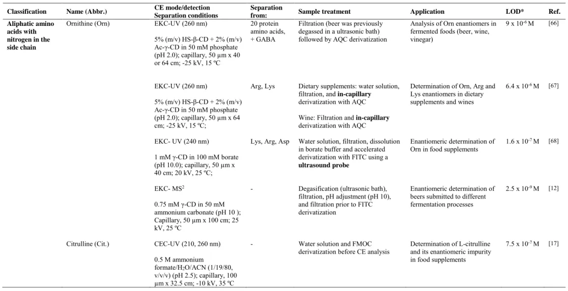
(38) Table 2. Characteristics of the analytical methodologies developed for the enantiomeric determination of non protein amino acids by CE. Classification Aliphatic amino acids with nitrogen in the side chain. Name (Abbr.) Ornithine (Orn). CE mode/detection Separation conditions EKC-UV (260 nm) 5% (m/v) HS-β-CD + 2% (m/v) Ac-γ-CD in 50 mM phosphate (pH 2.0); capillary, 50 µm x 40 or 64 cm; -25 kV, 15 ºC EKC-UV (260 nm). Separation from: 20 protein amino acids, + GABA. Arg, Lys. 5% (m/v) HS-β-CD + 2% (m/v) Ac-γ-CD in 50 mM phosphate (pH 2.0); capillary, 50 µm x 64 cm; -25 kV, 15 ºC; EKC- UV (240 nm). CEC-UV (210, 260 nm) 0.5 M ammonium formate/H2O/ACN (1/19/80, v/v/v) (pH 2.5); capillary, 100 µm x 32.5 cm; -10 kV, 35 ºC. LOD*. Ref.. 10-6 M. Filtration (beer was previously degassed in a ultrasonic bath) followed by AQC derivatization. Analysis of Orn enantiomers in fermented foods (beer, wine, vinegar). 9x. Dietary supplements: water solution, filtration, and in-capillary derivatization with AQC. Determination of Orn, Arg and Lys enantiomers in dietary supplements and wines. 6.4 x 10⁻⁶ M. [67]. [66]. Lys, Arg, Asp. Water solution, filtration, dissolution in borate buffer and accelerated derivatization with FITC using a ultrasound probe. Enantiomeric determination of Orn in food supplements. 1.6 x 10⁻⁷ M. [68]. -. Degasification (ultrasonic bath), filtration, pH adjustment (pH 10), and filtration prior to FITC derivatization. Enantiomeric determination of beers submitted to different fermentation processes. 2.5 x 10⁻⁹ M. [12]. -. Water solution and FMOC derivatization before CE analysis. Determination of L-citrulline and its enantiomeric impurity in food supplements. 7.5 x 10⁻⁷ M. [17]. 0.75 mM γ-CD in 50 mM ammonium carbonate (pH 10 ); Capillary, 50 µm x 100 cm; 25 kV, 25 ºC Citrulline (Cit.). Application. Wine: Filtration and in-capillary derivatization with AQC. 1 mM γ-CD in 100 mM borate (pH 10.0); capillary, 50 µm x 40 cm; 20 kV, 25 ºC; EKC- MS2. Sample treatment. 37.
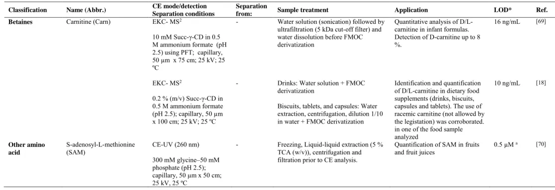
(39) Table 2. Continued Classification. Name (Abbr.). Betaines. Carnitine (Carn). CE mode/detection Separation conditions EKC- MS2. Separation from: -. 10 mM Succ-γ-CD in 0.5 M ammonium formate (pH 2.5) using PFT; capillary, 50 µm x 75 cm; 25 kV; 25 ºC EKC- MS2. -. 0.2 % (m/v) Succ-γ-CD in 0.5 M ammonium formate (pH 2.5); capillary, 50 µm x 100 cm; 25 kV; 25 ºC Other amino acid. S-adenosyl-L-methionine (SAM). CE-UV (260 nm) 300 mM glycine–50 mM phosphate (pH 2.5); capillary, 50 µm x 50 cm; 25 kV, 25 ºC. Sample treatment. Application. LOD*. Ref.. Water solution (sonication) followed by ultrafiltration (5 kDa cut-off filter) and water dissolution before FMOC derivatization. Quantitative analysis of D/Lcarnitine in infant formulas. Detection of D-carnitine up to 8 %.. 16 ng/mL. [69]. Drinks: Water solution + FMOC derivatization. Identification and quantification of D/L-carnitine in dietary food supplements (drinks, biscuits, capsules and tablets). The use of racemic carnitine (not allowed by the legistation) was corroborated. in one of the food sample analyzed Quantification of SAM in fruits and fruit juices. 10 ng/mL. [18]. 0.5 µM a. [70]. Biscuits, tablets, and capsules: Water extraction, centrifugation, dilution 1/10 in water + FMOC derivatization -. Freezing, Liquid-liquid extraction (5 % TCA (w/v)), centrifugation and filtration prior to CE analysis.. * LODs units expressed as in the original work. These LODs are referred to the injected solutions of standard samples except for a) in which LODs is referred to the injected solutions of food samples. **Capillary dimension expressed as internal diameter x effective length (cm to the detector). ACN, acetonitrile; Ac-γ-CD, acetylated-γ-CD; AQC, 6-aminoquinolyl-N-hydroxysuccinimidyl carbamate; Arg, arginine; Asp, asparagine; GABA, γ-aminobutiric acid; γ-CD, γ-cyclodextrin; FITC, fluorescein isothiocyanate; FMOC, 9-Fluorenylmethoxycarbonylchloride; HS-β-CD, highly sulfated-β-cyclodextrin; Lys, lysine; MS2, tandem mass spectrometry; PFT, partial filling technique; Succ- γ-CD, succinyl-γ-cyclodextrin; TCA, trichloroacetic acid. 38.
(40) 3.1 Aliphatic amino acids with nitrogen in the side chain Ornithine (Orn) and Citrulline (Cit) enantiomers were determined in foods by CE under the experimental conditions detailed in Table 2. Ornithine is a non-protein amino acid whose enantiomeric separation is receiving huge attention due to the beneficial properties of the L-enantiomers and the harmful effects of D-orn. L-Orn plays an important role in some biochemical processes such as fatty acid excess metabolism, human growth hormone synthesis, ammonia detoxification in urea cycle, synthesis of L-proline, etc. In contrast, D-enantiomer, which may occur during food processing or fermentation processes [71], can produce depletion in the urea synthesis giving toxic consequences. Humans can obtain L-orn through endogenous synthesis involving urea cycle, as well as from fermented foods (such as beer, wine, juices, cheese, etc), or functional foods (such as dietary supplements) [72]. To analyze Orn enantiomers in fermented foods, an EKC-UV method was developed by Martínez Girón et al. enabling LODs of 9 x 10⁻⁶ M. The method consisted of using AQC as off-line derivatizing reagent and 50 mM phosphate (pH 2.0) containing 5 % (m/v) HS-β-CD and 2 % (m/v) acetylated-γ-CD as BGE [66]. As shown in Figure 6, the methodology allowed the enantiomeric separation of ornithine (in less than 15 min) as well as the separation of the enantiomers of this amino acid from the enantiomers of the chiral amino acids contained in a mixture of the twenty protein amino acids and GABA in about 45 min. To short the analysis time and to increase CE method automatization, the same authors developed a second method using in-capillary derivatization [67] with the aim of determining Orn, arginine and lysine enantiomers in wine samples (this compounds have demonstrated to be responsible of wine’s organoleptic properties) and dietary supplements. The enantiomeric separation of Orn could also be performed with FITC as labeling reagent. To do that, Domínguez-Vega et al. [68] employed an ultrasound probe 39.
(41) which allowed to reduce derivatization time from 16 h to 10 min. The developed EKCUV method enabled the enantiomeric analysis of this amino acid with LODs of 1.6 x 10⁷. M, as well as the separation from the enantiomers of other protein amino acids such as. lysine, arginine and asparagine (see other CE conditions in Table 2) in food supplements. Moreover, to enhance the sensitivity and selectivity in the enantiomeric determination of ornithine in beers, an interesting EKC-MS2 method using the previous derivatization procedure was proposed. The method, based on the use of a 50 mM ammonium carbonate buffer at pH 10.0 containing 0.75 mM γ-CD as BGE, enabled to quantify the content of the Orn enantiomers in beers submitted to different fermentation processes with LODs of 2.5 x 10⁻⁹ M. The percentages for D-Orn in the analyzed samples ranged from 1.5 % to 10 %, the lowest value corresponding to a dietetic beer and the maximum to a double fermentation beer [12]. The four EKC methodologies described in the literature to carry out the chiral separation of ornithine show that this non-protein amino acid can be separated both under acid and basic conditions using different cyclodextrins as chiral selectors, depending on the characteristics of the analyzed samples or the requirements needed to perform the derivatization step. In any case, the lowest LODs for ornithine are achieved using a CE-MS2 methodology.. 40.
(42) Figure 6.. Fig. 6. A) Enantiomeric separation by EKC of a mixture of the 20 protein amino acids, Orn, and GABA (upper corner) divided in two migration zones: (a) first-migrating zone, and (b) second-migrating zone. B) Electropherograms corresponding to different fermented foods derivatized off-line with AQC (a) a rose wine (uncoated fused-silica, 50 µm × 72.5 cm; injection by pressure, 5066.25 Pa for 20 s of sample followed of 5 s of BGE; non-spiked sample and sample spiked with 2.5×10⁻⁵ M racemic Orn) and (b) a beer (uncoated fused-silica, 50 µm × 48.5 cm; injection by pressure, 5066.25 Pa for 5 s of sample followed of 5 s of BGE; non spiked sample and sample spiked with 5×10⁻⁴ M racemic Orn). EKC conditions: 50 mM phosphate buffer (pH 2.0) containing 5% (m/v) HS-γ-CD and 2% (m/v) acetylated-γ-CD; uncoated fused-silica, 50 µm × 72.5 cm; voltage, -25 KV; temperature, 15 ºC. (*) Unknown peaks. Reprinted from [66], copyright (2008) with permission from Elsevier. Citrulline is a non-protein amino acid which has also demonstrated to have different enantiomeric behavior. L-Cit, which is naturally occurring, is precursor of 41.
(43) protein amino acid Arginine, it is involved in urea cycle and it plays an important role in ammonia level decrease and NO cycle [73]. To carry out the enantiomeric determination of citrulline in food suplements, a CEC-UV method was developed using cellulose tris (3-chloro-4-methylphenylcarbamate chiral stationary phase (CSPs) as chiral selector, 9fluorenylmethoxycarbonylchloride (FMOC) as labeling reagent and 0.5 M ammonium formate as running buffer. The method, which demonstrated to be more efficient in comparison with the developed nano-LC method, gave rise to LODs of 7.5 x 10⁻⁷ M for citrulline and made possible to achieve the enantiomeric determination of nineteen more amino acids (among twenty three amino acids analyzed) [17].. 3.2 Betaines As above-mentioned, Carn is synthesized from lysine and methionine or it can be available to human through some dietary sources. It has shown to have different biological activities depending on its enantiomeric form. L-Carn has demonstrated to play an important role in long chain fatty acids metabolism while D-Carn posseses toxical properties [62,74]. For these reasons, the development of a method allowing to monitor the content of L/D-Carn in food samples is of great interest in order to carry out a correct quality control. With this aim, a CE-ESI-MS2 methodology was developed using FMOC as labeling reagent (derivatization at 45ºC for 60 min) and 0.5 M ammonium formate containing 10 mM Succ-γ-CD as running buffer. Firstly, a partial filling technique (PFT) was employed and the method gave rise to LODs of 0.1 µM of L-Carn. The analysis of carnitine enantiomers in 14 infant formulas supplemented with this amino acid enabled the detection of amounts as high as 8 % of the toxic enantiomer which exceeded by far the limits established by the European Pharmacopeia [69]. Afterwards, in order to avoid the use of the PFT technique, the method was optimized without this step by using 0.2 % 42.
(44) (m/v) Succ-γ-CD as chiral selector with a longer length capillary (see table 2) [18] improving the precision and sensitivity of the previous method (LODs of 10 ng/mL for D/L-Carn enantiomers were obtained enabling to detect enantomeric impurities up to 0.025 %). In this case, the optimized method was applied to the analysis of 22 dietary food supplements showing the use of L-Carn in 21 samples with enantiomeric impurities (D-Carn) up to 6 %. The use of racemic Carn (not allowed by the legislation) in one of the food supplements was corroborated which confirmed the need of analytical methodologies to ensure food quality [18].. 3.3 Other amino acids The only non-protein amino acid included in this group is S-Adenosyl-Lmethionine (SAM), which is a chiral compound involved in many biochemical pathways and the major methyl donor in living organisms [75]. It is synthesized from methionine in the presence of ATP [76]. In addition, two diastereoisomers of this compound exist, known as (S,S)- and (R,S)-S-adenosyl-L-methionine, among which only one has demonstrated to be biologically active (S,S-). Van de Poel et al. [70] demonstrated that SAM content varies depending on food processing (relationship between heat treatment and SAM content in tomato samples is shown in Figure 7A, 7B). The developed method consisted of optimizing CE-UV conditions to quantify SAM contents in different fruit tissue samples. The use of 300 mM glycine-50 mM phosphate (pH 2.5) was chosen as optimum running buffer, and the method, compared with the use of HPLC, made possible to enhance the enantiomeric separation (see Figure 7A) with minimum sample treatment, in half time and with LODs 2-fold lower than those obtained by the conventional methodology (see LOD and other CE conditions in Table 2). 43.
Figure
+5

Documento similar
