CE methods for the determination of non protein amino acids in foods: Review
15
0
0
Texto completo
(2) 4032. M. Castro-Puyana et al.. (the most used chromatographic technique in amino acid analysis in foods) where collaborative testing methodologies are needed. Moreover, since CE provides better resolution than HPLC, it is useful for the separation and/or rapid screening of low levels of amino acids from components of different chemistries in complex food matrixes during a single analysis [4]. However, a review devoted to the application of CE to non-protein amino acids analysis in foods has never been reported and thus has been the aim of this article. In order to describe the analytical methodologies developed by CE for the determination of non-protein amino acids in foods, these non-protein amino acids have been classified according to their structure. CE conditions employed, sample treatment, complexity of separations achieved, and samples analyzed are detailed. In addition, the application of microchip electrophoresis to the determination of non-protein amino acids in foodstuffs is also reported.. 2. Determination of non-protein amino acids in foods by CE. Table 1 summarizes the non-protein amino acids determined in foods using CE. They have been classified in different groups according to their structure and following a criterion based on the classification established by Hunt [1]. The name and structure of the different amino acids investigated as well as CE separation conditions, compounds separated from the amino acid studied, LODs achieved, sample treatment and samples analyzed are given in this table. Non-protein amino acids have mainly been determined by CE using two different separation modes: (i) CZE where a separation buffer without or with additives is used and the separation is based on the different electrophoretic mobilities of analytes at a certain pH, and (ii) EKC where a pseudophase (a micellar system, i.e. a surfactant at a concentration higher than its critical micelle concentration, usually SDS, and/or CD) is added to the electrolytic solution to perform the separation of non-protein amino acids in complex matrices (such as fishes, meats, vegetables, legumes, etc.) or their chiral separation based on the different interaction of amino acids with a chiral pseudophase. As shown in Table 1, UV absorption detection has mainly been used in the analysis of non-protein amino acids by CE in spite of the absence of strong chromophore groups in many of them (i.e. seleno and sulfur amino acids, aliphatic amino acids, cyloalkane amino acids and hydroxyl amino acids). This has been possible due to the use of different derivatization strategies with adequate probes and in most cases selecting low UV detection wavelength. Other detection systems employed have been LIF, ESI-MS, inductively coupled plasma MS (ICP-MS), and electrochemical detection. In the case of LIF detection, a previous derivatization step was necessary to provide fluorescent groups. Derivatization was also employed when using ESI-MS in order to obtain larger molecules providing better detection © 2007 WILEY-VCH Verlag GmbH & Co. KGaA, Weinheim. Electrophoresis 2007, 28, 4031–4045. sensitivity for the amino acids studied. ICP-MS and electrochemical detection enabled to avoid previous derivatization steps. Samples analyzed comprise beverages, seeds, vegetables and plants, food supplements, eggs, meats, fishes, and nuts. Usually, after the extraction of non-protein amino acids, a physical treatment (centrifugation, filtration) of samples has been performed. Moreover, protein hydrolysis has been employed in some cases such as (i) the determination of selenomethionine in yeast, and (ii) when the separation of non-protein amino acids from protein amino acids was performed (i.e., lysinoalanine in duck eggs, aromatic or isoxazolinone amino acids in seeds or heterocycle amino acids in seeds, dried milk, fishes, meats, vegetables, and nuts). In addition to these general aspects related with the determination of non-protein amino acids by CE, a more detailed description of the interest of their determination in food samples and of the CE methodology developed for each group of non-protein amino acids will be performed herein. 2.1 Seleno amino acids The selective qualitative and quantitative determination of particular species of selenium, including selenoamino acids, is vital in order to understand selenium’s metabolism and significance in biology, toxicology, clinical chemistry and nutrition [36]. At high concentrations, selenium is toxic, causing problems such as dermatitis, fatigue and hair loss. However, human nutritional studies have shown that appropriate doses of supplemental selenium enhance cellular defense against oxidative damage and may prevent certain types of cancer [13]. CE methods developed for the determination of selenoamino acids were devoted to the analysis of selenomethionine in selenized yeast, nutrition supplements, and human milk, and selenocystine in human milk. With respect to the determination of selenomethionine, the chiral speciation of this non-protein amino acid in selenized yeast has been performed by CE with UV and ICP-MS detection under aquiral conditions through the derivatization of the two enantiomers of selenomethionine with the Marfey’s reagent (1-fluoro-2,4-dinitrophenyl-5-L-alanine amide) to form diastereomers that were separated by CZE using 30 mM ammonium phosphate buffer at pH 3.3. Selenized yeast was enzymatically hydrolyzed with proteinase K prior to its analysis by UV and ICP-MS detection. Under these conditions, the identification of selenomethionine enantiomers (LOD , 1610–6 M for UV detection at 214 nm and , 7610–7 M for ICP-MS detection) in the sample was performed using pure L-selenomethionine standard. Due to the selectivity of ICP-MS detection, where only selenium species are detected, electropherograms obtained for the selenized yeast were simpler than those observed by UV detection. It is important to remark that racemization due to sample preparation and derivatization steps was not observed although other peaks at longer migration times www.electrophoresis-journal.com.
(3) © 2007 WILEY-VCH Verlag GmbH & Co. KGaA, Weinheim. Sulfur amino acids. Seleno-methionine (Se-Met). Seleno amino acids. Taurine. Selenocystine (Se-Cys). Name (Abbr.). Classification. Structure. CZE: 50 mM borate (pH 9.2) 25 mm 6 70cm; 30 kV; electrochemical detection. Medicinal herb, milk powder and health beverage. , 1 6 10–7 –. Human milk. –. –. Human milk. –. 4 - 6 6 10 –4 (CZE) Human milk 1 - 4 6 10–4 (CIEF). Nutrition supplement. , 1 6 10–5. Other arsenic and selenium species (see text) –. CZE: 15 mM phosphate buffer (pH 10.6) 75 mm x 64.5 cm (coated); 225 kV; UV-195 nm CZE: 100 mM phosphate (pH 2.5) or acetate (pH 1.9) 10 kV; 207C; UV-200 nm. Other selenium species (see text). Selenized yeast. , 1 6 10–6 (UV) , 7 6 10–7 (ICP-MS). –. CZE: 30 mM phosphate buffer (pH 3.3) 75 mm 6 50 cm; 237C; 225 kV; UV-214 nm and ICP-MS. CZE: 10 mM carbonate buffer (pH 11.5); CIEF: leading electrolyte: sodium hydroxide (pH 2); terminating electrolyte: phorphoric acid (pH 10) 50 mm 6 150 cm; 207C; 18 kV; ICP-MS CZE: 100 mM phosphate (pH 2.5) or acetate (pH 1.9) 10 kV; 207C; UV-200nm. Food sample. LOD (M). Separation from. CE conditions. [16]. [15]. [14]. [13]. Ref.. (Water dissolution and sonication/filtration) Buffer dissolution prior to CE injection. [5]. [15] Defatting, protein precipitation by centrifugation, SEC fractionation, drying and water dilution prior to CE injection. Protein hydrolysis (proteinase K), filtration and derivatization (Marfey’s reaction to form diastereomers) prior to CE injection Clean-up (C18 cartridge), dilution, centrifugation and filtration prior to CE injection Defatting, protein precipitation by centrifugation, SEC fractionation, drying and water dilution prior to CE injection Defatting, protein precipitation by centrifugation, preconcentration (drying) and dissolution prior to CE injection. Sample treatment. Table 1. Classification, name, structure, CE separation conditions, food sample and sample treatment of the different food samples where non-protein amino acids have been determined using CE as separation techniquea) Electrophoresis 2007, 28, 4031–4045. CE and CEC 4033. www.electrophoresis-journal.com.
(4) Name (Abbr.). © 2007 WILEY-VCH Verlag GmbH & Co. KGaA, Weinheim. Structure. Separation from. LOD (M). EKC: 25 mM phosphate (pH 7.0), 100 mM SDS, 5-6% methanol 50mm 6 50 cm; 257C; 14 kV, UV-200 nm CZE: 20 mM borax (pH 8.0) 50 mm 6 70 cm; 237C; 30 kV; UV-200 nm cITP: Leading electrolyte: 0.01 M HCl, 0.02 mM Tris, 0.05% HEC (pH 8.1). Terminating electrolyte: 0.01 mM DL-valine, barium hydroxide (pH 10) Conductimetric detection EKC: 250 mM borate (pH 9.5), 200 mM SDS, 75 mM b-CD (methanol) 75 mm 6 40 cm, 15 kV; 257C; UV-254 nm. CZE: 5 mM Tris, 7 mM phosphoric acid, 0.5 mM quinine 50 mm 6 25.5 cm; 15 kV; indirect UV-254 nm. , 2 6 10–5. , 4 6 10–4 Protein amino acids (Cys, Asp, Thr, Ser, Glu, Pro, Gly, Ala, Val, Met, Ile, Leu, Phe, Hys, Lys, and Arg). Tea cathechins, , 2 6 10–5 caffeine, gallic acid, and ascorbic acid Tea cathechins, – caffeine, and ascorbic acid – , 4 6 10–6. –. Enantiomers of , 2 6 10–8 EKC: 100 mM borate (pH 9.7), protein amino 30 mM SDS, 20 mM b-CD acids (Arg, Pro, 50 mm 6 50 cm; 257C; 20 kV; LIF: 488 Ala, Glu, Asp) nm (excitation) 520 nm (emission) Enantiomers of – EKC: 100 mM borate (pH 9.4), protein amino 30 mM SDS, 20 mM b-CD acids (Arg, Pro, 50 mm 6 50 cm; 257C; 23 kV; LIF: 488 Asn, Ser, Ala, nm (excitation) 520 nm (emission) Glu, Asp) – , 2 6 10–6 EKC: 100 mM ammonium acetate (pH 6.0), 5 mM b-CD 50 mm 6 87 cm (coated); 215 kV; ESI-MS Betaines, trigo- , 1 6 10–5 CZE: 80 mM phosphate buffer nelline, and (pH 3.0) carnitine 50 mm 6 70 cm; 15 kV; 257C; UV-214 nm. CE conditions. [20] Solvent extraction, centrifugation, drying, water dissolution, derivatization (p-bromophenacyl bromide) Dilution [21] Higher plants. Duck eggs. [24]. [23]. [6] Acid protein hydrolysis, water dissolution of hydrolysate and derivatization (DNS) prior to CE injection. Dried fresh tea Water extraction and filtration prior to CE leaf, black tea, injection bottled black tea Green tea Water extraction, filtration and dilution prior to CE injection Green teas and Water extraction, dilution food suppleand filtration prior to ments CE injection. [22]. Filtration, centrifugation and [19] derivatization (FITC) prior to CE injection. Orange juice. Food supplements. Centrifugation and deriva[18] tization (FITC) prior to CE injection. Orange juice. Ref.. Filtration and derivatization [17] (FITC) prior to CE injection. Sample treatment. Vinegars. Food sample. M. Castro-Puyana et al.. Lysinoalanine. Aliphatic amino Theanine acids with nitrogen in the side chain. Carnitine. Glycine betaine. g-aminobutyric Aliphatic acid (GABA) monoaminomonocarboxylic amino acids. Classification. Table 1. Continued. 4034 Electrophoresis 2007, 28, 4031–4045. www.electrophoresis-journal.com.
(5) 3,4-Dihydroxyphenylalanine (DOPA). 2-Carboxymethyl3-isoxazolin5-one (CMI). Phenyl amino acids. Isoxazolinone amino acids. © 2007 WILEY-VCH Verlag GmbH & Co. KGaA, Weinheim. g-Glutamylb-(isoxazolin5-on-2-yl)alanine (g-glu-BIA) 2-(g-Glutamylaminoethyl)isoxazolin5-one (g-glu-AEI) 2-(3-Amino-3carboxypropyl)isoxazolin5-one (ACI). b-(Isoxazolin-5on-2-yl)-alanine (BIA). Name (Abbr.). Classification. Table 1. Continued. Structure. CZE: 25 mM phosphate buffer (pH 7.5), 8% 1-propanol 75 mm 6 52.6 cm; 22.5 kV; 307C; UV-254 nm. Broad bean and lentil. , 3 6 10–6. , 3 6 10–5. BIP, CMI, BIA, g-glu-BIA„ and ACI. BIP, CMI, BIA, , 2 6 10–5 g-glu-BIA, and g-glu-AEI. , 1 6 10–5. BIP, CMI, BIA, g-glu-AEI, and ACI. Legume seedlings. Seeds of lentil, seedlings, legume seedlings. Food sample. LOD (M). b-(isoxazolin-5- , 3 6 10–5 on-2-yl)-propionitrile (BIP), BIA, g-glu-BIA, g-glu-AEI, and ACI BIP, CMI, g-glu- , 2 6 10–5 BIA, g-glu-AEI, and ACI. –. CZE: 35 mM phosphate (pH 4.55) 75 mm 6 47 cm; 17.5 kV; 307C; UV210nm. CZE: 25 mM phosphate buffer (pH 7.5), 8% 1-propanol 75 mm 6 52.6 cm; 22.5 kV; 307C; UV-254 nm. Separation from. CE conditions. Amino acid extraction, centrifugation and preconcentration (vacuum) prior to CE injection. Acid protein hydrolysis, amino acid extraction, centrifugation and preconcentration (vacuum) prior to CE injection. Ultrasonic extraction, centrifugation and filtration prior to CE injection. Sample treatment. [27]. [26] [27]. [25]. Ref.. Electrophoresis 2007, 28, 4031–4045. CE and CEC 4035. www.electrophoresis-journal.com.
(6) © 2007 WILEY-VCH Verlag GmbH & Co. KGaA, Weinheim. 1-Aminocyclopropane-1carboxylic acid (ACC). Pyroglutamic acid. e-N-Pyrrolylnorleucine. Structure. –. –. Short-chain Standards organic acids , 2.3 6 10–4 (see text) Samples , 1.2 6 10–4 Caffeine , 6.9 6 10–7. , 1 6 10–8 Protein amino acids (His, Tyr, Thr, Gly, Ala, Met, Val, Phe, Leu/Ile, Asn/Gln, Trp, Cys, Glu, Asp). CZE: 500 mM phosphoric acid (pH 6.25), 0.5 mM CTAB 50 mm 6 57 cm; 210 kV; 257 C, UV-200 nm EKC: 50 mM boric acid (pH 9.5), 130 mM SDS 50 mm 6 57 cm; 10 kV; 257 C, UV-200 nm EKC: 20 mM borate (pH 9.35), 40 mM SDS, 10 mM Brij 35 50 mm 6 50 cm; 25 kV; 257C; LIF: 488 nm (excitation) 560 nm (emission). – – CZE: 3-(N-morpholino-2-hydroxypropanesulfonic acid (pH 7.0) extended-path length capillary of 50 mm 6 57 cm, 25 kV, 207C, UV-280 nm – , 5 6 10–6 cITP-CZE: Leading electrolyte (preseparation capillary, 90 mm 6 0.8 mm id): 10 mM HCl, 20 mM b-alanine, 0.05 % HEC (pH 3.5); BGE (analytical capillary, 150 mm 6 0.3 mm id): 20 mM caproic acid, 20 mM b-alanine, 5% methanol, 0.1% HPMC Terminating electrolyte: 5 mM caproic acid, 5% methanol UV-242 nm – EKC: 100 mM borate buffer (pH 8.4), Protein amino acids (Arg, Lys) 100 mM SDS 75 mm 6 50 cm; 30 kV; 307C; UV-280 nm. Dried milk. , 2 6 10–6. –. CZE: 20 mM phosphoric acid (pH 2.5), 60 mM TMBD 75 mm 6 30 cm, 15 kV, 257C, UV-280 nm CZE: phosphoric acid (pH 8.0) 50 mm 6 72 cm, UV-280 nm. Sample treatment. Apples. Coffee. Coffee. Fishes, meats, vegetables, legumes and nuts. [31]. [9]. [30]. [29]. [28]. Ref.. Extraction, centrifugation and derivatization (FQ) prior to CE injection. [34]. Water ultrasonic extraction, [32] centrifugation and filtration prior to CE injection Water dilution, and filtration [33] prior to CE injection. Basic protein hydrolysis and derivatization (DEEMM) prior to CE injection. Acid protein hydrolysis, lyophilization and water dissolution prior to CE injection Milk and cheese Acid hydrolysis, lyophilization and buffer dissolution prior to CE injection Milk and cheese Acid hydrolysis, solidphase extraction and redissolved prior to CE injection Shellfish, food Filtration and dilution prior supplements to CE injection. Food sample. LOD (M). Separation from. CE conditions. M. Castro-Puyana et al.. Cycloalkane amino acids. e-N-2-Furoylmethyl-L-lysine (furosine). Heterocycle amino acids. Domoic acid (DA). Name (Abbr.). Classification. Table 1. Continued. 4036 Electrophoresis 2007, 28, 4031–4045. www.electrophoresis-journal.com.
(7) Hydroxyproline (Hy-pro) Hydroxyl amino acids. a) Marfey’s reagent, 1-fluoro-2,4-dinitrophenyl-5- L-alanine amide; FQ, 3-(2-furoyl)quinoline-2-carboxaldehyde; NDA, 2,3-naphthalenedicarboxaldehyde (producing cyanobenzoisoindole (CBI) derivatives); TMBD, N,N,N’,N’-tetramethyl-1,3-butanediamine; HP-a-CD, 2-hydroxypropyl-a-CD; HP-g-CD, 2-hydroxypropyl-g-CD; HEC, hydroxyethylcellulose; HPMC, hydroxypropylmethylcellulose; DEEMM, diethyl ethoxymethylenemalonate; CTAB, cetyltrimethylammonium bromide; glycine (Gly), alanine (Ala), valine (Val), leucine (Leu), isoleucine (Ile), serine (Ser), threonine (Thr), asparagine (Asn), glutamine (Gln), phenylalanine (Phe), cysteine (Cys), methionine (Met), aspartic acid (Asp), glutamic acid (Glu), tyrosine (Tyr), proline (Pro), tryptophan (Trp), histidine (His), lysine (lys), and arginine (Arg).. [35] Acid protein hydrolysis, drying, water dissolution, derivatization (DNS) prior to CE injection – Protein amino acids (Thr, Ser, Gln, Ala, Gly, Glu, Val, Asp, Pro, Met, Ile, Leu, Phe, Arg, Lys, Tyr)) EKC: 20 mM borax, 125 mM SDS 50 mm 6 50 cm; 107C; 25 kV; UV-214 nm. Corn seed flour. Separation from Name (Abbr.) Classification. Table 1. Continued. Structure. CE conditions. LOD (M). Food sample. Sample treatment. Ref.. Electrophoresis 2007, 28, 4031–4045. © 2007 WILEY-VCH Verlag GmbH & Co. KGaA, Weinheim. CE and CEC. 4037. were observed and preliminarily attributed to selenized dipeptides and tripeptides formed during hydrolysis (see Fig. 1) [13]. On the other hand, the separation of arsenic (arsenite, arsenate, monomethylarsonic acid, dimethylarsinic acid) and selenium species (selenate, selenite, selenocystine, selenomethionine and selenocystamine) was achieved by CZE when 15 mM phosphate buffer at pH 10.6 was used. These species were separated with negative separation voltage polarity in a capillary coated with poly(diallydimethylammonium chloride). Using direct UV detection at very low wavelengths (195 and 200 nm) in a capillary of 75 mm of ID and with normal stacking, LODs for standard samples were ,1610–5 M (expressed as selenium). This method was applied to the determination of selenomethionine in a selenium nutrition supplement. Due to the high concentration of inorganic ions, proteins, amino acids, food additives and sugar in this sample, cleanup (C18 cartridge) and dilution steps were necessary [14]. Different CZE methods for the separation of the two non-protein amino acids selenomethionine and selenocystine from other selenized molecules of similar relative molecular mass (glutathione and selenocystamine) and nearly equal mobility have been developed [15]. These methods have been used to identify and quantify low concentrated selenoamino acids in human milk. The comparison among migration times obtained by these methods was necessary for the identification of the seleno amino acids because migration times were different from sample to sample due to the different ionic strengths and compositions of samples. In this work, only selenocystine was clearly identified in breast milk. An on-line hyphenation of CE, using CZE or CIEF separation modes, with ICP-MS detection was applied to selenium speciation of six selenium species (Se(IV), Se(VI), selenium carrying glutathione, selenocystamine and the two. Figure 1. Electropherograms of a selenized yeast sample (enzymatically hydrolyzed with proteinase K) analyzed by CE-ICP-MS. Electrophoretic conditions: fused-silica capillary with a total length of 50 cm and 75 mm id; separation buffer, 30 mM phosphate (pH 3.3); separation temperature, 237C; applied voltage, 225 kV; sampling depth: 4.5 mm. ICP-MS detection (MicroMist nebulizer). Peak identification: 1, selenite; 2, L-selenomethionine; 3, D-selenomethionine; other peaks are unknown and probably due to selenized dipeptides and tripeptides. Reprinted from [13], with permission.. www.electrophoresis-journal.com.
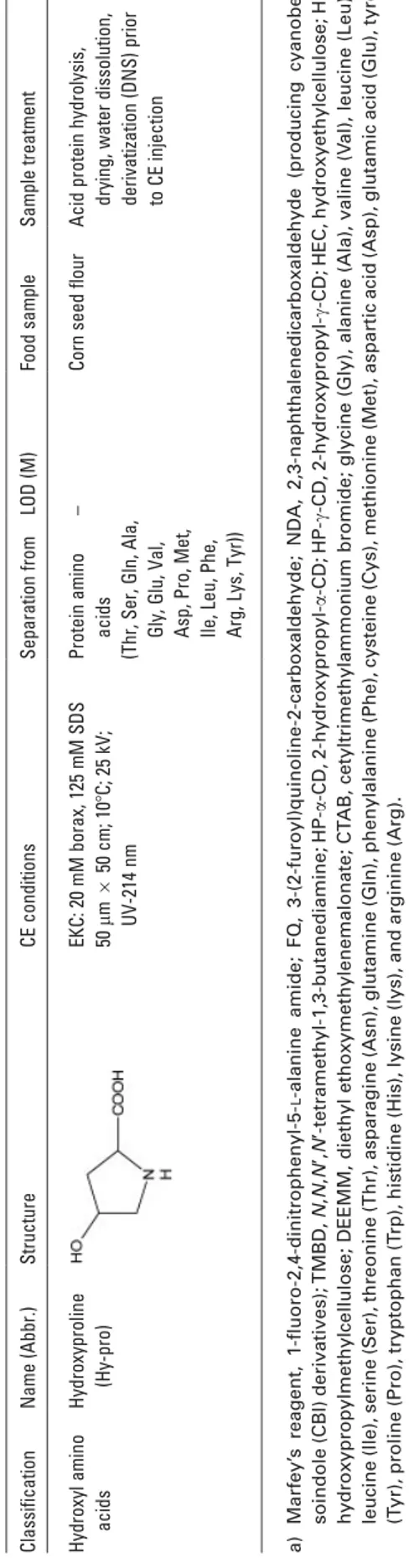
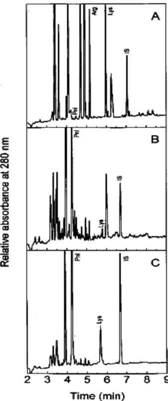
(8) 4038. M. Castro-Puyana et al.. seleno amino acids selenomethionine and selenocystine). The CZE method using an alkaline background electrolyte (10 mM sodium carbonate buffer adjusted to pH 11.5 with potassium hydroxide) separating the different species according to their different mobilities (charge to size ratio). The CIEF method used sodium hydroxide as the catholyte at the capillary inlet and phosphoric acid as the anolyte at the capillary outlet/nebulizer (pH range from 2 to 10) and the separation was produced according to the individual pI of the selenium species in the pH gradient generated after voltage application. Both coupling methods were very sensitive and highly resolving. However, LODs were slightly lower for CIEF-ICP-MS than for CZE-ICP-MS (see Table 1), due to higher sample intake and increased resolution of CIEF-ICPMS, while the CZE-ICP-MS method showed advantage in the number of species analyzed, six selenium species, including two inorganic ones. The two methods were applied separately to the analysis of human milk samples, being selenocystine the only non-protein amino acid found in samples submitted to a tenfold preconcentration step [16]. 2.2 Sulfur amino acids Taurine is the only sulfur amino acid analyzed in foods by CE. This compound is present in tissues in its free form and acts as physiological (i.e. maintenance of excitatory activity in muscle) and as a therapeutical (i.e. treatment of congestive heart failure) agent. However, the capacity for endogenous biosynthesis of taurine is insufficient being essential its assimilation from a dietary source [5]. A CZE method using 50 mM borate buffer at pH 9.2 was used for the determination of taurine in different foods. Due to the low sensitivity of UV or fluorescence detection of this non-protein amino acid (see its structure in Table 1), electrochemical detection with a carbon-disk as working electrode was used achieving LODs of , 10–7 M [5]. Figure 2 shows the typical electropherograms obtained for a Chinese medicinal herb (Lycium barbarum L. capsules), a health beverage (Lipovitan), and a milk powder using the CZE method developed. The authors concluded that, in terms of accuracy, sensitivity and reproducibility, the developed method was a useful tool to quantitate taurine in traditional Chinese medicines and beverages. 2.3 Aliphatic monoaminomonocarboxylic amino acids In this section, three aliphatic amino acids possessing one amino group and one carboxylic group are included. Another important characteristic of the three amino acids included in this group is that they are not a-amino acids. g-Aminobutyric acid (usually abbreviated as GABA) is a non-protein amino acid that together with other 14 protein amino acids forms a group of 15 amino acids responsible for more than 90% of the amino acidic content in vinegars [17] and orange juices [18, 19]. Its separation from other protein © 2007 WILEY-VCH Verlag GmbH & Co. KGaA, Weinheim. Electrophoresis 2007, 28, 4031–4045. Figure 2. Electropherograms of three food samples containing taurine (peak 1) obtained by CE with electrochemical detection. (A) Lycium barbarum L. capsules; (B) Lipovitan, and (C) milk powder. Electrophoretic conditions: fused-silica capillary with a total length of 70 cm and 25 mm id; separation buffer, 50 mM borate (pH 9.2); applied voltage, 30 kV; electrokinetic injection, 20 kV for 8 s. Electrochemical detection with a working electrode potential of 1.05 V. Reprinted from [5], with permission.. amino acids has been performed using an EKC system with SDS micelles and b-cyclodextrin (b-CD) (100 mM borate buffer at pH 9.5, 30 mM SDS, 20 mM b-CD). The use of this cyclodextrin (chiral selector) enabled the separation of the enantiomers of protein amino acids and was effective to separate them from GABA. To achieve the sensitive detection of GABA under these conditions (LODs in the nM range), derivatization with FITC prior to LIF detection was performed [17, 18]. Moreover, ESI-MS detection was also employed for a less sensitive detection (LODs in the mM range) of this non-protein amino acid and other protein amino acids, after derivatization with FITC, in orange juices [19] when an EKC system with a volatile buffer and a low concentration of the non-volatile b-CD was used. In addition, GABA (together with the protein amino acids L-arginine and L-aspartic acid) has been considered as an important parameter (its corrected peak area) for the characterization of orange juices (concentrated and pasteurized juices and nectars) [18]. Glycine betaine, named also trimethylglycine or betaine, is a quaternary ammonium non-protein amino acid with a widespread distribution in plants that has extensively been investigated since many plants accumulate it in response to environmental stress (such as salinity, high and low temperatures, etc.). In fact, glycine betaine is involved in osmotic adjustment, confers enzyme protection, and increases membrane stability under stress conditions. A CZE method based on the use of an 80 mM phosphate buffer at pH 3.0 was used to determine this compound in plant samples collected from high-salinity areas in North China and West www.electrophoresis-journal.com.
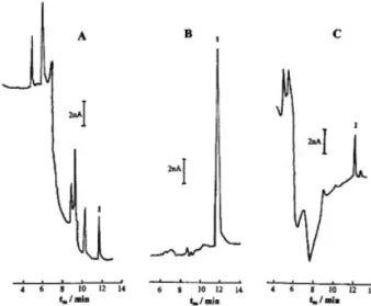
(9) Electrophoresis 2007, 28, 4031–4045. Australia (i.e. spinach) [20]. Prior to the CE injection, the amino acid was converted into their phenacyl ester to obtain a positive charged compound that at low pH migrates faster than other negative and neutral sample compounds which could interfere. The presence of glycine betaine in a leaf extract of a halophyte, Suaeda glauca, collected in a saline area in China showed that the interference caused by other amino acid esters in the determination of 1 mM glycine betaine ester was negligible. Finally, carnitine is a quaternary ammonium non-protein amino acid that participates in bioenergetic processes aiding the transformation of fats into energy, in the control of the mitochondrial acyl-CoA/CoA ratio, peroxisomal oxidation of fatty acids, and the production of ketone bodies. A deficiency of carnitine is known to have major deleterious effects on the central nervous system. It can be synthesized in the liver and kidney of adult people (from the protein amino acids lysine and methionine) or taken up in foods (meat, milk and vegetables). This amino acid has been analyzed in several food supplements by capillary isotachophoresis (cITP) with conductimetric detection and CZE with direct and indirect UV detection. cITP was achieved on an electrophoretic analyzer, CZE with direct detection was performed on a CE instrument, and CZE with indirect detection was carried out on both instruments. Since results obtained were similar by the above-mentioned methods, the comparison of them required to take into account the difficulty of sample preparation, instrumentation and automatic injection. The disadvantage of cITP and CZE with indirect UV detection performed on the electrophoretic analyzer was the impossibility of automatic injection. On the other hand, derivatization with FMOC was necessary for CZE with direct UV detection, which is not practical when analyzing series of samples. However, derivatization has the advantage of removing all interfering proteins and peptides. Finally, CZE with indirect UV detection achieved on the CE instrument was a practical option for simplicity and automation. This method consisted of a buffer with 5 mM Tris and 7 mM phosphoric acid containing 0.5 mM quinine as absorbing probe allowing LODs of , 2610–5 M. It was applied to determine carnitine in four different food supplements [21]. The results obtained by the CE methods for several manufactory samples with different concentrations of L-carnitine were compared with those obtained when using a validated HPLC method. Statistical differences among electrophoretic methods and HPLC were not found and the authors concluded that the CE analysis was faster, consumed less solvents and total running costs were lower than in HPLC [21]. 2.4 Aliphatic amino acids with nitrogen in the side chain The aliphatic non-protein amino acids containing nitrogen in the side chain are grouped in this section. The two amino acids included are a-amino acids with one (theanine) or two (lysinoalanine) asymmetric carbon atoms. © 2007 WILEY-VCH Verlag GmbH & Co. KGaA, Weinheim. CE and CEC. 4039. Theanine is a major component in tea related with its quality because it shows high correlation with the price of green tea. It has been separated from other tea catechins, caffeine, gallic acid and ascorbic acid by micellar EKC using 25 mM phosphate buffer (pH 7.0) containing 100 mM SDS and 6% methanol (for dried tea leaf and black tea samples) or 5% methanol (for bottled tea samples). This method together with direct UV detection at 200 nm enabled the detection up to 2610–5 M of theanine [22]. In addition, a CZE method using borax at pH 8.0 has also been used for the simultaneous separation of major components (theanine, catechins, caffeine, and ascorbic acid) in green tea infusions [23]. On the other hand, a cITP method using a conductivity detector was developed for the analysis of L-theanine in tea and food supplements. The leading electrolyte consisted of 0.01 M hydrochloric acid containing 0.02 mM TRIS and 0.05% hydroxyethylcellulose (pH 8.1), and the terminating electrolyte was 0.01 mM DL-valine with barium hydroxide (pH 10). After extraction with boiling water, theanine concentrations determined by this method ranged from 0.6 to 0.9% (m/m) in green tea (four different samples) and were about 90% (m/m) in food supplements (three samples) [24]. Lysinoalanine is a cross-linking amino acid formed in the reaction of lysine with dehydroalanine residues. It is indigestible by proteases and its formation implies a loss of lysine in proteins decreasing its nutritional value [6]. This non-protein amino acid has been determined in Pidan, a popular alkali-treated duck egg in Taiwan, and the relationship between the formation of lysinoalanine and the degradation of cysteine, serine, and threonine at various alkalitreating stages was investigated. A micellar EKC method using 250 mM borate buffer (pH 9.5) with 200 mM SDS and 75 mM b-CD and UV detection at 254 nm was used for the separation of this non-protein amino acid from most protein amino acids (see Table 1) [6]. Results obtained in this work indicated that the formation of lysinoalanine in albumen in the first stage of the pickling process was due to the speedy increase in the pH and the abundant formation of dehydroalanine from cysteine whereas the formation of lysinoalanine in the later pickling period was related much more to the alkali treating time than to the pH factor. In recent years, a growing interest in chemical miniaturization led to research and development of new methods and instrumentation on a microchip. Electrophoresis on microchips (usually named as chip or microchip electrophoresis) is a promising technology since it offers an easy integration of many steps including sample preparation, derivatization, and coupling of several separation procedures together [37, 38]. In addition, the use of microchips instead conventional CE systems leads to reduced analysis times. However, up to now very few applications to food samples have been performed by microchip electrophoresis. In fact, only one work where non-protein amino acids were determined in food samples by microchip electrophoresis has been reported in literature up to date. In this work, a plastic microchip with fluorescence detection was used to determine both non-prowww.electrophoresis-journal.com.
(10) 4040. M. Castro-Puyana et al.. tein (theanine) and protein amino acids (arginine and glutamine) in green tea [39]. The CE method used 5 mM phosphate buffer at pH 5.5 containing 0.05 mM SDS, and the derivatization of samples was carried out with 4-fluoro-7nitro-2,1,3-benzoxadiazole. Amino acid analysis was improved by removing polyphenols (catechins) using a polyvinylpolypyrrolidone pretreatment. Figure 3 shows the electropherogram corresponding to the separation of the amino acids theanine, arginine and glutamine found in Japanese green tea. Although arginine was not completely separated from unknown peaks, the separation of theanine and glutamine was good within 2 min [39].. Electrophoresis 2007, 28, 4031–4045. first isolated about one century ago from fruit of broad bean [41] and has been found in minute quantity in some beans [42]. A 35 mM phosphate buffer at pH 4.55 was used in the CZE method developed for the determination of levodopa in broad bean and lentil seeds. Under these conditions, and employing UV detection (210 nm), a LOD of , 3610–6 M was obtained [25]. Figure 4 shows the electropherograms obtained for the extracts of both samples using the described CZE method. A peak for levodopa was identified through the increase observed in the peak area after adding the standard of levodopa in the sample solution.. Figure 3. Electropherogram of Japanese green tea obtained using microchip electrophoresis with fluorescence detection. A polymethyl methacrylate (PMMA) microchip (85 mm650 mm) with three simple cross-channels of 100 mm width and 30 mm deep (detection point at 31 mm) was used; separation buffer, 5 mM phosphate (pH 5.5) containing 0.05 mM SDS; applied voltage, 1.7 kV; sample injection: 100 V for 60 s. Fluorescence detection: 460-490 nm (excitation wavelength) and 515 nm (emission wavelength). Reprinted from [39], with permission.. 2.5 Phenyl amino acids Among non-protein amino acids with a phenyl group in their structure, only the L-enantiomer of 3,4-dihydroxyphenylalanine (Levodopa or L-DOPA) has been analyzed under achiral conditions by CE in food samples. Levodopa is the precursor required by the brain to produce dopamine, a neurotransmitter (chemical messenger in the nervous system), thus it is used in the treatment of Parkinson disease [25]. However, auto-oxidation of levodopa generates toxic metabolites, such as free radicals, semi-quinones and quinones. In vitro, levodopa is a powerful toxin that is lethal to the culture of neurons, and a few animal studies have shown that levodopa can also be toxic [40]. Levodopa was © 2007 WILEY-VCH Verlag GmbH & Co. KGaA, Weinheim. Figure 4. Electropherograms of levodopa from broad bean (A) and lentil seed (B) sample solutions employing CE with UV detection. Electrophoretic conditions: fused-silica capillary with a total length of 47 cm (effective length of 40 cm) and 75 mm ID; separation buffer, 35 mM phosphate (pH 4.55); separation temperature, 307C; applied voltage, 17.5 kV; hydrodynamic injection (3447.4 Pa for 3 s). UV detection at 210 nm. Reprinted from [25], with permission.. 2.6 Isoxazolinone amino acids Isoxazolinone are N,O-heterocyclic natural compounds with high sensitivity to UV radiation and alkaline conditions. The amino acids included in this section are: 2-carboxymethyl-3isoxazolin-5-one, b-(isoxazolin-5-on-2-yl)-alanine, g-glutamylwww.electrophoresis-journal.com.
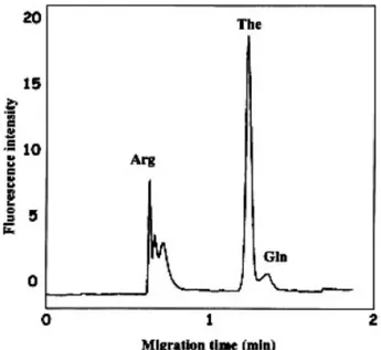
(11) Electrophoresis 2007, 28, 4031–4045. b-(isoxazolin-5-on-2-yl)-alanine, 2-(g-glutamylaminoethyl)isoxazolin-5-one, and 2-(3-amino-3-carboxypropyl)isoxazolin5-one. These compounds are present in legumes and seedlings, and especially in the seedlings of some Lathyrus species their concentration can be very high. They are biochemical precursors of a group of toxic metabolites present in some Lathyrus species and responsible for human diseases (i.e. neurotathysrism). Some of them have physiological activity on isolated neurones or cloned neuronal receptors or are of ecological importance [26, 27]. A CZE method using 25 mM phosphate buffer containing 8% 1-propanol at pH 7.5 was used to determine the five above-mentioned isoxazoline compounds in seedlings. This method allowed LOD ranging from 1610–5 to 3610–5 M [27]. Figure 5 shows the electropherograms of isoxazoline compounds in seedling extracts of grass pea (Lathyrus sativus) and of sweet pea (Lathyrus odoratus) using UV detection at 254 nm. Concentrations obtained by CE were in good agreement with those obtained by HPLC. 2.7 Heterocycle amino acids The non-protein amino acids containing a heterocycle group in the side chain are grouped in this section. The amino acids included here contain different heterocycles such as furane in furosine, pyrrolidine in domoic acid and pyroglutamic acid, and pyrrole in e-N-pyrrolylnorleucine (see structure in Table 1). Furosine is a non-protein amino acid formed from lysine, which is produced in milk and dairy products by the Maillard reaction (one of the most important reactions occurring in processed food during processing, sanitizing,. CE and CEC. 4041. packing and storage). Because furosine is formed during the acid hydrolysis of e-N-2-fructosylysine, its determination can be used to evaluate the extent of the Maillard reaction and the level of nutritionally unavailable lysine in processed food [28]. Furosine has been analyzed in dried milk and dairy products hydrolyzed with hydrochloric acid, by CZE with UV detection at 280 nm. This method consisted of 20 mM phosphoric acid buffer containing 60 mM N,N,N’N’-tetramethyl-1,3-butanediamine, and allowed an LOD of , 2610–6 M. This additive was used to avoid interactions of furosine with the capillary wall and to modulate its migration behavior by controlling the EOF [28]. Moreover, a previous method for the determination of furosine [29] has been improved for greater accuracy and sensitivity using a 3-(N-morpholino)-2-hydroxypropanesulfonic acid solution at pH 7.0 as running buffer together with an extended-path-length capillary enabled to obtain a CZE method whose repeatability and sensitivity fulfilled the requirements stated in the EU and Italian regulations for furosine determination in pasteurized milk and Mozzarella cheese. The data obtained when analyzing 48 different food samples including heat-treated milk, cheeses, and durum wheat products were comparable with those obtained by an HPLC method and proved to be accurate for furosine values up to at least 400 mg per 100 g protein [30]. Domoic acid is a neurotoxic tricarboxylic acid related to glutamic acid, which causes amnesic shellfish poisoning. In mammals, including human beings, it acts as a neurotoxin, causing short-term memory loss, brain damage, and death in severe cases [9]. A sensitive on-line coupled cITP-CZE method with UV detection at 242 nm has been developed to determine domoic acid in shellfish samples and food sup-. Figure 5. Electropherograms of isoxazoline compounds in 3-day-old seedlings of Lathyrus sativus (A) and Lathyrus odoratus (B) analyzed by CZE with UV detection. Electrophoretic conditions: coated fused-silica capillary with a total length of 60 cm (effective length of 52.6 cm) and 75 mm id; separation buffer, 25 mM phosphate containing 8% 1-propanol (pH 7.5); separation temperature, 307C; applied voltage, 22.5 kV; hydrostatic injection of 10 s. UV detection at 254 nm. BIP, b-(isoxazolin-5-on-2-yl)-propionitrile; ACI, 2-(3-amino-3-carboxypropyl)isoxazolin-5-one; BIA, b-(isoxazolin-5-on-2-yl)-alanine; g-glu-BIA, g-glutamyl-b-(isoxazolin-5-on-2-yl)-alanine; CMI, 2-carboxymethyl-3-isoxazolin-5-one; IS, internal standard. Reprinted from [27], with permission.. © 2007 WILEY-VCH Verlag GmbH & Co. KGaA, Weinheim. www.electrophoresis-journal.com.
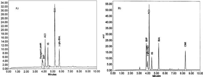
(12) 4042. M. Castro-Puyana et al.. plements containing algae extract. The conditions for cITPCZE analysis are detailed in Table 1, and enabled an LOD of ,4.8610–6 M [9]. e-N-Pyrrolylnorleucine is a product of the reaction between the lipid peroxidation product 4,5-(E)-epoxy-2-(E)-heptanol and the e-amino group of lysine [43]. This amino acid seems to be a normal component of many fresh food products, in which it may act as a natural antioxidant. In addition, the e-N-pyrrolylnorleucine/lysine ratio was directly correlated with lipid and iron contents and inversely correlated with the protein content in food [31]. A micellar EKC method based on the use of 100 mM borate buffer at pH 8.4 containing 100 mM SDS was employed to determine this compound in 22 different fresh food products hydrolyzed in basic conditions (5 fishes, 3 meats, 10 vegetables, and 4 nuts). To make easy the separation from other amino acids and the UV detection (280 nm), derivatization with diethyl ethoxymethylenemalonate prior to CE injection was performed. Figure 6 shows the electropherograms obtained for three different foods: salmon, spinach, and walnut where peaks corresponding to e-N-pyrrolylnorleucine, arginine, and lysine are marked [31]. The pyroglutamic acid content of coffee could provide it with some central activities previously associated with caffeine, such as improved attention and enhanced cognition by modulating dopamine and adenosine central functions. In fact, pyroglutamate is known to have a number of remarkable cognitive enhancing effects [32, 33]. It has been separated from other 16 short-chain organic acids (i.e. oxalic, formic, fumaric, mesaconic, succinic, maleic, malic, isocitric, citric, acetic, citraconic, glycolic, propionic, lactic, furanoic, and quinic acids) by CE with direct UV detection at 200 nm in coffee samples of different origins and with different treatments [32]. The method consisted of 500 mM phosphate buffer (pH 6.25) containing 0.5 mM CTAB, and allowed an LOD of , 1.2610–4 M in these samples. The organic acid profiles obtained for the samples showed important differences depending on industrial treatment of coffee. Moreover, pyroglutamate content was considerably higher in the lyophilized coffee than in the roasted one and the authors attributed these results to some stage of the industrial process [32]. On the other hand, an MEKC method using 50 mM borate buffer (pH 9.5) with 130 mM SDS, and using UV detection at 200 nm was also developed for the simultaneous determination of caffeine and pyroglutamate in ten soluble coffees acquired in the market. This method enabled LODs of ,6.9610–7 M. The coffees with the highest content in pyroglutamate (with or without caffeine) were preliminarily tested for sedative/stimulant properties and cognition enhancing effects in mice [33]. 2.8 Cycloalkane amino acids The 1-aminocyclopropane-1-carboxylic acid is a disubstituted cyclic amino acid, and it is the only cycloalkane amino acid that has been analyzed by CE in food samples. This amino © 2007 WILEY-VCH Verlag GmbH & Co. KGaA, Weinheim. Electrophoresis 2007, 28, 4031–4045. Figure 6. Electropherograms corresponding to the separation of e-N-pyrrolylnorleucine (Pnl), arginine (Arg), lysine (Lys), and internal standard (IS) in (A) salmon, (B) spinach, and (C) walnut by CE with UV detection after basic hydrolysis and derivatization with diethyl ethoxymethylenemalonate. Electrophoretic conditions: uncoated fused-silica capillary with a total length of 50 cm and 75 mm id; separation buffer, 100 mM borate (pH 8.4), 100 mM SDS; temperature, 307C; applied voltage, 30 kV; hydrodynamic injection (2 s). UV detection at 280 nm. Reprinted from [31], with permission.. www.electrophoresis-journal.com.
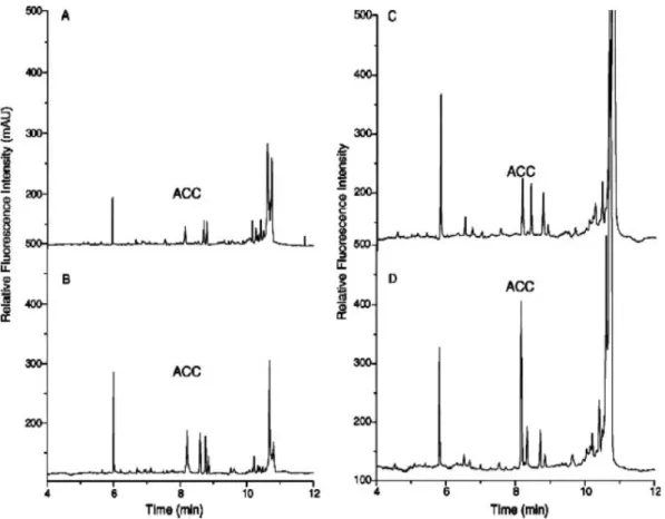
(13) CE and CEC. Electrophoresis 2007, 28, 4031–4045. acid plays an important role in the biosynthesis of ethylene, which is associated with many physiological processes in higher plants, including fruit ripening, senescence, and abscission of plant organs. In fact, 1-aminocyclopropane-1carboxylic acid was identified as an important immediate precursor of ethylene [34]. For this reason, the quantitative analysis of this non-protein amino acid is of interest in higher plants and their fruits. The separation of 16 common protein amino acids (see Table 1) and 1-aminocyclopropane-1-carboxylic acid in apple extracts was performed by an EKC method with a running buffer of 20 mM borate (pH 9.35), 40 mM SDS and 10 mM Brij 35 and employed LIF detection. Since the molecular structure of this non-protein amino acid does not have a fluorophore group, a derivatization with 3-(2-furoyl)quinoline-2-carboxaldehyde prior to LIF detection was necessary [34]. Figure 7 shows the electropherograms obtained for apple extracts using different extraction solvents with or without wounding treatment (this treatment is performed by nicking the apple with blade and then keeping the wounded apple at room temperature for 24 h). As it can be seen, the. 4043. content of 1-aminocyclopropane-1-carboxylic acid in apple is higher when employing an acid extraction with wounding treatment. This figure shows that the treatment of the sample is an important parameter since the efficiency of sample extraction can be affected by different solvents and can be improved under wounding treatment of apples. The mass LOD of 1-aminocyclopropane-1-carboxylic acid was 50 amol or 1610–8 M in terms of concentration. It is important to mention that the high sensitivity and selectivity of the method enabled the direct determination of this non-protein amino acid in crude extracts without further purification and concentration [34].. 2.9 Hydroxyl amino acids Hydroxyproline differs from proline in the presence of a hydroxyl group attached to the g-carbon. This non-protein amino acid is a major component of the protein collagen. It helps to provide stability to the triple-helical structure of collagen by forming hydrogen bonds.. Figure 7. Electropherograms of 1-aminocyclopropane-1-carboxylic acid (ACC) in apple extracts by CE with LIF detection, using aqueous methanol (80%, v/v) without wounding treatment (A), using aqueous methanol (80%, v/v) with wounding treatment (B), using 5% sulfosalicylic acid without wounding treatment (C), and using 5% sulfosalicylic acid with wounding treatment (D). Electrophoretic conditions: uncoated fused-silica capillary with a total length of 65 cm (effective length of 50 cm) and 50 mm id; separation buffer, 20 mM borate (pH 9.35), 40 mM SDS and 10 mM Brij 35; temperature, 257C; applied voltage, 25 kV; hydrodynamic injection (50 mbar for 5 s). LIF detection: 488 nm (excitation), 560 nm (emission). Reprinted from [34], with permission.. © 2007 WILEY-VCH Verlag GmbH & Co. KGaA, Weinheim. www.electrophoresis-journal.com.
(14) 4044. M. Castro-Puyana et al.. The separation of hydroxyproline from 16 protein amino acids (indicated in Table 1) was performed using a MEKC method based on the use of 20 mM borax containing 125 mM SDS with UV detection at 214 nm. Before UV detection, a preconcentration step of samples after derivatization with dansyl chloride (DNS) was necessary due to the low concentration of DNS-amino acids in the mixture [35]. The separation of the hydrolysis product of DNS (dansyl sulfonic acid) and DNShydroxyproline from corn seed flour using the MEKC method was achieved. It was observed that the concentration of SDS was an important parameter to avoid the coelution of the derivatization agent with the derivatized hydroxyproline.. Electrophoresis 2007, 28, 4031–4045. for the Ramón y Cajal program (RYC-2003-001). M. CastroPuyana thanks the University of Alcalá for her predoctoral grant.. 4. References. [1] Hunt, S., The Non-Protein Amino Acids, in: Barret, G. C. (Ed.), Chemistry and biotechnology of amino acids, 1985, 55–183. [2] Friedman, M., J. Agric. Food Chem. 1999, 47, 3457–3479. [3] Kvasnička, F., J. Sep. Sci. 2005, 28, 813–825. [4] Peace, R. W., Gilani, G. S., J. AOAC Int. 2005, 88, 877–887.. 3. Concluding remarks. This review is the first one reporting all the articles published in relation to the determination of non-protein amino acids in foods by CE. Since non-protein amino acids can be related with the quality and security of foods, their determination in these samples presents a high interest. Determination of non-protein amino acids by CE in foods was usually performed using CZE and EKC modes. Detection systems employed include direct and indirect UV absorption, fluorescence, electrochemical, and atomic and molecular MS. The most used were optical detectors, mainly UV absorption detection, which required a previous step of derivatization due to the low UV absorptivity of most non-protein amino acids. Derivatization was also employed when using molecular MS detection, in order to increase the molecular mass of the analytes to be determined. Although electrochemical detection did not require derivatization, it was very scarcely used. CE analytical methodologies with adequate sensitivity and selectivity were developed showing the big potential of this technique for the analysis of a great number of nonprotein amino acids in a considerable variety of samples of different complexity including beverages, seeds, vegetables and plants, food supplements, yeast, eggs, meats, fishes, and nuts. In fact, some problems in the food area not previously solved by other analytical separation techniques were solved by CE. Thus, for some of the non-protein amino acids included in this review, CE methodologies proposed are the only existing methodologies. As example, as far as we know, no HPLC methods were developed for the determination of taurine, glycine betaine, 3,4-dihydroxyphenylalanine, pyroglutamic acid and all the isoxazolinone amino acids included in Table 1. On the other hand, for those amino acids for which previous HPLC methodologies were recommended, CE was successful in the development of complementary methodologies that could be useful in carrying out collaborative testing needed in the food area.. The authors thank the Comunidad Autónoma de Madrid (Spain) (project S-0505/AGR/0312) for financial support. C. García-Ruiz also thanks the Ministry of Science and Technology © 2007 WILEY-VCH Verlag GmbH & Co. KGaA, Weinheim. [5] Cao, Y., Zhang, X., Chu, Q., Fang, Y., Ye, J., Electroanalysis 2003, 15, 898–902. [6] Chang, H.-M., Cheng-Fang, T., Chin-Fung, L., J. Agric. Food Chem. 1999, 47, 1495–1500. [7] Peng, H., Revell, D., McSweeney, C. S., Brooker, J. D., Anim. Feed Sci. Technol. 2005, 121, 139–146. [8] Kuo, Y.-H., Rozan, P., Lambein, F., Frias, J., Vidal-Valverde, C., Food Chem. 2004, 86, 537–545. [9] Kvasnička, F., Ševčik, R., Voldřich, M., J. Chromatogr. A 2006, 1113, 255–258. [10] Marina, M. L., Ríos, A., Valcárcel, M. (Eds.), Analysis and Detection by Capillary Electrophoresis, CAC, Elsevier, Amsterdam 2005. [11] Poinsot, W., Lacroix, M., Maury, D., Chataigne, G. et al., Electrophoresis 2006, 27, 176–194. [12] Cifuentes, A., Electrophoresis 2006, 27, 283–303. [13] Day, J. A., Kannamkumarath, S. S., Yanes, E. G., MontesBayón, M., Caruso, J. A., J. Anal. At. Spectrom. 2002, 17, 27– 31. [14] Sun, B., Macka, M., Haddad, P. R., J. Chromatogr. A 2004, 1039, 201–208. [15] Michalke, B., Fresenius’ J. Anal Chem. 1995, 351, 670–677. [16] Michalke, B., Schramel, P., J. Chromatogr. A 1998, 807, 71– 80. [17] Carlavilla, D., Moreno-Arribas, M. V., Fanali, S., Cifuentes, A., Electrophoresis 2006, 27, 2551–2557. [18] Simó, C., Martín-Alvarez, P. J., Barbas, C., Cifuentes, A., Electrophoresis 2004, 25, 2885–2891. [19] Simó, C., Rizzi, A., Barbas, C., Cifuentes, A., Electrophoresis 2005, 26, 1432–1441. [20] Zhang, J., Nishimura, N., Okubo, A., Yamazaki, S., Phytochem. Anal. 2002, 13, 189–194. [21] Prokorátová, V., Kvasnička, F., Ševčík, R., Voldřich, M., J. Chromatogr. A 2005, 1081, 60–64. [22] Aucamp, J. P., Hara, Y., Apostolides, Z., J. Chromatogr. A 2000, 876, 235–242. [23] Horie, H., Mukai, T., Kohata, K., J. Chromatogr. A 1997, 758, 332–335. [24] Kvasnička, F., Kratka, J., Central Eur. J. Chem. 2006, 4, 216– 222. [25] Chen, X., Zhang, J., Zhai, H., Chen, X., Hu, Z., Food Chem. 2005, 92, 381–386. [26] Rozan, P., Kuo, Y.-H., Lambein, F., Phytochemistry 2001, 58, 281–289.. www.electrophoresis-journal.com.
(15) Electrophoresis 2007, 28, 4031–4045. CE and CEC. 4045. [27] Chowdhurry, B., Rozan, P., Kuo, Y.-H., Sumino, M., Lambein, F., J. Chromatogr. A 2001, 933, 129–136.. [36] Uden, P. C., Boakye, H. T, Kahakachchi, C., Tyson, J. F., J. Chromatogr. A 2004, 1050, 85–93.. [28] Corradini, C., Cannarsa, G., Corradini, C., Nicoletti, I. et al., Electrophoresis 1996, 17, 120–124.. [37] Erickson, D., Li, D. Q., Anal. Chim. Acta 2004, 507, 11–26.. [29] Tirelli, A., Pellegrino, L., Italian J. Food Sci. 1995, 7, 379–385. [30] Tirelli, A., J. Food Protection 1998, 61, 1400–1404. [31] Zamora, R., Alaiz, M., Hidalgo, F. J., J. Agric. Food Chem. 1999, 47, 1942–1947. [32] Galli, V., Barbas, C., J. Chromatogr. A 2004, 1032, 299–304. [33] Maeso, N., del Castillo, C., Cornejo, L., García-Acicollar et al., J. Pharm. Biomed. Anal. 2006, 41, 1095–1100. [34] Liu, X., Li, D.-F., Wang, Y., Lu, Y.-T., J. Chromatogr. A 2004, 1061, 99–104. [35] Skočir, E., Prošek, M., Chromatographia 1995, 41, 638–644.. © 2007 WILEY-VCH Verlag GmbH & Co. KGaA, Weinheim. [38] Bilitewski, U., Genrich, M., Kadow, S., Marsal, G., Anal. Bioanal. Chem. 2003, 377, 556–569. [39] Kato, M., Gyoten, Y., Sakai-Kato, K., Toyo’oka, T., J. Chromatogr. A 2003, 1013, 183–189. [40] Melamed, E., Offen, D., Shirvan, A., Ziv, I., J. Neurol. 2000, 247, 135–139. [41] Brain, K. R., Plant Sci. Lett. 1976, 7, 157–161. [42] Vadivel, V., Janardhanan, K., Plant Foods Hum. Nutr. 2002, 57, 151–164. [43] Zamora, R, Navarro, J. L., Hidalgo, F. J., Lipids 1995, 30, 477– 483.. www.electrophoresis-journal.com.
(16)
Figure
+3
Documento similar
