Percutaneous shunt reduction for
the management of TIPS-induced acute liver
decompensation: A follow-up study
Bart De Keyzer,* Frederik Nevens,** Annouschka Laenen,*** Sam Heye,* Wim Laleman,** Chris Verslype,** Schalk van der Merwe,** Geert Maleux*
* Department of Radiology, University Hospitals Leuven, Department of Imaging & Pathology, KU Leuven, Belgium. ** Department of Gastroenterology and Hepatology, University Hospitals Leuven, Belgium. *** Interuniversity Centre for Biostatistics and Statistical Bioinformatics, Catholic University of Leuven and Hasselt University, Belgium.
A B S T R A C T A B S T R A C T A B S T R A C T A B S T R A C T A B S T R A C T
Background and rationale for the study. Background and rationale for the study.Background and rationale for the study. Background and rationale for the study.
Background and rationale for the study. The purpose of this study was to assess the technical and clinical outcomes of transjugular intrahepatic portosystemic shunt (TIPS) reduction for the management of TIPS-induced acute liver decompensation. Be-tween August 2000 and November 2013, 347 patients underwent a TIPS procedure in the authors’ institution; 21/347 (6%) de-veloped post-TIPS acute liver decompensation which was managed using a percutaneous shunt reduction technique. Patient demographics, laboratory tests before and after initial TIPS and TIPS reduction, procedural data and clinical follow-up data were ana-lysed. Results.Results.Results.Results.Results. Twenty-one patients (mean age 63 years) who underwent an initial TIPS procedure for variceal bleeding (n = 7; 33%) or refractory ascites (n = 14; 67%) successfully underwent shunt reduction ten days (3-34 days) after the initial TIPS pro-cedure. The portosystemic pressure gradient (PSPG) increased from 8 (3-17) mmHg before reduction to 12 (7-23) mmHg after shunt reduction. Survival at one and six months follow-up was 15 (71%) and 11 patients (52%), respectively. The international normalised ratio (INR) (1.7 vs. 1.5; p = 0.044) was significantly different after TIPS reduction in the non-survival group compared to the sur-vival group. In conclusion, TIPS reduction for the management of TIPS-induced acute liver decompensation is technically feasible and is associated with a one and six-month mortality rate of 29% and 48%, respectively. Higher post-TIPS-reduction INR values may be associated with higher risk of early mortality.
Key words. Key words.Key words. Key words.
Key words. Interventional radiology. Transjugular intrahepatic portosystemic shunt. Acute liver decompensation. Shunt reduction. November-December, Vol. 15 No. 6, 2016: 911-917
INTRODUCTION
The creation of a transjugular intrahepatic portosystemic shunt (TIPS) may be associated with several complica-tions, both local procedural, including puncture-related, and more general shunt-related, including hepatic encepha-lopathy and acute liver decompensation.1
Acute liver decompensation in patients with pre-exist-ing liver cirrhosis is defined as an acute deterioration of liver function and subsequently the functioning of other end organs over a period of weeks following a precipitat-ing event, either indirect (variceal haemorrhage, sepsis) or direct (drug-induced, liver resection, hepatotoxic factor and TIPS placement), in a patient with previously
well-compensated or reasonably well-well-compensated chronic liv-er disease.2-4
When transplantation is not an option, TIPS occlu-sion or reduction may ameliorate the degree of liver in-sufficiency.5,6 The rationale of TIPS reduction is to
narrow the shunt flow lumen while preserving its paten-cy, thereby increasing portal blood perfusion of the liver. The technique of TIPS reduction is the same as de-scribed for the interventional radiological management of TIPS-induced hepatic encephalopathy.7-10 The
pur-pose of this study was to characterise the technical, haemodynamic, laboratory and clinical outcomes of TIPS reduction in the treatment of TIPS-induced acute liver decompensation.
The Official Journal of the Mexican Association of Hepatology, the Latin-American Association for Study of the Liver and
the Canadian Association for the Study of the Liver
Manuscript received: Manuscript received:Manuscript received:
MATERIAL AND METHODS
Study design
An electronic search in the institution’s interventional radiology database was performed to identify patients who underwent an initial TIPS procedure between Au-gust 2000 and November 2013 and, owing to the develop-ment of TIPS-induced acute liver decompensation, were later referred for an interventional procedure for TIPS reduction. TIPS-induced acute liver decompensation was defined as liver decompensation associated with the development of jaundice (≥ 50% increase in bilirubin levels compared to pre-TIPS) and coagulopathy (≥ 50% increase in International Normalised Ratio (INR) com-pared to pre-TIPS), complicated by ascites or encepha-lopathy or both ≤ 4 weeks after a TIPS procedure. This definition excludes liver decompensation elicited by in-fection or gastrointestinal bleeding. Clinical success was defined as improvement of patient’s general and bio-chemical status, allowing discharge from the intensive care unit.
Pre and post-interventional clinical, laboratory and radiological data for these patients were collected ret-rospectively using patients’ electronic medical and radi-ological hospital records. Approval by the local Ethics Committee was obtained for retrospective data analysis.
Interventional radiological technique for initial TIPS and TIPS reduction procedure:
In all 21 patients, the initial portosystemic shunt was created as previously described.11 Briefly, percutaneous
access to the right internal jugular vein was obtained under general anaesthesia. Pressure measurements were per-formed in the inferior vena cava using a pigtail catheter and in the right hepatic vein using an occlusion balloon catheter (wedged hepatic vein pressure measurement) to calculate the PSPG. Carbon dioxide (CO2) wedged por-tography was performed to guide the puncture from the right hepatic vein into the right proximal portal vein using a coaxial Rösch-Uchida puncture set (Cook Medical, Bloomington, IN, USA). Following predilatation of the shunt tract (Wanda 5 mm diameter angioplasty balloon catheter, Boston Scientific, Natick, MA, USA), an ex-panded-polytetraethylene (ePTFE)-covered Nitinol stent graft (Viatorr, W. L. Gore & Associates, Flagstaff, AZ, USA) was inserted, with a nominal diameter of 10 mm and a length covering the intrahepatic parenchymal tract as far as the inferior vena cava. Afterwards, the stent graft was postdilated using a 10 mm diameter angioplasty balloon (Wanda; Boston Scientific, Natick, MA, USA) in the case of variceal bleeding and using an 8 mm angioplasty balloon (Wanda; Boston Scientific) in the case of refractory ascites or hepatic hydrothorax. Finally, pressure measurements
were performed in the inferior vena cava and in the portal vein main branch, respectively.
The TIPS reduction procedure was performed using the parallel technique.9,10 Briefly, depending on the
pa-tient’s general condition, the percutaneous TIPS reduc-tion procedure was performed under either general or local anaesthesia. Percutaneous venous access was ob-tained to the right jugular and right common femoral vein, followed by catheterisation of the TIPS tract from both the jugular and femoral approach. The PSPG was calculat-ed bascalculat-ed on pressure measurements in the proximal infe-rior vena cava and in the portal vein main branch. Iodised contrast portography was performed to evaluate TIPS Stent graft patency. Shunt reduction was achieved by plac-ing a stainless steel balloon-expandable 6 mm diameter and 17 mm long stent mounted on an 0.035 inch catheter system (Express Vascular, Boston Scientific, Natick,
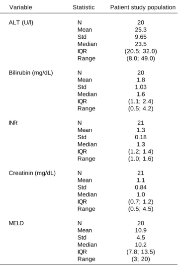
Table 1. Laboratory values before initial TIPS placement.
Variable Statistic Patient study population
ALT (U/l) N 20
Mean 25.3
Std 9.65
Median 23.5
IQR (20.5; 32.0)
Range (8.0; 49.0)
Bilirubin (mg/dL) N 20
Mean 1.8
Std 1.03
Median 1.6
IQR (1.1; 2.4)
Range (0.5; 4.2)
INR N 21
Mean 1.3
Std 0.18
Median 1.3
IQR (1.2; 1.4)
Range (1.0; 1.6)
Creatinin (mg/dL) N 21
Mean 1.1
Std 0.84
Median 1.0
IQR (0.7; 1.2)
Range (0.5; 4.5)
MELD N 20
Mean 10.9
Std 4.5
Median 10.2
IQR (7.8; 13.5)
Range (3; 20)
MA,USA), which was deployed in the middle third of the initial TIPS tract, in parallel with an ePTFE-covered stent graft (Viatorr, W.L. Gore & Associates) with a nominal di-ameter of 10 mm and the same length as the initial stent graft. Afterwards, completion portography and PSPG measurements were performed. In the event of reopacifi-cation of any variceal collaterals immediately after shunt reduction, these collaterals were preventively coil-embol-ised using 0.035 inch stainless steel coils (MR eye coils, Cook Medical, Bloomington, IN, USA).
Statistical analysis
The Mann-Whitney U test was used to compare survi-vors and non-survisurvi-vors on continuous variables. All tests were two-sided. A 5% significance level was assumed for all tests. All analyses were performed using SAS software, version 9.4 of the SAS system for Windows (SAS Institute Inc., Cary, NC, USA).
RESULTS
21/347 patients (6%), with a median age of 63 years (range 47-84 years) including 11 male patients (53%) who had undergone a TIPS procedure between August 2000 and January 2013 developed TIPS-induced acute liver decom-pensation. Pre-interventional duplex ultrasound revealed a fully patent hepatic artery, portal vein and stented TIPS tract in all patients.
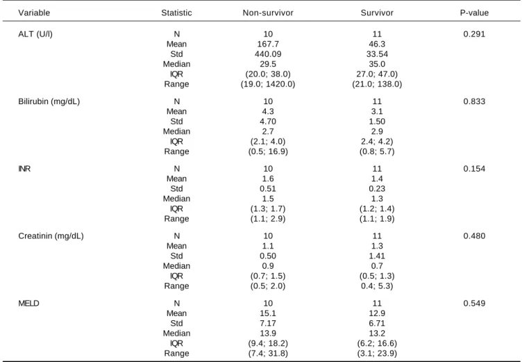
The underlying liver diseases were alcoholic cirrhosis (n = 12; 57%); non-alcoholic steatohepatitis (n = 3; 14%); alpha-1-antitrypsin deficiency (n = 1; 5%); hepatitis C (n = 1; 5%); chronic auto-immune cirrhosis (n = 1; 5%); cryptogenic cirrhosis (n = 1; 5%); glycogenosis (n = 1; 5%) and auto-immune hepatitis (n = 1; 5%). Indications for TIPS placement were variceal bleeding (n = 6; 29%), hepatic hydrothorax (n = 1; 5%), ascites (n = 12; 57%) or a combination of ascites and hepatic hydrothorax (n = 2; 10%). The laboratory values for the Table 2. Laboratory values immediately before TIPS reduction.
Variable Statistic Non-survivor Survivor P-value
ALT (U/l) N 10 11 0.291
Mean 167.7 46.3
Std 440.09 33.54
Median 29.5 35.0
IQR (20.0; 38.0) 27.0; 47.0)
Range (19.0; 1420.0) (21.0; 138.0)
Bilirubin (mg/dL) N 10 11 0.833
Mean 4.3 3.1
Std 4.70 1.50
Median 2.7 2.9
IQR (2.1; 4.0) 2.4; 4.2)
Range (0.5; 16.9) (0.8; 5.7)
INR N 10 11 0.154
Mean 1.6 1.4
Std 0.51 0.23
Median 1.5 1.3
IQR (1.3; 1.7) (1.2; 1.4)
Range (1.1; 2.9) (1.1; 1.9)
Creatinin (mg/dL) N 10 11 0.480
Mean 1.1 1.3
Std 0.50 1.41
Median 0.9 0.7
IQR (0.7; 1.5) (0.5; 1.3)
Range (0.5; 2.0) 0.4; 5.3)
MELD N 10 11 0.549
Mean 15.1 12.9
Std 7.17 6.71
Median 13.9 13.2
IQR (9.4; 18.2) (6.2; 16.6)
Range (7.4; 31.8) (3.1; 23.9)
21 patients prior to the initial TIPS procedure are sum-marised in table 1.
The mean PSPG before shunt creation was 15 mmHg (range 8-18 mmHg). The time interval between TIPS placement and reduction was 14 days (range 2-44 days).
The indication for shunt reduction was a twofold or more increase in bilirubin levels (n = 12; 57%) and/or a 50% increase in INR (n = 3; 14%) associated with clinical symptoms of grade 3-4 hepatic encephalopathy (n = 18, 88%).
Depending on the general status of the patient, the shunt reduction procedure was performed under local (n = 17; 81%) or general (n = 4; 19%) anaesthesia. The laboratory values before and after the shunt reduction pro-cedure are summarised in tables 2 and 3.
The shunt reduction procedure was technically suc-cessful in all cases. Pressure measurements in the inferior vena cava and main portal vein, as well as the assessment of the PSPG and MELD scores for both survivors and
non-survivors are summarised in table 4. The median time in-terval between TIPS placement and TIPS reduction was 10 (3-34) days in surviving patients and 20 (2-44) days in non-surviving patients (p-value = 0.1).
In all cases, angiographic evaluation before shunt reduc-tion demonstrated a fully patent TIPS stent graft without opacification of any intrahepatic portal vein end branches; after shunt reduction there was antegrade reopacification of the intrahepatic portal vein branches in 13 patients (62%), i.e. in eight (73%) survivors and in five (50%) non-survivors.
In 5/21 (24%) patients additional coil embolisation of reopacified varices was performed.
During follow-up, reduced shunt occlusion occurred in two patients, resulting in recurrent refractory ascites in both patients. The occluded and reduced TIPS shunt was reopened using interventional techniques.
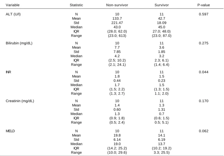
All patients were monitored until death (n = 13; 62%), liver transplantation (n = 4; 19%), or time of data analysis Table 3. Peak laboratory data ten days after TIPS reduction.
Variable Statistic Non-survivor Survivor P-value
ALT (U/l) N 10 11 0.597
Mean 133.7 42.7
Std 221.47 18.09
Median 43.0 45.0
IQR (28.0; 62.0) 27.0; 48.0)
Range (23.0; 613) (23.0; 87.0)
Bilirubin (mg/dL) N 10 11 0.275
Mean 7.7 3.6
Std 7.85 1.85
Median 4.2 3.2
IQR (2.5; 10.2) 2.3; 6.1)
Range (2.1; 24.1) (1.4; 6.4)
INR N 10 11 0.044
Mean 1.8 1.5
Std 0.44 0.23
Median 1.7 1.5
IQR (1.5; 2.2) (1.3; 1.5)
Range (1.3; 2.7) 1.1; 2.0)
Creatinin (mg/dL) N 10 11 0.170
Mean 1.4 1.3
Std 0.60 1.31
Median 1.3 0.7
IQR (0.9; 1.8) (0.6; 1.5)
Range (0.5; 2.4) 0.5; 5.1)
MELD N 10 11 0.062
Mean 19.8 14.1
Std 6.14 6.19
Median 19.0 13.7
IQR (14.2; 25.2) (10.2; 19.2)
Range (10.0; 29.6) 3.3; 25.5)
Table 4. Pressure measurements before and after TIPS reduction.
Variable Statistic Non-survivor Survivor P-value
PSPG before TIPS reduction (mmHg) N 10 11 0.523
Std 3.21 4.49
Median 7.5 9.0
IQR (5.0; 10.0) 5.0; 13.0)
Range (3.0; 13.0) (3.0; 17.0)
Pressure in the inferior vena cava N 10 11 0.158
before TIPS reduction (mmHg) Mean 10.8 4.9
Std 8.56 3.48
Median 13.0 4.0
IQR (3.0; 17.0) 2.0; 7.0)
Range (-2.0; 23.0) (1.0; 12.0)
Pressure in the main portal vein before N 10 11 0.168
TIPS reduction (mmHg) Mean 18.3 13.3
Std 10.44 4.86
Median 20.0 13.0
IQR (10.0; 27.0) 9.0; 19.0)
Range (3.0; 31.0) 7.0; 20.0)
PSPS after TIPS reduction (mmHg) N 9 11 0.252
Mean 12.0 15.1
Std 4.90 5.56
Median 11.0 15.0
IQR (8.0; 15.0) 11.0; 21.0)
Range (7.0: 22.0) 7.0; 23.0)
Pressure in the inferior vena cava after N 9 11 0.039
TIPS reduction (mmHg) Mean 11.1 5.0
Std 8.13 4.43
Median 12.0 6.0
IQR (7.0; 17.0) (1.0; 7.0)
Range (-5.0; 22.0) (-1.0; 12.0)
Pressure in the main portal vein after N 9 11 0.401
TIPS reduction (mmHg) Mean 23.1 19.3
Std 11.37 5.37
Median 21.0 18.0
IQR (15.0; 31.0) 15.0; 24.0)
Range (3.0; 40.0) 13.0; 29.0)
Time interval between TIPS and shunt N 10 11 0.27
reduction, days days 20 (2-44) 10 (3-14)
PSPG: portosystemic pressure gradient.
(n = 4; 19%). A total of 11 out of 21 patients (52%) were able to be discharged from the intensive care unit after a median of 10.6 days (range: 3-25 days). The overall one and six-month survival rates were 15/21 (71%) and 11/21 (52%), respectively. Four of the surviving patients (19%) under-went liver transplantation, respectively at 55, 56, 105 and 510 days after the shunt reduction procedure.
DISCUSSION
This study confirms the technical feasibility and repro-ducibility of the parallel technique to reduce the diameter of a portosystemic shunt in patients suffering from
TIPS-induced acute liver decompensation, as previously dem-onstrated for the management of TIPS-induced hepatic encephalopathy.7-10 In line with previous reports,9,10 we
also found quite a low reocclusion rate of 10% during fol-low-up; however, the follow-up period was relatively short, mainly due to the high early mortality rate in this pa-tient population. In the case of reduced shunt occlusion, it is still technically feasible to recanalise and reopen the oc-cluded shunt and reline with another ePTFE-covered stent graft.10
under 70% and 50%, respectively, despite maximum medi-cal and interventional management. TIPS-induced acute liver decompensation is a rare disease, with an incidence of 6% in this series, and might be considered as another form of acute-on-chronic liver failure (ACLF): all patients suffered from underlying liver cirrhosis with severe clini-cal signs of portal hypertension, and shortly after TIPS placement these patients developed acute liver decompen-sation with typical signs of ascites, encephalopathy, jaun-dice and other laboratory findings indicating liver failure.2-4 It is still unclear what the overall survival rate in
patients with post-TIPS acute liver decompensation would be without interventional shunt-reduction treat-ment: no reports on the natural outcome of this disease exist in the literature. Moreau, et al.3 analysed the
out-comes for a patient population suffering from ACLF not related to TIPS insertion and treated through medical management, and found a transplant-free mortality of 34% and 51% after 30 and 90 days, respectively, which is no dif-ferent from our results after TIPS shunt reduction. Al-though a prospective multicentre study randomising patients between interventional shunt reduction and max-imised conservative treatment should provide an answer to this question, such a study design seems to be very diffi-cult in terms of recruiting patients, not only due to the rarity of the disease, but also because most of the patients will favour an interventional treatment as shunt reduction/ closure might return them to their pre-TIPS status. It is also clear that several pre-TIPS factors are prognostic for early post-TIPS mortality, such as a high MELD score,12-14
or Child-Pugh score.14 However, we did not identify any
prognostic pre-reduction TIPS factors for improved sur-vival once a patient had developed TIPS-induced acute liver decompensation. Conversely, increased INR values post-TIPS-reduction (p = 0.044) seem to be predictive of worse outcomes. There was also a tendency (p = 0.06) to-wards higher post-reduction MELD scores in the non-surviving group (MELD score 19.8) compared to the surviving group (MELD score 14.1). In a larger study population, this trend might potentially be significant. Another point of interest is the management of patients with post-TIPS severe liver function disturbances with-out clear clinical signs of liver failure, including ascites and hepatic encephalopathy. We encountered 3 patients with severe liver function disturbances post-TIPS and de-cided to reduce the TIPS in order to avoid later clinical complications of liver failure, although it is not clear if these clinical symptoms may occur later on. Last, we also found a post-TIPS-reduction increase in measured inferi-or vena cava pressure as another prognostic factinferi-or finferi-or early death. These data might be explained by the multi-organ failure status of most of these patients at the time of the shunt reduction procedure.
In conclusion, this study shows high one-month and six-month mortality rates of 30% and 50%, respectively in patients undergoing TIPS reduction for TIPS-induced acute liver decompensation. Increased levels of INR after TIPS reduction seem to be the most important prognostic parameter for early death.
REFERENCES
1. Suhocki P, Lungren M, Kapoor B, Kim C. Transjugular intra-hepatic portosystemic shunt complication: prevention and management. Semin Intervent Radiol 2015; 32: 123-32. 2 Laleman W, Verbeke L, Meersseman P, Wauters J, van Pelt
J, Cassiman D, Wilmer A, et al. Acute-on-chronic liver fail-ure: current concepts on definition,pathogenesis, clinical manifestations and potential therapeutic interventions. Ex-pert Rev Gastroenterol Hepatol 2011; 5: 523-37.
3. Moreau R, Jalan R, Gines P, Pavesi M, Angeli P, Cordoba J, Durand F, et al; CANONIC Study Investigators of the EASL-CLIF Consortium. Acute-on-Chronic Liver Failure is a Distinct Syndrome That Develops in Patients With Acute Decompen-sation of Cirrhosis. Gastroenterology 2013; 144: 1426-37. 4. Verbeke L, Nevens F, Laleman W. Bench-to-bedside review:
Acute-on-chronic liver failure – linking the gut, liver and sys-temic circulation. Critical Care 2011; 15: 233-44.
5. Haskal Z, Cope C, Soulen M, Shlansky-Goldberg R, Baum R, Redd D. Intentional reversible thrombosis of transjugular in-trahepatic portosystemic shunts. Radiology 1995; 195: 485-8.
6. Hauenstein K, Haag K, Ochs A, Langer M, Rössle M. The re-ducing stent: treatment for transjugular intrahepatic shunt-in-duced refractory hepatic encephalopathy and liver failure.
Radiology 1995; 194: 175-9.
7. Sze D, Hwang G, Kao J, Frisoli J, Kee S, Razavi M, Ahmed A. Bidirectionally adjustable TIPS reduction by parallel stent and stent-graft deployment. J Vasc Interv Radiol 2008; 19: 1653-8.
8. Maleux G, Verslype C, Heye S, Wilms G, Marchal G, Nevens F. Endovascular shunt reduction in the management of tran-sjugular portosystemic shunt-induced hepatic encephalopa-thy: Preliminary experience with reduction stents and stent-grafts. AJR Am J Roentgenol 2007; 188: 659-64. 9. Cookson DT, Zaman Z, Gordon-Smith J, Ireland H, Hayes P.
Management of transjugular intrahepatic portosystemic shunt (TIPS)-associated refractory hepatic encephalopathy by shunt reduction using the parallel technique: Outcomes of a retrospective case series. Cardiovasc Intervent Radiol
2011; 34: 92-9.
10. Maleux G, Heye S, Verslype C, Nevens F. Management of transjugular intrahepatic portosystemic shunt-induced re-fractory hepatic encephalopathy with the parallel technique: Results of a clinical follow-up study. J Vasc Interv Radiol
2007; 18: 986-93.
11. Maleux G, Nevens F, Wilmer A, Heye S, Verslype C, Thijs M, Wilms G. Early and long-term clinical and radiological follow-up results of expanded-polytetrafluoroethylene-covered stent-grafts for transjugular intrahepatic portosystemic shunt procedures. Eur Radiol 2004; 14: 1842-50.
12. Casadaban LC, Parvinian A, Couture PM, Minocha J, Knuttin-en MG, Bui JT, Gaba RC. Characterization of liver function parameter alterations after transjugular intrahepatic porto-systemic shunt creation and association with early mortality.
13. Casadaban L, Gabra M, Parvinian A, Minocha J, Knuttinen M, Bui J, Gaba R. Impact of transjugular intrahepatic portosys-temic shunt creation on intermediate-term model for end-stage liver disease score progression. Transplant Proc
2014; 46: 1384-8.
14. Gaba R, Couture P, Bui J, Knuttinen M, Walzer N, Kallwitz E, Berkes J, et al. Prognostic capability of different liver dis-ease scoring systems for prediction of early mortality after transjugular intrahepatic portosystemic shunt creation. J Vasc Interv Radiol 2013; 24: 411-20, 420.e1-4; quiz 421.
Correspondence and reprint request: Geert Maleux, M.D., Ph.D.
Department of Radiology, University Hospitals Leuven. Herestraat 49, B-3000 Leuven. Belgium. Tel.: ++32 16 343782. Fax: ++32 16 343765