A genome-wide association study in
multiple system atrophy
ABSTRACT
Objective:
To identify genetic variants that play a role in the pathogenesis of multiple system
atro-phy (MSA), we undertook a genome-wide association study (GWAS).
Methods:
We performed a GWAS with
.5 million genotyped and imputed single nucleotide
poly-morphisms (SNPs) in 918 patients with MSA of European ancestry and 3,864 controls. MSA
cases were collected from North American and European centers, one third of which were
neuro-pathologically confirmed.
Results:
We found no significant loci after stringent multiple testing correction. A number of
re-gions emerged as potentially interesting for follow-up at
p , 1 3 10
26, including SNPs in the
genes
FBXO47, ELOVL7, EDN1, and MAPT. Contrary to previous reports, we found no
associ-ation of the genes
SNCA and COQ2 with MSA.
Conclusions:
We present a GWAS in MSA. We have identified several potentially interesting gene
loci, including the
MAPT locus, whose significance will have to be evaluated in a larger sample set.
Common genetic variation in
SNCA and COQ2 does not seem to be associated with MSA. In the
future, additional samples of well-characterized patients with MSA will need to be collected to
perform a larger MSA GWAS, but this initial study forms the basis for these next steps.
Neurology® 2016;87:1591–1598GLOSSARY
GWAS5 genome-wide association study; MAF 5 minor allele frequency; MSA 5 multiple system atrophy; OR 5 odds ratio; PD5 Parkinson disease; SNP 5 single nucleotide polymorphism.
Multiple system atrophy (MSA) is an adult-onset neurodegenerative disorder of unknown cause.
Clinical features include parkinsonism, cerebellar ataxia, pyramidal signs, and dysautonomia. MSA
typically presents as a sporadic disease, although rare reports of familial occurrences have been
described,
1–3and there is a greater risk of parkinsonian disorders in relatives of patients with MSA.
4The pathogenesis of MSA is largely unknown. Identification of
a-synuclein-positive glial
cytoplasmic inclusions as a neuropathologic hallmark provided the first clue that abnormal protein
accumulation is involved in the development of MSA.
5Furthermore, it suggested a link to other
neurodegenerative diseases that are characterized by
a-synuclein deposition, commonly referred to
as synucleinopathies. These include Parkinson disease (PD), MSA, dementia with Lewy bodies,
pure autonomic failure, and neurodegeneration with brain iron accumulation type I.
6Additional evidence in support of shared pathogenic mechanisms among synucleinopathies
was suggested by recent genetic findings demonstrating that variants at the SNCA locus, coding
for the deposited
a-synuclein protein, are associated with increased risk for PD and MSA.
7,8In
addition, the (MAPT) H1 haplotype has been associated with MSA.
7,9More recently, the
COQ2 gene was reported to harbor mutations in familial MSA cases from Japan.
10However,
follow-up studies in non-Asian ethnic cohorts were unable to replicate this finding.
11,12Thus,
many prior observations require further evaluation.
Anna Sailer, MD, PhD
Sonja W. Scholz, MD,
PhD
Michael A. Nalls, PhD
Claudia Schulte, PhD
Monica Federoff, MS
T. Ryan Price, MS
Andrew Lees, MD
Owen A. Ross, PhD
Dennis W. Dickson, MD
Kin Mok, PhD
Niccolo E. Mencacci, MD
Lucia Schottlaender, MD
Viorica Chelban, MD
Helen Ling, MD, PhD
Sean S. O’Sullivan, MD,
PhD
Nicholas W. Wood, MD,
PhD
Bryan J. Traynor, MD,
PhD
Luigi Ferrucci, MD, PhD
Howard J. Federoff, MD,
PhD
Timothy R. Mhyre, PhD
Huw R. Morris, PhD
Günther Deuschl, MD
Niall Quinn, MD
Hakan Widner, MD,
PhD
Alberto Albanese, MD
Jon Infante, MD
Kailash P. Bhatia, MD
Werner Poewe, MD
Wolfgang Oertel, MD
Günter U. Höglinger,
MD
Ullrich Wüllner, MD
Stefano Goldwurm, MD,
PhD
Maria Teresa Pellecchia,
MD
Joaquim Ferreira, MD
Eduardo Tolosa, MD
Author list continued on next pageAuthors’ affiliations are listed at the end of the article. Coinvestigators are listed at Neurology.org.
Go to Neurology.org for full disclosures. Funding information and disclosures deemed relevant by the authors, if any, are provided at the end of the article. The Article Processing Charge was paid by Wellcome Trust.
This is an open access article distributed under the terms of the Creative Commons Attribution License 4.0 (CC BY), which permits unrestricted use, distribution, and reproduction in any medium, provided the original work is properly cited.
To investigate whether common genetic
risk factors play a role in MSA, we performed
a genome-wide association study (GWAS)
including 918 patients with MSA and 3,864
controls.
METHODS
Study design.
We performed a multicenter GWAS with patients with MSA of European ancestry and region-ally matched controls. Collection sites for this study included brain banks and clinical centers in Germany, Austria, Netherlands, Denmark, the United Kingdom, Italy, Portugal, Spain, and the United States (table e-1 at Neurology.org). Since patients were recruited in different geographic regions across Europe and the United States, we matched cases with regional controls. Genotype data of healthy individuals from previous GWAS were obtained from Germany, the United Kingdom, the United States, and Italy. The MSA cases were grouped into 4 geographic regions and each case was matched with 4 controls from the same geographic region: UK and Irish MSA cases were matched with UK controls; Northern European MSA cases from Germany, Netherlands, Belgium, Austria, Denmark, and Sweden were matched with German controls; Southern European cases from Italy and Spain were matched with Italian controls; American MSA cases were matched with American controls. Matching was carried out using multidimensional scaling based on the first 2-component vectors for each subpopulation, derived from common variants assayed in both cases and controls (minor allele frequency [MAF] . 0.05). A flowchart of studied populations and quality control steps is shown in figure e-1.Study participants.
DNA samples from a total of 1,030 pa-tients with MSA were collected for this study (details for each population are shown in table 1, demographics are listed in table e-2). Patients were clinically diagnosed with possible or probable MSA (n5 699) by movement disorders specialists, or patholog-ically with definite MSA (n5 331) by neuropathologists accord-ing to Gilman criteria.13For controls, we used genotype data from 3,864 previously published neurologically normal individuals from the United Kingdom (n5 936 samples), Germany (n 5 944 samples), United States (n5 794 samples), and Italy (n 5 1,190samples) (table 1). All controls were genotyped on Illumina Bead-Chips (Illumina, San Diego, CA, USA). Details about sample col-lection and genotyping procedures in control cohorts have been described elsewhere.7,14–16
Standard protocol approvals, registrations, and patient
consents.
The appropriate institutional review boards approved the study, and written informed consent was obtained for each participant.Genotyping.
Samples from 1,030 MSA cases were genotyped on 1 of 3 Illumina genotyping BeadChips (Human610-Quad v1, Human660W-Quad v1, or HumanOmniExpress-12 v1). Control samples were genotyped on the following BeadChips: UK WTCCC on Illumina Human1.2M-DuoCustom BeadChip; US, German, and Italian controls on Illumina BeadChips 550K version 1 chips or version 3.7,14,16 These chip versions have 343,783 unique single nucleotide polymorphisms (SNPs) in common, and only shared SNPs were used for downstream analyses.Statistical analysis.
Genotype calling. For each of the 3 geno-typing platforms, raw data were imported to GenomeStudio (v2010.1, genotyping module v1.6.3, Illumina) and genotype calls were reclustered using a no-call threshold of,0.15.Quality control. For quality control, we excluded samples with a genotyping call rate,95%, duplicate samples, cryptically related individuals (pi-hat threshold.0.2), individuals in whom the reported sex did not match the genotypic sex, and individuals with non-European ancestry as determined by multiple dimen-sional scaling analysis (samples deviating.6 SDs from the CEU/ TSI population were excluded; see figure e-2). Next, we excluded SNPs with inaccurate cluster separation (cluster separation score ,0.3), SNPs that were not shared among all 3 genotyping chip versions, and SNPs with an individual SNP call rate,95%. Following this step, we excluded SNPs with missingness by haplotype or phenotype that exceeded a significance of p value, 0.0001 or a significant deviation from Hardy-Weinberg equi-librium with a p value, 0.00001. Of the remaining SNPs, only those with a MAF.0.01 were used for downstream analyses.
Power calculations and heritability analysis. Power calcu-lations were performed for a GWAS testing 918 cases and 3,864 controls under a log-additive and a recessive model (QUANTO software v1.2.3). A minor allele frequency.1% and a 2-sided a 5 5 3 1028 were assumed. Under the log-additive model (figure e-3A), power analysis for this study indicated greater than 80% power to detect associated loci with an odds ratio greater than 1.8 at risk allele frequencies between 7% and 87%. We recently also performed a heritability analysis based on our GWAS data demonstrating that the heritability for MSA due to common coding variants is estimated to be between 2.1% and 6.7%.17
Genotype imputation. About 4,903,804 SNPs were imputed based on haplotype reference data from the 1000 Genomes project (1000genomes.org/; December 2010 version). These data were derived from studying the genomic sequence of 104 participants of European ancestry. Imputations were performed for each of the 4 cohorts separately using MACH software as described elsewhere (version 1.0.16).18,19SNPs with an R2,0.3 and MAF ,0.01 were excluded from further analysis as imputed genotypes below this threshold are likely to be of poor quality.
Association tests. Due to the relatively diverse ancestries of European and US cohorts included in this study, we used princi-pal component vectors 1 to 10 from a multidimensional scaling analysis generated in PLINK as covariates to adjust for possible population substructure in the logistic regression model used for the GWAS (figure e-4). The genomic inflation factor was
Bastiaan R. Bloem, MD,
PhD
Olivier Rascol, MD
Wassilios G. Meissner, MD,
PhD
John A. Hardy, PhD, MD
(Hon)
Tamas Revesz, MD
Janice L. Holton, MD, PhD
Thomas Gasser, MD
Gregor K. Wenning, MD,
PhD
Andrew B. Singleton, PhD
Henry Houlden, MD, PhD
On behalf of the European
Multiple System Atrophy
Study Group and the UK
Multiple System Atrophy
Study Group
Correspondence to Dr. Houlden: [email protected] or Dr. Scholz: [email protected] Editorial, page 1530 Supplemental data at Neurology.orgTable 1 Samples included
Cohorts No. of participants % Female Definite/clinically diagnosed Cases United Kingdom 282 36.8 161/121 United States 148 47.6 126/22 Germany/Austria/Netherlands/Denmark 344 52.5 29/315 Italy/Spain/Portugal 256 53.5 15/241 Total 1,030 47.8 331/699 Controls United Kingdom 936 47.9 NA United States 794 57.8 NA Germany 944 46.8 NA Italy 1,190 54.2 NA Total 3,864 52.2 NA
l 5 1.057. For each SNP, p values and odds ratios under the additive models were calculated using Mach2dat software.18,19 Only SNPs exceeding the conservative Bonferroni threshold for multiple testing (p, 5 3 1028) were considered genome-wide significant. We also performed a subanalysis for association in pathologically confirmed MSA cases (n5 295 cases after quality control) vs 3,864 controls. The genomic inflation factor in this subgroup wasl 5 1.024.
SNCA SNP genotyping using a restriction enzyme assay. SNP rs11931074, located downstream of the SNCA gene, was re-genotyped using a restriction enzyme assay. Primers were designed to amplify the SNP and surrounding region. Restriction enzyme Bsr1 (New England Biolabs, Ipswich, MA) was used to cut the PCR product. The major allele was cut, whereas the minor allele remained uncut. Disease association was tested with ax2test.
Analysis of brain tissue quantitative trait loci. For SNPs of interest, we attempted to infer functional consequences in frontal cortex and cerebellar tissue samples from neurologically normal individuals that were assayed for both genome-wide methyla-tion and expression levels.20These analyses may shed light on potential disease mechanisms for follow-up in future studies. We tested cis associations (any methylation or expression probes61 Mb from each SNP) in each of the datasets.21
RESULTS
Genotyping, imputation, and quality control.
After quality control procedures, the total study
con-sisted of 918 cases and 3,864 controls. Of the MSA
cases, 291 had a pathologically confirmed diagnosis.
Samples were successfully genotyped for 267,998
SNPs and imputed to 4,903,804 SNPs. After removal
of extreme ancestry outliers and using
multidimen-sional scaling component vectors as covariates in
regression models, only mild population stratification
was evident based on genomic inflation factor
calcula-tions (l 5 1.057) (figure e-2).
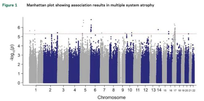
Association results.
Statistical analysis of association
under an additive model was performed in the full
case-control dataset (figure 1) and in the subset of
MSA
cases
with
neuropathologically
confirmed
diagnosis. Loci with the lowest p values (,1 3 10
26)
in the full dataset and pathology confirmed cases are
shown in tables 2 and e-3, respectively. In the full
dataset, we identified 4 loci for future follow-up in
a larger sample series at a p value
,1 3 10
27including the genes FBXO47, ELOVL7, EDN1, and
MAPT. None of the tested SNPs surpassed the
Bonferroni threshold for multiple testing (p value
,
5
3 10
28) in the full dataset or in the subanalysis of
pathologically confirmed cases.
COQ2 in the MSA GWAS data.
Recently, homozygous
(or compound heterozygous) mutations in COQ2
were reported in familial cases of MSA in Japan.
10While coding variants were also found at a higher
fre-quency in sporadic MSA compared to controls, this
finding did not replicate outside of Japanese MSA
samples. We investigated the COQ2 locus for common
variation in our GWAS data. A total of 453 SNPs in
the COQ2 gene and the flanking region
6100 kb
(build 36.3 positions) were tested for association.
The most associated SNP was chr4:84473327 with
a p value of 0.02169, which is far from genome-wide
or even regional Bonferroni-adjusted significance. We
therefore lack evidence that common genetic variation
in the COQ2 locus plays a major role in MSA risk in
the European/Northern American population.
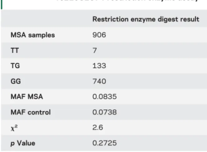
SNCA rs11931074 SNP genotyping.
SNP rs11931074 in
the SNCA locus (or rs3822086, which is in close linkage
disequilibrium) has been linked to MSA in several smaller
studies.
8,22,23This SNP is not represented on the
Om-niExpress Bead arrays; hence, it was not genotyped in any
samples run on this array (n
5 703) and consequently
was not in the dataset of genotyped SNPs in common.
Instead, the allele frequency was calculated
postimputa-tion, demonstrating that it was neither significantly
asso-ciated in the entire dataset (p value
5 0.4722) nor in the
subgroup of definite MSA cases (p value
5 0.4407). We
Figure 1 Manhattan plot showing association results in multiple system atrophy
p Values under the additive association model are log transformed (y-axis) and plotted against the chromosomal position (x-axis). The dotted line indicates the threshold of potentially interesting single nucleotide polymorphisms.
decided to regenotype SNP rs11931074 in the MSA
GWAS cohort using a restriction enzyme to exclude
technical or imputation uncertainty (despite a high
impu-tation score of 0.9670). Within the GWAS cohort, 906
MSA samples were available for regenotyping (table 3).
In controls, data for this SNP were available from the
bead array data. The minor allele of SNP rs11931074
was slightly more common in MSA cases vs controls
(8.3% vs 7.3%) but again no significant association
was found with MSA (
x
25 2.6; p value 5 0.27). This
is in concordance with the imputed GWAS finding, but
contradicts previous reports in the literature.
8,22,23Analysis of brain tissue quantitative trait loci in candidate
loci.
Twenty-one out of 24 SNPs of interest (p value
, 1 3 10
25) passed quality control in the mRNA
expression datasets, and 23 SNPs of interest passed
quality control in the CpG methylation datasets. We
tested multiple probes per SNP in each set of analyses.
A total of 168 unique SNP
–probe pairs were tested in
the frontal cortex mRNA expression dataset, 165 pairs
in the cerebellar mRNA expression dataset, and 391
pairs in the frontal cortex and cerebellar CpG
methyl-ation dataset. Associmethyl-ations were tested using linear
regression adjusting for appropriate covariates and
re-sulting p values were adjusted based on the
false-discovery rate correction as previously described.
21After correcting for multiple testing, we found 8
significant associations between SNPs of interest
and either CpG methylation or mRNA expression
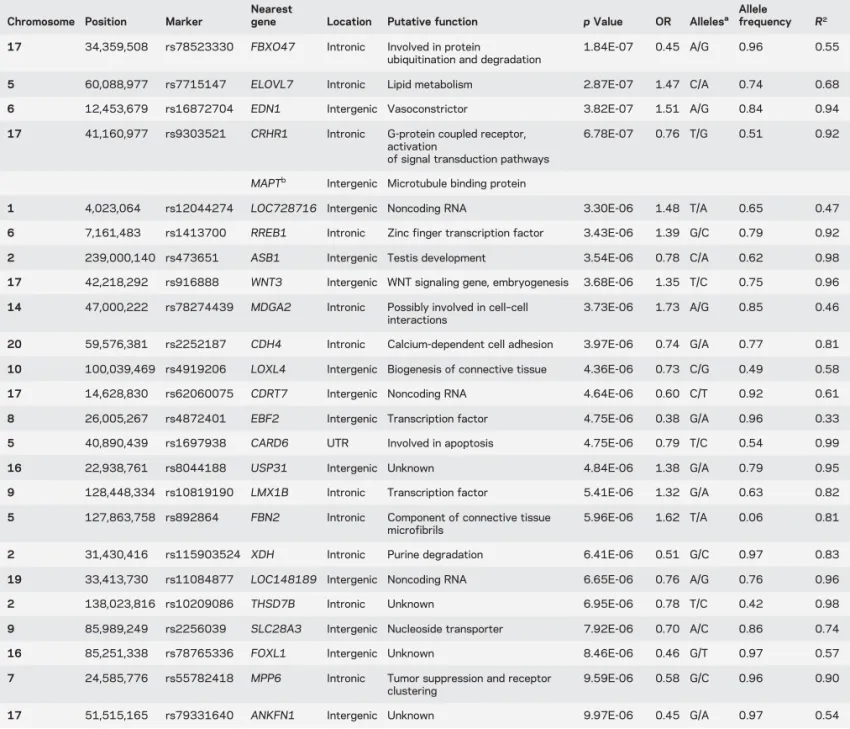
Table 2 Summary of loci with a p value below 1E-6
Chromosome Position Marker
Nearest
gene Location Putative function p Value OR Allelesa Allelefrequency R2
17 34,359,508 rs78523330 FBXO47 Intronic Involved in protein
ubiquitination and degradation
1.84E-07 0.45 A/G 0.96 0.55
5 60,088,977 rs7715147 ELOVL7 Intronic Lipid metabolism 2.87E-07 1.47 C/A 0.74 0.68 6 12,453,679 rs16872704 EDN1 Intergenic Vasoconstrictor 3.82E-07 1.51 A/G 0.84 0.94 17 41,160,977 rs9303521 CRHR1 Intronic G-protein coupled receptor,
activation
of signal transduction pathways
6.78E-07 0.76 T/G 0.51 0.92
MAPTb Intergenic Microtubule binding protein
1 4,023,064 rs12044274 LOC728716 Intergenic Noncoding RNA 3.30E-06 1.48 T/A 0.65 0.47 6 7,161,483 rs1413700 RREB1 Intronic Zinc finger transcription factor 3.43E-06 1.39 G/C 0.79 0.92 2 239,000,140 rs473651 ASB1 Intergenic Testis development 3.54E-06 0.78 C/A 0.62 0.98 17 42,218,292 rs916888 WNT3 Intergenic WNT signaling gene, embryogenesis 3.68E-06 1.35 T/C 0.75 0.96 14 47,000,222 rs78274439 MDGA2 Intronic Possibly involved in cell–cell
interactions
3.73E-06 1.73 A/G 0.85 0.46
20 59,576,381 rs2252187 CDH4 Intronic Calcium-dependent cell adhesion 3.97E-06 0.74 G/A 0.77 0.81 10 100,039,469 rs4919206 LOXL4 Intergenic Biogenesis of connective tissue 4.36E-06 0.73 C/G 0.49 0.58 17 14,628,830 rs62060075 CDRT7 Intergenic Noncoding RNA 4.64E-06 0.60 C/T 0.92 0.61 8 26,005,267 rs4872401 EBF2 Intergenic Transcription factor 4.75E-06 0.38 G/A 0.96 0.33 5 40,890,439 rs1697938 CARD6 UTR Involved in apoptosis 4.75E-06 0.79 T/C 0.54 0.99 16 22,938,761 rs8044188 USP31 Intergenic Unknown 4.84E-06 1.38 G/A 0.79 0.95 9 128,448,334 rs10819190 LMX1B Intronic Transcription factor 5.41E-06 1.32 G/A 0.63 0.82 5 127,863,758 rs892864 FBN2 Intronic Component of connective tissue
microfibrils
5.96E-06 1.62 T/A 0.06 0.81
2 31,430,416 rs115903524 XDH Intronic Purine degradation 6.41E-06 0.51 G/C 0.97 0.83 19 33,413,730 rs11084877 LOC148189 Intergenic Noncoding RNA 6.65E-06 0.76 A/G 0.76 0.96 2 138,023,816 rs10209086 THSD7B Intronic Unknown 6.95E-06 0.78 T/C 0.42 0.98 9 85,989,249 rs2256039 SLC28A3 Intergenic Nucleoside transporter 7.92E-06 0.70 A/C 0.86 0.74 16 85,251,338 rs78765336 FOXL1 Intergenic Unknown 8.46E-06 0.46 G/T 0.97 0.57 7 24,585,776 rs55782418 MPP6 Intronic Tumor suppression and receptor
clustering
9.59E-06 0.58 G/C 0.96 0.90
17 51,515,165 rs79331640 ANKFN1 Intergenic Unknown 9.97E-06 0.45 G/A 0.97 0.54
Abbreviations: OR5 odds ratio; UTR 5 untranslated region.
Results for association tests in multiple system atrophy cases vs controls are shown.
aFirst allele refers to effect allele.
(table e-4). All of these were located on chromosome 17
and associated with 3 SNPs (rs78523330, rs9303521,
and rs916888) in a region spanning 8 MB. SNP
rs9393521 is located only 174 Kb upstream of MAPT,
a gene previously suggested as a risk gene for MSA, and
it is within a large 900 kb inversion polymorphism
sur-rounding the MAPT locus.
DISCUSSION
The contribution of common genetic
factors to disease development has been established
for many neurodegenerative conditions, which are
complex, often sporadic conditions such as PD and
progressive supranuclear palsy. In MSA, the identity
of these contributing genetic factors remains almost
entirely unknown. Here, we describe a GWAS
inves-tigating common genetic markers in 918 MSA cases
and 3,864 controls for disease association.
Although none of the tested SNPs surpassed the
stringent Bonferroni threshold, we identified 4
MSA regions with p values
,1 3 10
26: FBXO47,
ELOVL7, EDN1, and MAPT. These loci are most
promising for further follow-up.
Among the most highly associated regions is the
MAPT locus. MAPT is an intriguing candidate as it
has already been implicated in a number of
neurode-generative diseases, particularly in tauopathies such as
progressive supranuclear palsy, frontotemporal
degen-eration, and Alzheimer dementia.
24,25Although tau is
not considered a key protein in MSA neuropathology,
PD
—which is also an a-synucleinopathy—has been
found to be consistently associated with common
var-iation in the MAPT locus.
7Furthermore, MSA was
also previously found to be associated with the MAPT
H1 haplotype in a small cohort of American MSA
cases.
9These results, however, will need to be verified
in a larger MSA cohort.
Located on chromosome 6, EDN1 is part of the
endothelin gene family, which functions in the
main-tenance of vascular tone. Studies have demonstrated
the presence of endothelin peptides in nonvascular
struc-tures including epithelial cells, glia, and neurons.
26,27Given the phenotype of autonomic dysfunction or
fail-ure in patients with MSA, one may hypothesize that
such pathophysiology may be associated with variation
in EDN1.
ELOVL7, originating on chromosome 5, is involved
in transferase activity. Chiefly, transferase is a
condens-ing enzyme that acts as a catalyst for the synthesis of
saturated and polyunsaturated very long chain fatty
acids.
28,29FBXO47 has a known association with papillary
renal cell carcinoma.
30Located on chromosome 17,
FBXO47 promotes protein ubiquitination and
degra-dation via phosphorylation, which are critical
mech-anisms in many neurodegenerative diseases.
30,31This
locus could provide an important mechanism to MSA
pathology in light of the oligodendroglial
a-synuclein
burden, but requires further study to draw any
conclusions.
Common variation in the COQ2 gene (recently
found to be mutated in familial and sporadic probable
MSA cases in Japan) did not show a significant
asso-ciation in the MSA GWAS data. This suggests that
common genetic variation in this gene does not play
a major role in disease pathogenesis in patients with
MSA of European descent.
We were unable to replicate the previously reported
association of variants at the SNCA.
8,22,23A Korean
study was also unable to reproduce the association
indicating possible differences between populations
or an actual null result.
32The previously associated
SNP rs11931074 has considerable variation in
fre-quency among different populations. The reported risk
allele has a frequency of about 7%–8% in our different
European controls and in the European HapMap
data.
33In contrast, the same allele is much more
abun-dant in Asian and African populations (e.g., 58% in
Japanese and 68% in Yorubans). Our study attempts
to account for both interpopulation and
intrapopula-tion heterogeneity through principal component
anal-ysis, and interpopulation heterogeneity of SNCA is
a plausible explanation for the previous findings.
The quantitative trait analysis data suggest a
com-plex risk locus on chromosome 17. This includes
sev-eral SNPs in the region associated with multiple
expression or methylation effects. Several of these
genes are involved in pathways potentially relevant
to MSA pathology: namely, the ribosylation protein
(ARL17A), the ribosome protein (RPL19), and the
proteasome protein (PSMB3).
The disease pathology of several
neurodegenera-tive disorders illustrates a common theme of
disrup-ted proteasomal protein clearance and degradation.
Notably, PSMB3 may be involved in trinucleotide
repeat expansion—a phenomenon seen in several
Table 3 Results from the SNCA SNP
rs11931074 restriction enzyme assay
Restriction enzyme digest result MSA samples 906 TT 7 TG 133 GG 740 MAF MSA 0.0835 MAF control 0.0738 x2 2.6 p Value 0.2725
Abbreviations: MAF5 minor allele frequency, MSA 5 multiple system atrophy.
hereditary neurologic disorders (in particular
autoso-mal dominant ataxias)—and it may be driving the
tri-nucleotide expansion process.
34Supporting this
hypothesis, there is considerable clinical overlap
between MSA and several hereditary ataxias.
35,36Sim-ilarly, ribosomal dysfunction with aberrant protein
synthesis may promote neurodegeneration.
37Interest-ingly, no alteration in expression of MAPT
—the most
obvious candidate gene in the chromosome 17 locus
—
was found. All but one associated QTL in MSA were
found in cerebellar tissue (table e-4). Since the
cerebel-lum is among the most disturbed brain regions in
MSA, differences in expression in this region are
con-sistent with an etiologic role in MSA.
There are a number of limitations to this study.
First, although this represents by far the largest
collec-tion of MSA samples to date, the number of MSA
cases is still relatively small for a GWAS. GWAS are
designed to identify disease variants of relatively high
frequency (MAF
.1%) in a population operating
under the common disease/common variant
hypoth-esis.
38In PD, most identified risk factors have an odds
ratio (OR) between 0.8 and 1.5.
39Similarly, in
Alz-heimer disease
—apart from APOE (OR
;4)—com-mon genetic risk factors have low ORs ranging from
0.8 to 1.2. In these diseases, successful identification
of significant loci was only feasible with considerably
larger sample sizes than our study. However, with our
current number we can reasonably exclude the
exis-tence of common risk alleles of large effect. A second
limitation is the use of population control cohorts
rather than age-matched controls, which increases
the chance for population heterogeneity. We applied
stringent corrections, including principal components
from multidimensional scaling as covariates in our
statistical model to address this concern. Third, the
misdiagnosis rate of clinically ascertained patients, in
particular those in early disease stages, can be as high
as 38%.
40We addressed this concern by including
a large number of pathology-proven cases (;
one-third of all patients in this study). The inclusion of
pathology-confirmed cases is particularly important
for future follow-up studies in MSA.
Finally, a major challenge is that currently no
additional large cohorts of MSA cases with a similar
genetic background are available. A comparison of
European and non-European cohorts may be
infor-mative with regard to interpopulation heterogeneity.
None of the studied variants were statistically
sig-nificant after appropriate correction for multiple
test-ing. However, we identified 4 promising loci that
could reveal important insight into the disease
path-ways of MSA. Increasing the sample size of our
GWAS will be key to the successful confirmation of
these candidate loci and the identification of other
risk genes.
AUTHOR AFFILIATIONS
From the Departments of Molecular Neuroscience and Movement Disor-ders and The Reta Lila Weston Institute (A.S., A.L., K.M., N.E.M., L. S., V.C., H.L., S.S.O., N.W.W., J.A.H., T.R., J.L.H., H.H.), Department of Clinical Neuroscience (H.R.M.), and Sobell Department of Motor Neu-roscience and Movement Disorders (N.Q., K.P.B.), UCL Institute of Neurology, London, UK; Klinik für Neurologie (A.S.), Universitätsklinik der Johann-Wolfgang-Goethe Universität Frankfurt, Germany; Laboratory of Neurogenetics, National Institute on Aging (S.W.S., M.A.N., M.F., T.R. P., B.J.T., A.B.S.), and Neurodegenerative Diseases Research Unit, National Institute of Neurological Disorders and Stroke (S.W.S.), National Institutes of Health, Bethesda, MD; Department of Neuroscience (S.W.S.), George-town University, Washington, DC; Department of Neurology (S.W.S.), Johns Hopkins Hospital, Baltimore, MD; Department of Neurodegenera-tive Diseases (C.S., T.G.), Hertie Institute for Clinical Brain Research, and German Center for Neurodegenerative diseases (DZNE), Tübingen, Germany; Department of Neuroscience (O.A.R., D.W.D.), Mayo Clinic, Jacksonville, FL; Division of Life Science (K.M.), Hong Kong University of Science and Technology, Hong Kong SAR, China; National Institute on Aging (L.F.), National Institutes of Health, Baltimore, MD; Department of Neuroscience (H.J.F., T.R.M.), Georgetown University, Washington, DC; Department of Neurology (G.D.), UKSH, Kiel Campus, Christian-Albrechts University Kiel, Germany; Division of Neurology (H.W.), Department of Clinical Sciences, University Hospital, Lund, Sweden; Isti-tuto Clinico Humanitas (A.A.), Università Cattolica del Sacro Cuore, Mi-lan, Italy; Service of Neurology (J.I.), University Hospital Marqués de Valdecilla (IDIVAL), University of Cantabria (UC), Santander, Spain; Insti-tute of Neurology (W.P., G.K.W.), Medical University Innsbruck, Austria; Centre of Nervous Diseases (W.O.), Philipps-University of Marburg; Department of Neurology (G.U.H.), Technische Universität München; German Center for Neurodegenerative Diseases (DZNE) (G.U.H.), Mu-nich; Department of Neurology (U.W.) and German Center for Neurode-generative Diseases (U.W.), University Hospital of Bonn Medical Center, Germany; Parkinson Institute (S.G.), Istituti Clinici di Perfezionamento, Milan; Center for Neurodegenerative Diseases (M.T.P.), Department of Medicine and Surgery, Neuroscience Section, University of Salerno, Italy; Clinical Pharmacological Unit (J.F.), Instituto de Medicina Molecular, Fac-ulty of Medicine, University of Lisbon, Portugal; Hospital Clinic (E.T.), University of Barcelona, Spain; Department of Neurology (B.R.B.), Donders Institute for Brain, Cognition and Behaviour, Radboud University Medical Centre, Nijmegen, Netherlands; Clinical Investigation Center CIC1436 (O.R.), Departments of Clinical Pharmacology and Neuroscien-ces, INSERM and University Hospital of Toulouse, Faculty of Medicine; Centre de Référence Atrophie Multisystématisée (W.G.M.), CHU de Bor-deaux, Pessac; and Université de Bordeaux (W.G.M.), Institut des Maladies Neurodégénératives, UMR 5293, Bordeaux, France.
AUTHOR CONTRIBUTIONS
Patient recruitment: H.H., A.S., N.W., G.K.W., A.L., W.P., W.O., U.W., S.G., M.T.P., T.G., C.S., O.R., D.D., H.L., S.O., A.L., L.F., H.M., G.D., N.Q., H.W., A.A., J.I., K.B., W.P., W.O., G.U.H., U.W., S.G., M.T.P., J.F., E.T., B.B., J.A.H., T.R., J.H., D.B., V.K., P.C., H.R., J.S., A.S., E.R., G.M., C.C., P.B., L.D.T., O.R., R.D., T.G., A.A., W.M., K.O., E.D., N.P., M.J.L., I.S., G.C.-B., S.C., L.S., T.K., F.T., A.F., S.A.S., A.L. R.P., C.N., O.H., V.R., F.D., H.B., J.K., A.R., C.P., G.F., F.D., M.S., M.I.P., A.P., M.S., D.B., O.R., N.R., N.G., Y.B., H.S., Y.M., M.P., and R.B. Sample preparations: A.S., J.L.H., T.R., S.W.S., C.S., H.H., and O.R. Genotyping: A.S., S.W.S, H.H., A.B.S., and N.E.M. Writ-ing: A.S., S.W.S, M.A.N., H.H., A.B.S, M.F., R.P., and L.V.S. Conceptu-alization and design: H.H., A.B.S, J.A.H, A.S., S.W.S, and M.A.N. Critical review of manuscript (all authors); Supervision: H.H., A.B.S., and J.A.H.
ACKNOWLEDGMENT
DNA and brain samples: The authors used DNA samples and phenotype data from the NINDS Human Genetics Resource Center DNA and Cell Line Repository at Coriell (Newark, NJ; ccr.coriell.org/ninds), and thank the patients and the submitters who contributed samples to this repository. Human tissue was obtained from the Queen Square Brain Bank (London, UK), the Institute of Psychiatry Brain Bank, King’s College (London, UK), the UK Parkinson’s disease tissue bank at Imperial College (London, UK),
Newcastle Brain Tissue Resource at Newcastle University (Newcastle, UK), the Manchester Brain Bank at the University of Manchester (Manchester, UK), Jacksonville Brain Bank for Alzheimer’s, Parkinson’s and Related Disorders at the Mayo Clinic (Jacksonville, FL), the NICHD Brain and Tissue Bank for Developmental Disorders at the University of Maryland (Baltimore, MD), the New York Brain Bank of the Taub Institute at Columbia University (New York, NY), the Human Brain and Spinal Fluid Resource Center (Los Angeles, CA), the Miami Brain Bank (Miami, FL), the Center for Neurodegenerative Disease Research at the University of Pennsylvania (Philadelphia, PA), the Harvard Brain Bank (Boston, MA), the Emory University Alzheimer’s Disease Research Center Brain Bank (Atlanta, GA), Neurobiobank München at the Ludwig-Maximilians-Universität (Munich, Germany), Brain Bank Center Würzburg (Würzburg, Germany), the Netherlands Brain Bank at the Netherlands Institute for Neuroscience (Amsterdam, Netherlands), and the Neurological Tissue Bank at the University of Barcelona (Barcelona, Spain). The Italian samples were obtained from the Parkinson Institute Biobank (parkinsonbiobank.com), member of the Telethon Network of Genetic Biobank (project n. GTB12001) funded by TELETHON Italy, and supported by“Fondazione Grigioni per il Morbo di Parkinson.”
STUDY FUNDING
No targeted funding reported.
DISCLOSURE
A. Sailer was supported in part by the Intramural Research Program of the National Institutes of Health (National Institute of Neurological Disorders and Stroke, National Institute on Aging; project number: Z01 AG000949). She also received funding support from the Multiple System Atrophy Trust, the Medical Research Council UK, and Wellcome Trust. S. Scholz was sup-ported in part by the Intramural Research Program of the National Insti-tutes of Health (National Institute of Neurological Disorders and Stroke, National Institute on Aging; project number: Z01 AG000949). She also received a R25 career development grant by the National Institute of Neu-rological Disorders and Stroke (grant number: R25 NS065729) and a Rapid Response Innovation Award by the Michael J. Fox Foundation. M. Nalls was supported in part by the Intramural Research Program of the National Institutes of Health (National Institute on Aging; project number: Z01 AG000949). C. Schulte reports no disclosures relevant to the manuscript. M. Federoff was supported in part by the Intramural Research Program of the National Institutes of Health (National Institute on Aging; project number: Z01 AG000949). T. Price was supported in part by the Intramural Research Program of the National Institutes of Health (National Institute of Neurological Disorders and Stroke, National Institute on Aging; project number: Z01 AG000949). A. Lees received funding support from the Mul-tiple System Atrophy Trust, the Medical Research Council UK, Wellcome Trust, and the Reta Lila Weston Institute for Neurological Studies. O. Ross, D. Dickson, K. Mok, N. Mencacci, L. Schottlaender, and V. Chelban report no disclosures relevant to the manuscript. H. Ling receives funding from a CBD Solutions Research Grant and is employed by Reta Lila West-on Institute for Neurological Studies. S. O’Sullivan reports no disclosures relevant to the manuscript. N. Wood received funding support from the Multiple System Atrophy Trust, the Medical Research Council UK, Well-come Trust, and the Reta Lila Weston Institute for Neurological Studies. B. Traynor was supported in part by the Intramural Research Program of the National Institutes of Health (National Institute on Aging; project number: Z01 AG000949). L. Ferrucci was supported in part by the Intra-mural Research Program of the National Institutes of Health (National Institute of Neurological Disorders and Stroke, National Institute on Aging; project number: Z01 AG000949). H. Federoff reports no disclosures rele-vant to the manuscript. T. Mhyre reports no disclosures relerele-vant to the manuscript. H. Morris reports grants from Parkinson’s UK and Medical Research Council UK during the conduct of the study and grants from Welsh Assembly Government, personal fees from Teva, personal fees from Abbvie, personal fees from Teva, personal fees from UCB, personal fees from Boehringer-Ingelheim, personal fees from GSK, nonfinancial support from Teva, grants from Ipsen Fund, nonfinancial support from Medtronic, grants from MNDA, grants from PSP Association, grants from CBD Sol-utions, grants from Drake Foundation, and personal fees from Acorda, outside the submitted work; in addition, Dr. Morris has a patent related to C9ORF72. H. R. M. is a co-applicant on a patent application related to
C9ORF72: Method for diagnosing a neurodegenerative disease (PCT/ GB2012/052140) pending. G. Deuschl, N. Quinn, H. Widner, A. Alba-nese, J. Infante, K. Bhatia, and W. Poewe report no disclosures relevant to the manuscript. W. Oertel has served as a consultant for Mundipharma, Novartis, and UCB Pharma; on advisory boards for Boehringer Ingelheim, Merck, Sharp & Dohme, Medtronic, Mundipharma, Neuropore, Novartis, UCB Pharma, and Teva; and received honoraria for presenting lectures from Abbvie, Desitin, Boehringer Ingelheim, Mundipharma, Novartis, Orion Pharma, Schwarz Pharma Neuroscience/UCB, and Teva. He has received scientific grants from the German Ministry of Education and Health, the German Research Foundation, the Charitable Hertie Founda-tion, the Internaal ParkinsonFonds, the Michael J. Fox FoundaFounda-tion, and Novartis Pharma Germany, and holds shares in Merck, Medigene, and Roche. G. Höglinger has served on the advisory boards for Abbvie, Asce-neuron, Bristol-Myers Squibb, Roche, Sellas, and UCB; has received hon-oraria for scientific presentations from Abbvie, Roche, and UCB; has received research support from CurePSP, the International Parkinson Fonds, the German Academic Exchange Service (DAAD), German Research Foundation (DFG) and the German Ministry of Education and Research (BMBF), and the Sellas Life Sciences Group; and has received institutional support from the German Center for Neurodegenerative Dis-eases (DZNE). U. Wüllner, S. Goldwurm, M. Pellecchia, J. Ferreira, E. Tolosa, B. Bloem, O. Rascol, and W. Meissner report no disclosures rele-vant to the manuscript. J. Hardy received funding support from the Mul-tiple System Atrophy Trust, the Medical Research Council UK, and Wellcome Trust. T. Revesz received funding support from the Multiple System Atrophy Trust, the Medical Research Council UK, Wellcome Trust, and the Reta Lila Weston Institute for Neurological Studies. J. Holton received funding support from the Multiple System Atrophy Trust, the Medical Research Council UK, Wellcome Trust, and the Reta Lila Weston Institute for Neurological Studies. T. Gasser and G. Wenning report no disclosures relevant to the manuscript. A. Singleton was supported in part by the Intramural Research Program of the National Institutes of Health (National Institute on Aging; project number: Z01 AG000949). H. Houlden received funding support from the Multiple System Atrophy Trust, the Medical Research Council UK, the Michael J. Fox Foundation, Wellcome Trust, and the Reta Lila Weston Institute for Neurological Stud-ies. Go to Neurology.org for full disclosures.
Received January 17, 2016. Accepted in final form June 15, 2016.
REFERENCES
1. Hara K, Momose Y, Tokiguchi S, et al. Multiplex families with multiple system atrophy. Arch Neurol 2007;64:545– 551.
2. Soma H, Yabe I, Takei A, Fujiki N, Yanagihara T, Sasaki H. Heredity in multiple system atrophy. J Neurol Sci 2006;240:107–110.
3. Wullner U, Schmitt I, Kammal M, Kretzschmar HA,
Neumann M. Definite multiple system atrophy in a Ger-man family. J Neurol Neurosurg Psychiatry 2009;80:449– 450.
4. Vidal JS, Vidailhet M, Derkinderen P, Tzourio C, Alperovitch A. Familial aggregation in atypical Parkinson’s disease: a case control study in multiple system atrophy and progressive supranuclear palsy. J Neurol 2010;257: 1388–1393.
5. Wakabayashi K, Yoshimoto M, Tsuji S, Takahashi H. Alpha-synuclein immunoreactivity in glial cytoplasmic in-clusions in multiple system atrophy. Neurosci Lett 1998; 249:180–182.
6. Galvin JE, Lee VM, Trojanowski JQ. Synucleinopathies: clinical and pathological implications. Arch Neurol 2001; 58:186–190.
7. Simon-Sanchez J, Schulte C, Bras JM, et al. Genome-wide association study reveals genetic risk underlying Parkinson’s disease. Nat Genet 2009;15:15.
8. Scholz SW, Houlden H, Schulte C, et al. SNCA variants are associated with increased risk for multiple system atro-phy. Ann Neurol 2009;65:610–614.
9. Vilarino-Guell C, Soto-Ortolaza AI, Rajput A, et al. MAPT H1 haplotype is a risk factor for essential tremor and multiple system atrophy. Neurology 2011;76:670– 672.
10. Multiple-System Atrophy Research Collaboration. Muta-tions in COQ2 in familial and sporadic multiple-system atrophy. N Engl J Med 2013;369:233–244.
11. Sharma M, Wenning G, Kruger R; European Multiple-System Atrophy Study Group. Mutant COQ2 in multiple-system atrophy. N Engl J Med 2014;371:80–81. 12. Schottlaender LV, Houlden H; Multiple-System Atrophy Brain
Bank Collaboration. Mutant COQ2 in multiple-system atro-phy. N Engl J Med 2014;371:81.
13. Gilman S, Wenning GK, Low PA, et al. Second consensus statement on the diagnosis of multiple system atrophy. Neurology 2008;71:670–676.
14. Wellcome Trust Case Control Consortium. Genome-wide association study of 14,000 cases of seven common dis-eases and 3,000 shared controls. Nature 2007;447:661– 678.
15. Ferrucci L, Bandinelli S, Benvenuti E, et al. Subsystems contributing to the decline in ability to walk: bridging the gap between epidemiology and geriatric practice in the InCHIANTI study. J Am Geriatr Soc 2000;48:1618– 1625.
16. Chio A, Schymick JC, Restagno G, et al. A two-stage genome-wide association study of sporadic amyotrophic lateral sclerosis. Hum Mol Genet 2009;18:1524–1532.
17. Federoff M, Price TR, Sailer A, et al. Genome-wide estimate of the heritability of multiple system atrophy. Parkinsonism Relat Disord 2016;22:35–41.
18. Li Y, Willer C, Sanna S, Abecasis G. Genotype imputa-tion. Annu Rev Genomics Hum Genet 2009;10:387–406. 19. Li Y, Willer CJ, Ding J, Scheet P, Abecasis GR. MaCH: using sequence and genotype data to estimate haplotypes and unobserved genotypes. Genet Epidemiol 2010;34: 816–834.
20. Gibbs JR, van der Brug MP, Hernandez DG, et al. Abun-dant quantitative trait loci exist for DNA methylation and gene expression in human brain. PLoS Genet 2010;6: e1000952.
21. Nalls MA, Pankratz N, Lill CM, et al. Large-scale meta-analysis of genome-wide association data identifies six new risk loci for Parkinson’s disease. Nat Genet 2014;46:989– 993.
22. Al-Chalabi A, Durr A, Wood NW, et al. Genetic variants of the alpha-synuclein gene SNCA are associated with multiple system atrophy. PLoS One 2009;4:e7114.
23. Ross OA, Vilarino-Guell C, Wszolek ZK, Farrer MJ, Dickson DW. Reply to: SNCA variants are associated with increased risk of multiple system atrophy. Ann Neurol 2010;67:414–415.
24. Hoglinger GU, Melhem NM, Dickson DW, et al. Identi-fication of common variants influencing risk of the
tauopathy progressive supranuclear palsy. Nat Genet 2011; 43:699–705.
25. Hollingworth P, Harold D, Sims R, et al. Common var-iants at ABCA7, MS4A6A/MS4A4E, EPHA1, CD33 and CD2AP are associated with Alzheimer’s disease. Nat Genet 2011;43:429–435.
26. Arinami T, Ishikawa M, Inoue A, et al. Chromosomal assignments of the human endothelin family genes: the 1 gene (EDN1) to 6p23-p24, the endothelin-2 gene (EDNendothelin-2) to 1p34, and the endothelin-3 gene (EDN3) to 20q13.2-q13.3. Am J Hum Genet 1991;48: 990–996.
27. MacCumber MW, Ross CA, Snyder SH. Endothelin in brain: receptors, mitogenesis, and biosynthesis in glial cells. Proc Natl Acad Sci USA 1990;87:2359–2363. 28. Leonard AE, Pereira SL, Sprecher H, Huang YS. Elongation
of long-chain fatty acids. Prog Lipid Res 2004;43:36–54. 29. Janssen RJ, Distelmaier F, Smeets R, et al. Contiguous
gene deletion of ELOVL7, ERCC8 and NDUFAF2 in a patient with a fatal multisystem disorder. Hum Mol Genet 2009;18:3365–3374.
30. Simon-Kayser B, Scoul C, Renaudin K, et al. Molecular cloning and characterization of FBXO47, a novel gene containing an F-box domain, located in the 17q12 band deleted in papillary renal cell carcinoma. Genes Chromo-somes Cancer 2005;43:83–94.
31. Deger JM, Gerson JE, Kayed R. The interrelationship of proteasome impairment and oligomeric intermediates in neurodegeneration. Aging Cell 2015;14:715–724. 32. Yun JY, Lee WW, Lee JY, Kim HJ, Park SS, Jeon BS.
SNCA variants and multiple system atrophy. Ann Neurol 2010;67:554–555.
33. International HapMap Consortium. A haplotype map of the human genome. Nature 2005;437:1299–1320. 34. Concannon C, Lahue RS. The 26S proteasome drives
tri-nucleotide repeat expansions. Nucleic Acids Res 2013;41: 6098–6108.
35. Kim HJ, Jeon BS, Shin J, et al. Should genetic testing for SCAs be included in the diagnostic workup for MSA? Neurology 2014;83:1733–1738.
36. Lin IS, Wu RM, Lee-Chen GJ, Shan DE,
Gwinn-Hardy K. The SCA17 phenotype can include features of MSA-C, PSP and cognitive impairment. Parkinsonism Re-lat Disord 2007;13:246–249.
37. Martin I, Kim JW, Lee BD, et al. Ribosomal protein s15 phosphorylation mediates LRRK2 neurodegeneration in Parkinson’s disease. Cell 2014;157:472–485.
38. Risch N, Merikangas K. The future of genetic studies of complex human diseases. Science 1996;273:1516–1517. 39. International Parkinson Disease Genomics Consortium,
Nalls MA, Plagnol V, Hernandez DG, et al. Imputation of sequence variants for identification of genetic risks for Par-kinson’s disease: a meta-analysis of genome-wide association studies. Lancet 2011;377:641–649.
40. Koga S, Aoki N, Uitti RJ, et al. When DLB, PD, and PSP masquerade as MSA: an autopsy study of 134 patients. Neurology 2015;85:404–412.