Nonalcoholic Fatty Liver Disease Progresses
into Severe NASH when Physiological Mechanisms
of Tissue Homeostasis Collapse
Silvia Sookoian,*
,** Carlos J. Pirola*
,***
* University of Buenos Aires, Institute of Medical Research A Lanari, Buenos Aires, Argentina. ** National Scientific and Technical Research Council (CONICET) - University of Buenos Aires, Institute of Medical Research (IDIM), Department of Clinical and Molecular Hepatology, Buenos Aires, Argentina. *** National Scientific and Technical Research Council (CONICET)-University of Buenos Aires, Institute of Medical Research (IDIM), Department of Molecular Genetics and Biology of Complex Diseases, Buenos Aires, Argentina.
March-April, Vol. 17 No. 2, 2018: 182-186
Opinion Article on the MS published in
the Journal of Clinical Investigation entitled:
Hepatic neuregulin 4 signaling defines an
endocrine checkpoint for steatosis-to-NASH progression.
Nonalcoholic fatty liver disease (NAFLD) is regarded
as the most prevalent chronic liver disease worldwide.
1Although the disease is considered benign and having a
relatively good prognosis when the diagnosis is made at
earlier histological stages, the progression into severe
clinical forms, including nonalcoholic steatohepatitis
(NASH), NASH-fibrosis and NASH-cirrhosis, imposes
tremendous medical challenges.
1For these reasons,
exten-sive body of research aimed at the search for factors and or
causes that modify the natural history of NAFLD is
cur-rently being conducted.
The complexity of the NAFLD-associated clinical
pic-ture, specifically the association with co-morbidities such
as type 2 diabetes (T2D),
1obesity and cardiovascular
dis-ease,
2-4suggests the presence of an even more intricate
network of pathogenic mechanisms that not only interact
with each other but also increase the chances of having a
more dramatic and severe phenotype. In this regard, the
cross-talk between NAFLD and adipose tissue has gained
particular attention of researchers and practitioners
be-cause cytokines derived from either white fat-WAT
(adi-pokines) or brown fat-BAT (batokines) play a remarkable
role in the development and maintenance of NAFLD
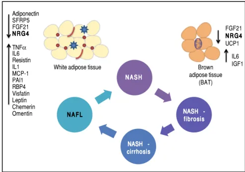
dis-ease severity. Figure 1 illustrates the concept of
phenotyp-ic modulation of NAFLD severity by molecules derived
from adipose tissue that act as paracrine and endocrine
hormones.
5,6Pathways involved in organ damage and injury,
includ-ing cellular apoptosis or other process associated with cell
death, as well as persistent tissue inflammation and the
consequent fibrogenesis, are all triggered by multiple
fac-tors, including genetic predisposition and epigenetic
modifiers.
7-9Nevertheless, the exact mechanism/s that
The Official Journal of the Mexican Association of Hepatology, the Latin-American Association for Study of the Liver and
the Canadian Association for the Study of the Liver
Manuscript received: Manuscript received: Manuscript received: Manuscript received:
Manuscript received: January 23, 2018. Manuscript accepted:Manuscript accepted:Manuscript accepted:Manuscript accepted:Manuscript accepted: January 23, 2018.
DOI:10.5604/01.3001.0010.8631
A B S T R A C T A B S T R A C T A B S T R A C T A B S T R A C T A B S T R A C T
Phenotypic modulation of NAFLD-severity by molecules derived from white (adipokines) and brown (batokines) adipose tissue may be important in inducing or protecting against the progression of the disease. Adipose tissue-derived factors can promote the pro-gression of NAFLD towards severe histological stages (NASH-fibrosis and NASHcirrhosis).
This effect can be modulated by the release of adipokines or batokines that directly trigger an inflammatory response in the liver tissue or indirectly modulate related phenotypes, such as insulin resistance. Metabolically dysfunctional adipose tissue, which is often infiltrated by macrophages and crown-like histological structures, may also show impaired production of anti-inflammatory cytokines, which may favor NAFLD progression into aggressive phenotypes by preventing its protective effects on the liver tissue.
Key words. Key words.Key words. Key words.
promote the transition of NAFL (nonalcoholic fatty liver)
to NASH remain(s) elusive.
Guo, et al. recently reported the role of Neuregulin 4
(Nrg4), an adipose tissue-enriched endocrine factor, in the
development of NASH.
10Specifically, the authors used a
mouse model of high-fat-fructose diet-induced NASH
that recapitulates main histological features of human
NASH.
10Key molecular findings associated with the
pres-ence of NASH in mice were:
• Cell death, which was associated with both increased
phosphorylation of the member of the MAP-kinase
family JNK1/2 and reduced protein levels of the
inhib-itor of apoptotic and necroptotic cell death c-FLIPL
(Caspase 8 and FADD-Like Apoptosis Regulatory
Pro-tein, CFLAR), and
• Significantly decreased mRNA expression of Nrg4 in
epididymal WAT and BAT.
10Subsequent Nrg4 gain- and loss-of-function studies in
mouse models showed that restoration of Nrg4 signaling
protects animals from NASH; specifically, Nrg4 signaling
protects animals from stress-induced cell death through
interaction with c-FLIP.
10Notably, genes associated with
fibrogenesis (Col1a1, Acta2, Tgfb1, and Mmp13) and
inflam-mation (Tnfa, Il1b, Il12b, Nos2, Ccl2, Ccl5, and Adgre1) were
upregulated in mice lacking Nrg4, suggesting that Nrg4
de-ficiency exacerbated liver inflammation and fibrosis
fol-lowing NASH-diet feeding.
10It is worth noting that a
similar profile in human NAFLD was described by our
research group.
2Considering the aforementioned findings collectively,
it is plausible to hypothesize that NAFLD progression is a
process in which physiological mechanisms of tissue
homeostasis collapse. There is compelling evidence from
human studies supporting this hypothesis. For instance,
ballooning degeneration —a hallmark histological feature
of NAFLD severity and NASH progression— is
associat-ed with down-regulation of liver HSP27 gene and protein
expression.
11HSP27 is a member of the heat shock family
of proteins also known as stress-responsive protein 27.
Furthermore, NASH development is associated with
mi-tochondrial dysfunction, increased mimi-tochondrial DNA
(mtDNA) genetic diversity, down-regulation of liver
ex-pression of genes of the oxidative phosphorylation
(OX-PHOS) chain, and aberrant patterns of mtDNA
methylation.
7,8,12,13The first question that immediately emerges is what
factor/s trigger this complex picture?
Robust evidence supports the notion that NAFLD
severity is associated with metabolic stress. In fact, it
has been linked with increased transamination reactions
that would promote coping with the liver metabolic
de-rangement.
14This, in turn, leads to changes in the
amounts of amino acids released into the circulation,
particularly in pathways related to glutamic acid.
13,14As
reported recently, glutaminolysis controls
accumula-tion of myofibroblast hepatic stellate cells.
15Deregulat-ed liver metabolism also leads to aberrant patters of
hedgehog signaling
16,17and insufficient ketogenesis,
which result in extensive hepatocyte injury and
inflam-mation, decreased glycemia, and deranged hepatic TCA
Figure 1. Figure 1.Figure 1. Figure 1.
Figure 1. Phenotypic modulation of NAFLD-se-verity by molecules derived from white (adipok-ines) and brown (batok(adipok-ines) adipose tissue by inducing or protecting against the progression of the disease. Adipose tissue-derived factors can promote the progression of NAFLD towards se-vere histological stages (fibrosis and NASH-cirrhosis). This effect can be modulated by the release of adipokines that directly trigger an in-flammatory response in the liver tissue or indirectly modulate related phenotypes, such as insulin re-sistance (up-arrows). Metabolically dysfunctional adipose tissue, which is often infiltrated by macro-phages and crown-like histological structures, may also show impaired production of anti-inflammato-ry adipokines, which may favor NAFLD progres-sion into aggressive phenotypes by preventing its protective effects on the liver tissue (down-ar-rows). SFRP5: secreted frizzled-related protein 5. UCP1: uncoupling protein 1. FGF21: fibroblast growth factor 21. TNFα: tumor necrosis factor α. IL1 and IL6: interleukin 1 and 6. MCP-1: mono-cyte chemotactic protien-1.
Adiponectin SFRP5 FGF21 NRG 4 NRG 4NRG 4 NRG 4NRG 4
TNFα IL6 Resistin IL1 MCP-1 PAI1 RBP4 Visfatin Leptin Chemerin Omentin
White adipose tissue
N A S H N A S HN A S H N A S HN A S H
NASH -NASH -NASH NASH NASH -fibrosis fibrosisfibrosis fibrosis fibrosis
NASH -NASH -NASH NASH NASH -cirrhosis cirrhosiscirrhosis cirrhosiscirrhosis
N A F L N A F L N A F L N A F L N A F L
Brown adipose tissue
(BAT)
FGF21 NRG 4 NRG 4NRG 4 NRG 4 NRG 4 UCP1
(tricarboxylic acid) cycle intermediate
concentra-tions.
14,18The second, and even more difficult to answer
ques-tion is why a brown fat-derived secreted factor is
impor-tant in maintaining liver tissue homeostasis?
It is well known that systemic metabolic homeostasis
is regulated by the “in concert” action of a myriad of
tis-sue-derived factors, which originate from different
sources, including adipose tissue, gut, muscle, liver and
bone. These hormones / growth factors / bioactive
mole-cules basically sense global metabolism and energy
homeostasis. These mechanisms may eventually fail to
ensure cell / tissue physiological functions. Alternatively,
they can simply malfunction because the affected cell /
tissue has abnormally increased its metabolic demands,
which might occur in common diseases like NAFLD and
obesity, as well as in cancer. In the latter case, epidermal
growth factors (EGFs) are critically implicated in cancer
growth and survival by engaging a number of proteins via
the ErbB (Tyrosine Kinase-Type Cell Surface Receptor
HER2) family of proteins. In this regard, the
neuregu-lins, including NRG4, activate type-1 growth factor
re-ceptors involved in initiating cell-to-cell signaling
through tyrosine phosphorylation.
19Neuregulins
trans-duce signals via activation of the ErbB receptors,
specifi-cally ErbB4.
Evidence supporting the role of
Nrg4
in NAFLD and
metabolic syndrome, while presently limited, is promising.
For example, it has been shown that
Nrg4
attenuates hepatic
lipogenic signaling and preserves glucose and lipid
homeos-tasis in experimental models of obesity.
20In this particular
study, Wang,
et al.
demonstrated in mice that
Nrg4
negatively
regulates
de novo
lipogenesis mediated by Lxr (liver X
recep-tor) and Srebp1c (sterol regulatory element binding
tran-scription factor 1) in a cell-autonomous manner.
20Data yielded by human studies is not only insufficient,
but is also inconclusive. Kang,
et al.
found that the
circulat-ing NRG4 levels are increased in patients with type 2
dia-betes and positively correlate with serum glucose level,
HOMA-IR, and serum triglycerides;
21however, opposite
results were reported by Yan,
et al.
22Controversial
evi-dence has also been derived from studies of gestational
di-abetes.
23,24For example, Cai,
et al.
reported that NRG4
might protect against metabolic syndrome development.
25Similarly, Dai,
et al.
found that patients with NAFLD also
show reduced levels of serum NRG4.
26The question that still remains to be answered is
whether the role of
Nrg4
in protecting hepatocytes from
apoptosis and necroptosis triggered by metabolic stress is
mediated by a circulating endocrine signaling released
from WAT or BAT, or if it is a truly liver-specific derived
Nrg4
. NRG4 human liver gene and protein expression
lev-els are considerably low (http://www.proteinatlas.org/
ENSG00000169752-NRG4/tissue). Moreover, it is
pres-ently unclear whether
Nrg4
signaling is mediated by
inter-action with hepatocytes or whether the actual effector/s of
Nrg4
are non-liver related resident cells found in NAFLD
and NASH. Hence, what exactly determines that
Nrg4
is a
key molecule in protecting the liver from severe NAFLD
is uncertain. There are many possibilities, including the
fact that
Nrg4
indeed controls insulin sensitivity and
se-cretion, as
Nrg4
seems to be a potent insulin releaser.
27or
macrophages migration into the liver. Still, Guo,
et al.
took
the first steps toward unraveling the mechanisms
involv-ing the role of
Nrg4
in protecting fatty liver from severe
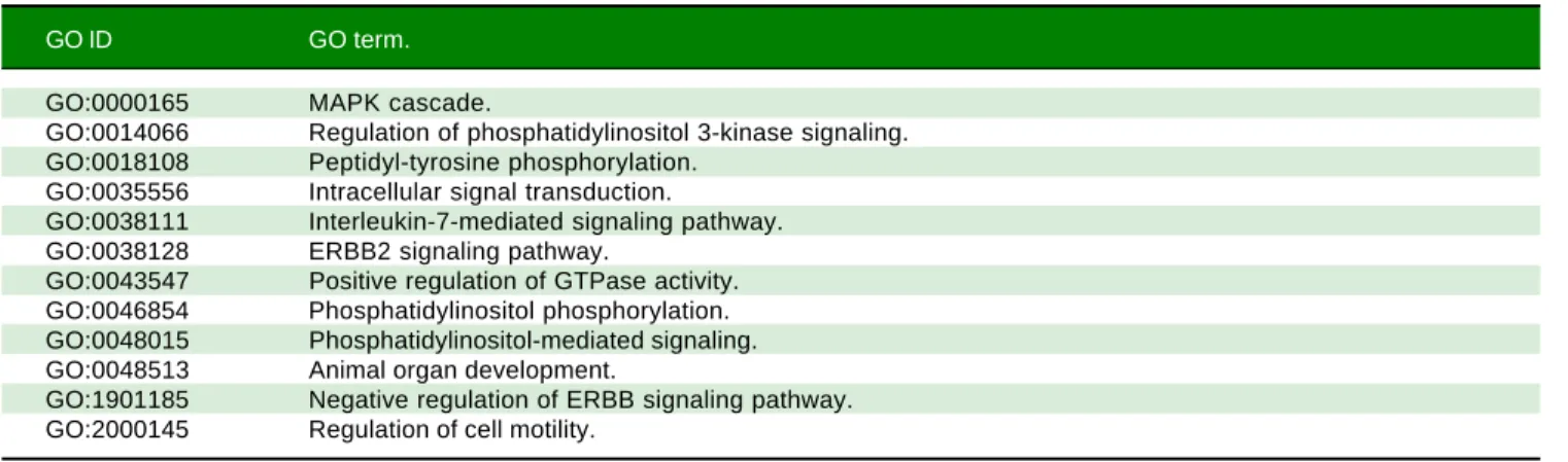
NASH. A list of Gene Ontology (GO) terms and
biologi-Table 1. Gene Ontology (GO) - Biological Process for NRG4 Gene.
GO ID GO term.
GO:0000165 MAPK cascade.
GO:0014066 Regulation of phosphatidylinositol 3-kinase signaling. GO:0018108 Peptidyl-tyrosine phosphorylation.
GO:0035556 Intracellular signal transduction.
GO:0038111 Interleukin-7-mediated signaling pathway. GO:0038128 ERBB2 signaling pathway.
GO:0043547 Positive regulation of GTPase activity. GO:0046854 Phosphatidylinositol phosphorylation. GO:0048015 Phosphatidylinositol-mediated signaling. GO:0048513 Animal organ development.
GO:1901185 Negative regulation of ERBB signaling pathway. GO:2000145 Regulation of cell motility.
cal process for NRG4 gene and its interaction network are
shown in table 1 and figure 2, respectively. It is clear that
this molecule seems to play a key role in ensuring
effec-tive protection against the progression of NAFLD by
opposing caspase-mediated cell death. Interestingly,
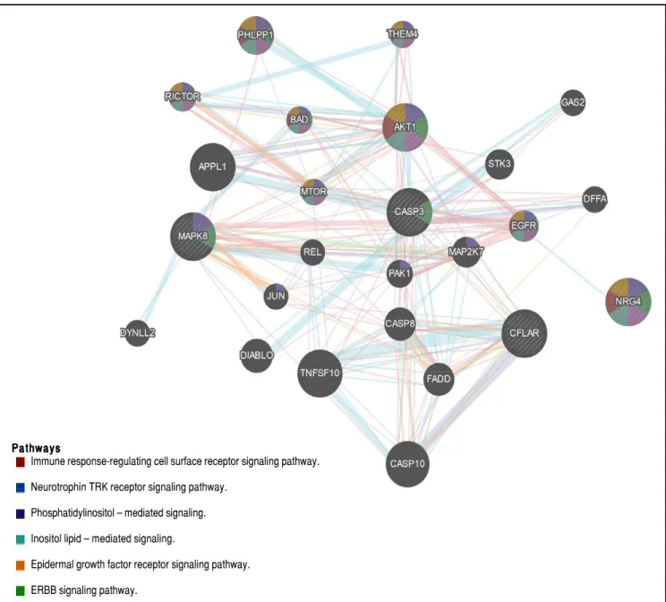
hu-Figure 2. Figure 2.Figure 2.
Figure 2.Figure 2. NRG4 interaction network. Gene-gene functional network construction was done using GeneMANIA, available at: https://github.com/GeneMANIA/ genemania; visualization was performed by Cytoscape (http://apps.cytoscape.org/apps/GeneMania). NRG4: neuregulin 4; CFLAR CASP8 and FADD like ap-optosis regulator; MAPK8: mitogen-activated protein kinase 8. CASP3: caspase 3. AKT1: AKT serine/threonine kinase 1. APPL1: adaptor protein, phosphoty-rosine interacting with PH domain and leucine zipper 1. TNFSF10: tumor necrosis factor superfamily member 10. CASP10: caspase 10. PHLPP1: PH domain and leucine rich repeat protein phosphatase 1. DIABLO: diablo IAP-binding mitochondrial protein. CASP8: caspase 8. FADD: Fas associated via death do-main. EGFR: epidermal growth factor receptor. STK3: serine/threonine kinase 3. MAP2K7: mitogen-activated protein kinase kinase 7. RICTOR: RPTOR inde-pendent companion of MTOR complex 2. PAK1: p21 (RAC1) activated kinase 1. BAD: BCL2 associated agonist of cell death. THEM4: th ioesterase superfamily member 4. MTOR: mechanistic target of rapamycin. DFFA: DNA fragmentation factor subunit alpha. DYNLL2: dynein light chain LC8-type 2. GAS2: growth arrest specific 2. JUN: Jun proto-oncogene. AP-1 transcription factor subunit. REL REL proto-oncogene. NF-κB subunit.
Pathways Pathways Pathways Pathways Pathways
Immune response-regulating cell surface receptor signaling pathway.
Neurotrophin TRK receptor signaling pathway.
Phosphatidylinositol – mediated signaling.
Inositol lipid – mediated signaling.
Epidermal growth factor receptor signaling pathway.
ERBB signaling pathway.
man “knock-out” for NRG4 has been identified; however,
its phenotypic manifestation in the liver remains to be
characterized.
28homeostasis modulates the natural history of NAFLD.
Both their upregulation and deficiency can either cause or
exacerbate an adverse phenotype by evading from
protec-tive mechanisms against liver damage (Figure 1). Hence,
the evolution of NAFLD into severe NASH is mediated
by loss of systemic and/ or local protective mechanisms of
organ damage.
REFERENCES
1. Brunt EM, Wong VW, Nobili V, Day CP, Sookoian S, Maher JJ, Bugianesi E, et al. Nonalcoholic fatty liver disease. Nat Rev
Dis Primers 2015; 17: 15080.
2. Sookoian S, Gianotti TF, Rosselli MS, Burgueno AL, Castano GO, Pirola CJ. Liver transcriptional profile of atherosclero-sis-related genes in human nonalcoholic fatty liver disease.
Atherosclerosis 2011; 218: 378-85.
3. Sookoian S, Castano GO, Burgueno AL, Rosselli MS, Gianotti TF, Mallardi P, Martino JS, et al. Circulating levels and hepatic expression of molecular mediators of atherosclerosis in non-alcoholic fatty liver disease. Atherosclerosis 2010; 209: 585-91.
4. Sookoian S, Pirola CJ. Non-alcoholic fatty liver disease is strongly associated with carotid atherosclerosis: a system-atic review. J Hepatol 2008; 49: 600-7.
5. Adolph TE, Grander C, Grabherr F, Tilg H. Adipokines and Non-Alcoholic Fatty Liver Disease: Multiple Interactions. Int J
Mol Sci 2017 29; 18(8).
6. Polyzos SA, Kountouras J, Mantzoros CS. Adipokines in nonalcoholic fatty liver disease. Metabolism 2016; 65: 1062-79.
7. Sookoian S, Rosselli MS, Gemma C, Burgueno AL, Fernan-dez GT, Castano GO, Pirola CJ. Epigenetic regulation of insu-lin resistance in nonalcoholic fatty liver disease: impact of liver methylation of the peroxisome proliferator-activated re-ceptor gamma coactivator 1alpha promoter. Hepatology 2010; 52: 1992-2000.
8. Pirola CJ, Scian R, Gianotti TF, Dopazo H, Rohr C, Martino JS, Castano GO, et al. Epigenetic Modifications in the Biology of Nonalcoholic Fatty Liver Disease: The Role of DNA Hy-droxymethylation and TET Proteins. Medicine (Baltimore) 2015; 94: e1480.
9. Sookoian S, Pirola CJ. Genetic predisposition in nonalcoholic fatty liver disease. Clin Mol Hepatol 2017; 23: 1-12. 10. Guo L, Zhang P, Chen Z, Xia H, Li S, Zhang Y, Kobberup S,
et al. Hepatic neuregulin 4 signaling defines an endocrine checkpoint for steatosis-to-NASH progression. J Clin Invest 2017 November 6 [Epub ahead of print].
11. Sookoian S, Castano GO, Scian R, San MJ, Pirola CJ. Heat Shock Protein 27 is down-regulated in Ballooned Hepato-cytes of Patients with Nonalcoholic Steatohepatitis (NASH).
Sci Rep 2016; 3:22528.
12. Sookoian S, Flichman D, Scian R, Rohr C, Dopazo H, Gianotti TF, Martino JS, et al. Mitochondrial genome architecture in non-alcoholic fatty liver disease. J Pathol 2016; 240: 437-49.
13. Sookoian S, Pirola CJ. The NASH Metabotype: Imbalance of Circulating Amino Acids and Transamination Reactions Re-flect Impaired Mitochondrial Function. Hepatology 2017 [Epub ahead of print]
14. Sookoian S, Castano GO, Scian R, Fernandez GT, Dopazo H, Rohr C, Gaj G, et al. Serum aminotransferases in
nonalcohol-ic fatty liver disease are a signature of liver metabolnonalcohol-ic per-turbations at the amino acid and Krebs cycle level. Am J Clin Nutr 2016; 103: 422-34.
15. Du K, Hyun J, Premont RT, Choi SS, Michelotti GA, Swiders-ka-Syn M, Dalton GD, et al. Hedgehog-YAP Signaling Path-way Regulates Glutaminolysis to Control Hepatic Stellate Cell Activation. Gastroenterology 2018 [Epub ahead of print]. 16. Chen Y, Choi SS, Michelotti GA, Chan IS, Swiderska-Syn M,
Karaca GF, Xie G, et al. Hedgehog controls hepatic stellate cell fate by regulating metabolism. Gastroenterology 2012; 143: 1319-29.
17. Verdelho MM, Diehl AM. Role of Hedgehog Signaling Path-way in NASH. Int J Mol Sci 2016; 17(6).
18. Cotter DG, Ercal B, Huang X, Leid JM, d’Avignon DA, Graham MJ, Dietzen DJ, et al. Ketogenesis prevents diet-induced fat-ty liver injury and hyperglycemia. J Clin Invest 2014; 124: 5175-90.
19. Harari D, Tzahar E, Romano J, Shelly M, Pierce JH, Andrews GC, Yarden Y. Neuregulin-4: a novel growth factor that acts through the ErbB-4 receptor tyrosine kinase. Oncogene 1999; 18: 2681-9.
20. Wang GX, Zhao XY, Meng ZX, Kern M, Dietrich A, Chen Z, Cozacov Z, et al. The brown fat-enriched secreted factor Nrg4 preserves metabolic homeostasis through attenuation of hepatic lipogenesis. Nat Med 2014; 20(12): 1436-43. 21. Kang YE, Kim JM, Choung S, Joung KH, Lee JH, Kim HJ, Ku
BJ. Comparison of serum Neuregulin 4 (Nrg4) levels in adults with newly diagnosed type 2 diabetes mellitus and controls without diabetes. Diabetes Res Clin Pract 2016; 117: 1-3. 22. Yan PJ, Xu Y, Wan Q, Feng J, Li H, Gao CL, Yang J, et al.
Decreased plasma neuregulin 4 concentration is associated with increased high-sensitivity C-reactive protein in newly diagnosed type 2 diabetes mellitus patients: a cross-section-al study. Acta Diabetol 2017; 54: 1091-9.
23. Kralisch S, Hoffmann A, Kratzsch J, Bluher M, Stumvoll M, Fasshauer M, Ebert T. The brown-fat-secreted adipokine neuregulin 4 is decreased in gestational diabetes mellitus.
Diabetes Metab 2017 [Epub ahead of print].
24. Kurek EM, Yayla AC, Sahin EG, Altun ET, Pekin O, Cevik O. Clinical significance of neuregulin 4 (NRG4) in gestational di-abetes mellitus. Gynecol Endocrinol 2017; 28: 1-4.
25. Cai C, Lin M, Xu Y, Li X, Yang S, Zhang H. Association of circulating neuregulin 4 with metabolic syndrome in obese adults: a cross-sectional study. BMC Med 2016; 14: 165. 26. Dai YN, Zhu JZ, Fang ZY, Zhao DJ, Wan XY, Zhu HT, Yu
CH, et al. A case-control study: Association between serum neuregulin 4 level and non-alcoholic fatty liver disease.
Me-tabolism 2015; 64: 1667-73.
27. South JC, Blackburn E, Brown IR, Gullick WJ. The neuregulin system of ligands and their receptors in rat islets of langer-hans. Endocrinology 2013; 154: 2385-92.
28. Saleheen D, Natarajan P, Armean IM, Zhao W, Rasheed A, Khetarpal SA, Won HH, et al. Human knockouts and pheno-typic analysis in a cohort with a high rate of consanguinity.
Nature 2017; 544: 235-9.
Correspondence and reprint request: Silvia Sookoian, M.D., Ph.D.
Instituto de Investigaciones Médicas, IDIM-CONICET Combatientes de Malvinas 3150, CABA-1427, Argentina.
Tel.: 54-11-52873905