Salidroside and Curcumin Formula Prevents Liver Injury
in Nonalcoholic Fatty Liver Disease in Rats
Hong-Shan Li,*,** Hao Ying,* Zhe-Yun He** Department of Hepatology, Ningbo No.2 Hospital, Ningbo, Zhejiang, China. ** Animal Lab Center, Medical School of Ningbo University, Ningbo, Zhejiang, China.
INTRODUCTION
Nonalcoholic fatty liver disease (NAFLD) is a nonal-coholic-related liver disease manifested by lipid deposi-tion and liver pathological changes.1 With the changes of life pattern and conditions, the incidence of NAFLD is in a rising trend. The incidence of NAFLD is 25-30% in USA, 12.6-50% in Asia countries, and 3-10% in children globally.2-6 Therefore, unravelling the pathogenetic mech-anisms of NAFLD and developing novel therapeutic strategies for NAFLD are key imperatives.
NAFLD has the common features as obesity, dyslipi-demia and diabetes.7 Similar to other diseases, such as hy-pertension, atherosclerosis and coronary heart disease, NAFLD also belongs to metabolic syndromes.8 Multiple
factors could contribute to the aggravation of the disease, including inflammation reaction, lipid peroxidation inju-ry, insulin resistance, etc.9 The chemical compound with single therapeutic target is inefficient to treat the metabol-ic diseases. The drugs with multiple pharmacologmetabol-ical po-tential and combination therapy are supposed as the potential candidate treatment for HAFLD.10
Traditional Chinese medicine (TCM) is an alternation for the treatment of diseases because its multiple-target and -signaling pathway efficacy.11 TCM- salidroside and curcumin (SC) formula is composed of salidroside and curcumin, which are the major active components of TCM rhodiola rosea and rhizoma curcumae longae, respectively. Previous study revealed that SC formula had excellent ad-vantages in ameliorating lipid deposition in NALFD rats.12 The Official Journal of the Mexican Association of Hepatology,
the Latin-American Association for Study of the Liver and the Canadian Association for the Study of the Liver
Manuscript received: Manuscript received:Manuscript received:
Manuscript received:Manuscript received: September 11, 2017. Manuscript accepted:Manuscript accepted:Manuscript accepted:Manuscript accepted:Manuscript accepted: October 31, 2017.
DOI:10.5604/01.3001.0012.3135
A B S T R A C T A B S T R A C T A B S T R A C T A B S T R A C T A B S T R A C T
Introduction and aim. Introduction and aim.Introduction and aim. Introduction and aim.
Introduction and aim. Salidroside and curcumin (SC) formula could alleviate lipid deposition in high fat diet-induced nonalcoholic fatty liver disease (NAFLD). However, the mechanisms are still unknown, and the magnitude of potential therapeutic benefit remains understudied. Material and methods.Material and methods.Material and methods.Material and methods.Material and methods. The rats were treated with high fat diet for 14 weeks to induce NAFLD. The experiment was divided into control, model (NAFLD), SC formula and rosiglitazone groups (n = 7 in each group). Hematoxylin-eosin (H&E) staining was applied to detect liver morphological changes. Biochemical, metabolic indices and inflammation factors in liver tissue and serum were detected. Additionally, the activities of related enzymes were detected by enzyme-linked immunosorbent assay. Results.
Results.Results. Results.
Results. In the established rat model, typical lipid deposition and liver steatosis were observed. Liver triglyceride, free fatty acids, sera alanine aminotransferase, aspartate aminotransferase, gamma-glutamyl transferase, fasting insulin, fasting blood glucose and homeostasis model assessment of insulin resistance were elevated in model group. Liver malondialdehyde was significantly elevated, while superoxide dismutase was significantly decreased in model group, compared with control. Moreover, tumor necrosis factor-α
and Interleukin-1 were significantly produced in model group, compared with control. As a mechanism, high fat diet decreased tissue AMP-activated protein kinase (AMPK), phosphorylated AMPK, carnitine palmitoyltransferase 1 and increased inacetyl-CoA carboxy-lase (ACCase), phosphorylated ACCase. Importantly, these abnormal changes caused by high fat diet were reduced by SC formula administration. Conclusion.Conclusion.Conclusion.Conclusion.Conclusion. SC formula could ameliorate the injury caused by high fat diet. The effect was likely mediated via its influence on insulin resistance, lipid peroxidation injury and AMPK signaling pathway.
Key words. Key words.Key words. Key words.
Compared with the single component, SC formula dis-plays synergistic and additional effects.12 However, the magnitude of potential therapeutic benefit, as well as the mechanisms involved, remain uncertain.
In this study, we established NALFD model and evalu-ated the preventive effects of SC formula on high fat diet-induced liver injury. The potential mechanisms, including insulin resistance, lipid oxidative stress and the changes of AMPK signaling pathways were screened.
MATERIAL AND METHODS
Preparation of SC formula
TCM-SC formula was prepared as described previ-ously.12 The ingredients of SC formula include salidro-side and curcumin, and the dose of SC formula was salidroside 5.77 mg/kg/d and curcumin 21.76 mg/kg/d, which were referred to the literature12 and based on the Pharmacopoeia of the People’s Republic of China. In our study, salidroside (98%, Batch No. ZL20160504HJT) and curcumin (98%, Batch No. ZL20160309JHS) were pur-chased from Nanjing Zelang Medical Technology Co., Ltd. Rosiglitazone (Batch No.: 150109, Chengdu Hengrui Pharmaceutical Co., Ltd.) was applied as positive control for the treatment of NALFD. The usage dose of rosiglita-zone for a 60-kg (body weight) adult was 4 mg/d. After calculation according to the conversion formula between humans and rats (1:6), a dose of 0.4 mg/kg/d was applied in rats, which was also referred to the literature.13
Animals and modeling
28 SD rats (male, 100-120 g, clean grade) were pur-chased from Laboratory Animal Center of Zhejiang Acade-my of Medical Sciences (SCXK(Zhe)2014-0001) and raised in Animal Center of Ningbo University with free access to water.
The rats were randomly divided into control group (n = 7), NALFD model group (n = 7), SC treatment group (n = 7) and rosiglitazone treatment group (n = 7). NALFD was established by administration of high fat diet as previously described.13 Briefly, the NALFD rats were fed with high fat diet (10% lard, 2% Cholesterol and 88% basal diet) for 14 weeks. The high fat diet was purchased from Trophic Animal Feed High-Tech Co., Ltd., China. Control rats were fed with basal diet. From the ninth week after modeling, the rats in SC treatment, and rosigli-tazone treatment groups received SC formula (27.53 mg/ kg/d) and rosiglitazone (0.4 mg/kg/d) for consecutive six weeks through intragavage administration, respectively, while the rats in control and model groups received simi-lar volume of potable water.
Specimen collection
14 weeks after modeling or administration, the rats were fasted for 12 h and anesthetized by 2% pentobarbital sodium (3 mL/kg, i.p.). Veinal blood was collected and liver tissues were isolated for hematoxylin-eosin staining (H&E) staining and biochemical detection.
H&E staining
Liver tissues were fixed in 10% neutral formaldehyde solution. After fixation, the tissues were dehydrated by the automatic dehydration machine, and embedded in paraf-fin. The tissues were then sectioned into 4-μm thickness slices and underwent routine H&E staining. The slides were observed under light microscope. According to Nonalcoholic fatty liver disease diagnosis and treatment guidelines established by the Chinese Medical Associa-tion liver disease branch,14 the degree of liver steatosis is divided into five grades (F0-F4) as follows:
• F0, liver steatosis < 5%. • F1, liver steatosis (5%-30%). • F2, liver steatosis (31%-50%). • F3, liver steatosis (51%-75%). • F4, liver steatosis > 75%.
Biochemical detection
Liver triglyceride (TG) and free fatty acids (FFA) were detected as previously described.13 Sera alanine ami-notransferase (ALT), aspartate amiami-notransferase (AST), gamma-glutamyl transferase (GGT) were detected as pre-viously described.15
Fasting insulin (FINS) was detected as previously de-scribed.13 Fasting blood glucose test (FBG) was also de-tected. Homeostasis model assessment of insulin resistance (HOMA-IR) was calculated based on the for-mula: HOMA-IR = FBG x FINS/22.5.
Liver superoxide dismutase (SOD) (Batch No. 20170106) and malondialdehyde (MDA) (Batch No. 20170111) were detected by biochemical method accord-ing to the instructions of the kits as previously de-scribed.16
Enzyme-linked immunosorbent assay (ELISA)
carboxylase (ACCase), phosphorylated acetyl-CoA cat-boxylase (pACCase), carnitine palmitoyltransferase 1 (CPT-1), were also detected by ELISA method according to the instructions of the kits (Shanghai Yuanye Biotech, Shanghai, China). Liver tissue homogenate was prepared as previously described.15
Statistical analysis
All the data were in normal distribution, and expressed as mean and standard deviation, and analyzed by SPSS 16.0. The difference among groups was analyzed by ANOVA followed by LSD. P value less than 0.05 was considered as significant difference.
RESULTS
SC formula alleviates the high fat diet-induced liver injury
Compared with normal control, body weight, liver weight and liver index were significantly elevated in the
rats from model group. By contrast, SC formula, as well as rosiglitazone significantly decreased high fat diet-induced increase of body weight, liver weight and liver index. Moreover, liver weight and index in SC formula treat-ment group were significantly lower than those in rosigli-tazone group (Figure 1).
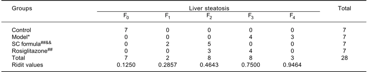
Morphological changes of liver tissue were also com-pared in different groups. As shown in figure 2, hepatic cells in control group had a normal morphologic. Lipid deposition was not observed in cytoplasm of the normal cells. There was no cell infiltration in the liver tissue (Figure 2). By contrast, lipid deposition was found in he-patic cells in model group. Nucleus shift was observed in some of the cells. Mild infiltration and necrosis were found in some of the hepatic cells. However, SC formula and rosiglitazone treatment remarkably mitigated high fat diet-caused liver injury. Additionally, the injury in SC for-mula was much milder, compared with rosiglitazone group. Liver steatosis was analyzed in different groups. As shown in table 1, liver steatosis was not observable in con-trol group, while high steatosis was found in model group. SC formula and rosiglitazone treatment remarkably
re-Figure 1. Figure 1.Figure 1.
Figure 1.Figure 1. SC formula decreases high fat diet-induced increase of body weight (AAAAA), liver weight (BBBBB) and liver index (CCCCC). *P<0.05 compared with control;
#P < 0.05, ##P < 0.01 compared with model; &P < 0.05, &&P < 0.01, compared with Rosiglitazone.
Table 1. Liver steatosis quantified from HE images in each group.
Groups Liver steatosis Total
F0 F1 F2 F3 F4
Control 7 0 0 0 0 7
Model* 0 0 0 4 3 7
SC formula##&& 0 2 5 0 0 7
Rosiglitazone## 0 0 3 4 0 7
Total 7 2 8 8 3 28
Ridit values 0.1250 0.2857 0.4643 0.7500 0.9464
* P < 0.05 compared with control. ## P < 0.01 compared with model. && P < 0.01 compared with Rosiglitazone.
Body weigth (g)
600
400
200
0
Control Model
SC formula
Rosiglitazone
Liver weight (g)
30
20
10
0
Control Model
SC formula
Rosiglitazone
Liver index (%)
6
4
2
0
Control Model
SC formula
Rosiglitazone
AAAAA BBBBB CCCCC
# ## *
## *
&& ## ##
*
duced high fat diet-caused liver steatosis. Additionally, the steatosis in SC formula was much milder, compared with rosiglitazone group.
Effects of
SC formula on liver TG and FFA
Compared with control, liver TG and FFA levels in model group were significantly elevated (Figure 3). SC formula, as well as rosiglitazone significantly decreased TG and FFA, compared with model group. Additionally, SC formula was more effective to alleviate TG and FFA compared with rosiglitazone group.
Effects of
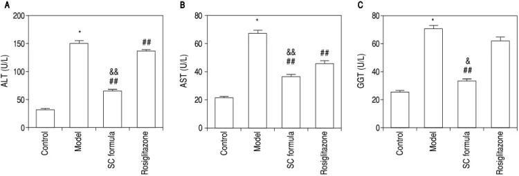
SC formula on sera ALT, AST and GGT
The liver function related enzymes, ALT, AST and GGT were detected. As compared to control, sera ALT, AST and GGT in model group were significantly elevated (Figure 4). By contrast, SC formula alleviated the high fat
diet-induced increase of ALT, AST and GGT. By contrast, rosiglitazone alleviated ALT and AST, but not influenced GGT.
Effects of SC formula on sera TNF-ααααα and IL-1
The effects of SC formula on inflammation factors were also evaluated. As shown in figure 5, high fat diet promoted sera TNF-α and IL-1 levels. By contrast, SC formula reduced high fat diet-induced inflammation fac-tors, while rosiglitazone did not influence TNF-α and IL-1 levels compared with model group.
Effects of SC formula on sera FINS, FBG and HOMA-IR
The effects of SC formula on FINS, FBG and HOMA-IR were also evaluated. As shown in figure 6, high fat diet elevated FINS, FBG and HOMA-IR levels, which were attenuated by SC formula, as well as by rosiglitazone.
Ad-Figure 2. Figure 2. Figure 2. Figure 2.
Figure 2. Histopathological changes in liver tissues of rats in various study groups. A, B, C and D are representative images from control, model, SC formula and rosiglitazone groups (400 x).
AAAAA BBBBB
C CC
Figure 3. Figure 3.Figure 3.
Figure 3.Figure 3. SC formula decreases high fat diet-induced increase of liver TG (AAAAA) and liver FFA (BBBBB). *P < 0.05 compared with control; ##P < 0.01 compared with
model; &&P < 0.01 compared with Rosiglitazone.
Figure 4. Figure 4.Figure 4.
Figure 4.Figure 4. SC formula decreases high fat diet-induced increase of liver ALT (AAAAA), AST (BBBBB) and GGT (CCCCC). *P < 0.05 compared with control; ##P < 0.01
com-pared with model; &&P < 0.01 compared with Rosiglitazone.
Figure 5. Figure 5.Figure 5.
Figure 5.Figure 5. SC formula decreases high fat diet-induced increase of TNF-α (AAAAA) and IL-1 (BBBBB). *P < 0.05 compared with control; ##P < 0.01 compared with
mod-el; &&P < 0.01 compared with Rosiglitazone.
ALT (U/L)
200
150
100
50
0
Control Model
SC formula
Rosiglitazone
AST
(U/L)
80
60
40
20
0
Control Model
SC formula
Rosiglitazone
GGT (U/L)
80
60
40
20
0
Control Model
SC formula
Rosiglitazone
AAAAA BBBBB CCCCC
*
## *
&& ##
## *
& ##
&&
##
Control Model
SC formula
Rosiglitazone
AAAAA BBBBB
Control Model
SC formula
Rosiglitazone
Li
ve
r T
G
(m
g/
g)
150
100
50
0
Liver FFA (
μ
mol/gprot)
300
200
100
0 *
## &&
##
*
## &&
##
Control Model
SC formula
Rosiglitazone
AAAAA BBBBB
Control Model
SC formula
Rosiglitazone
TN
F-α
(ng/L)
200
150
100
50
0
IL-I (ng/L)
150
100
50
0 *
##
&& *
ditionally, SC formula was more effective to reduce FINS, FBG and HOMA-IR compared with rosiglitazone group.
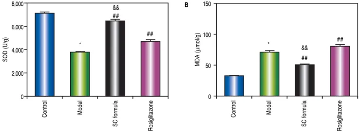
Effects of SC formula on tissue MDA and SOD
As shown in figure 7, high fat diet decreased SOD and elevated MDA levels. By contrast, SC formula, as well as rosiglitazone alleviated the effects of high fat diet on MDA and SOD. Moreover, the effect of SC formula was prior to rosiglitazone.
Figure 6. Figure 6. Figure 6. Figure 6.
Figure 6. SC formula decreases high fat diet-induced increase of FINS (AAAAA), FBG (BBBBB) and HOMA-IR (CCCCC). *P < 0.05 compared with control; ##P < 0.01
com-pared with model; &&P < 0.01 compared with Rosiglitazone.
Figure 7. Figure 7. Figure 7. Figure 7.
Figure 7. SC formula decreases high fat diet-induced decrease of SOD (AAAAA) and increase of MDA (BBBBB). *P < 0.05 compared with control; ##P < 0.01 compared
with model; &&P < 0.01 compared with Rosiglitazone.
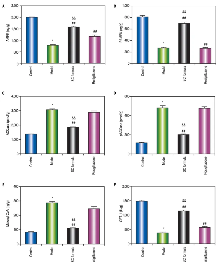
Effects of SC formula on tissue AMPK, pAMPK, ACCase, pACCase, Malonyl CoA and CPT-1
We also detected AMPK signaling pathway in our study. As shown in figure 8, high fat diet decreased AMPK, pAMPK, and CPT-1, while increased ACCase, pACCase and Malonyl CoA. SC formula reduced the effect of high fat diet induced decrease of AMPK, pAMPK, and CPT-1, and increase of ACCase, pACCase and malonyl CoA, while osiglitazone only targeted AMPK and CPT-1. These data implicate that SC formula was prior to rosiglitazone to influence the AMPK signaling pathway.
FINS (mLU/L)
40
30
20
10
0
Control Model
SC formula
Rosiglitazone
FBG (mmol/L)
8
6
4
2
0
Control Model
SC formula
Rosiglitazone
HOMAR-IR
10
8
6
4
2
0
Control Model
SC formula
Rosiglitazone
AAAAA BBBBB CCCCC
*
## *
&& ##
## *
& ##
&& ##
Control Model
SC formula
Rosiglitazone
AAAAA BBBBB
Control Model
SC formula
Rosiglitazone
SOD (U/g)
8,000
6,000
4,000
2,000
0
MDA (
μ
mol/g)
150
100
50
0 *
## &&
*
## &&
##
##
Figure 8. Figure 8.Figure 8.
Figure 8.Figure 8. SC formula decreases high fat diet-induced inactivation of AMPK signaling pathway. A.A.A.A. AMPK. B.A. B.B.B.B. pAMPK. C.C. ACCase. D.C.C.C. D.D.D.D. pACCase. E.E.E.E.E. Malo-nyl CoA. F.F.F.F.F. CPT-1. * P < 0.05 compared with control. ##P < 0.01 compared with model; &&P < 0.01 compared with Rosiglitazone.
Control Model
SC formula
Rosiglitazone
AAAAA BBBBB
Control Model
SC formula
Rosiglitazone
AMPK (ng/g)
2,500
2,000
1,500
1,000
500
0
PAMPK (ng/g)
1,000
800
600
400
200
0 *
## &&
*
## &&
##
##
Control Model
SC formula
Rosiglitazone
Control Model
SC formula
Rosiglitazone
ACCase (pmol/g)
4,000
3,000
2,000
1,000
0
pACCase (pmol/g)
600
400
200
0 *
## &&
*
## &&
Control Model
SC formula
Rosiglitazone
Control Model
SC formula
Rosiglitazone
M
al
on
yl C
oA
(n
g/
g)
400
300
200
100
0
CPT-1 (U/g)
2,000
1,500
1,000
500
0 *
## &&
*
## &&
## C
CC
CC DDDDD
DISCUSSION
In this study, we evidenced that SC formula alleviated high fat diet-induced liver injury. Besides its effects on liver morphological changes, SC formula also ameliorated liver function biochemically. Additionally, SC formula prevented inflammation reaction, enhanced anti-oxidative ability. The hepatoprotective effect of SC formula ap-peared to be mediated via its effects on the AMPK signal-ing pathway.
NAFLD shares a lot of common symptoms with obesi-ty, dyslipidemia and diabetes.7,8 The common pathological changes include the disorders of glucose and lipid metab-olism, and insulin resistance.9 Liver steatosis can also pro-mote the development and progression of type 2 diabetes mellitus and atherosclerosis. Therefore, NAFLD is a complex and systemic metabolic disease, and it is difficult to design ideal drugs for single target. Treatment aimed at multiple targets or combination therapy has recently be-come the main stream to ameliorate the symptoms of NAFLD. For examples, vitamin E has the ability to de-crease the activity of apoptosis-associated enzymes and has potential synergistic effects in combination with caspase (Caspase) inhibitors.17 Vit E and probiotics/prebiotics can synergistically increase their antioxidant stress and anti-inflammatory efficacy.18 carnitine with antioxidant or L-carnitine with statins/fenofibrate can improve oxidative damage of mitochondrial (MT) and promote MT biogen-esis.19 These studies provide certain reference to promote clinical and translational research of combination therapy for NAFLD.
In this study, we investigated the therapeutic effects of SC formula on NAFLD. SC formula is composed of sal-idroside and curcumin, two components obtained from TCM rhodiola rosea and rhizoma curcumae longae. A previous study has demonstrated that SC formula has good activity in inhibiting lipid deposition in the liver of NAFLD rats.12 We established NAFLD rat model by administrating high fat diet for 14 weeks. We further confirmed the efficacy of SC in the prevention and treatment of NAFLD. Liver TG and FFA were reduced after SC formula treat-ment. Additionally, sera ALT, AST, TNF-α, GGT activity and TNF-α, IL-1 content were alleviated in NAFLD after SC formula treatment.
The pathogenesis of NAFLD is still not clear, and the two “hits” theory proposed by Day et al has become a rela-tively recognized NAFLD pathogenesis.20 According to this theory, insulin resistance, formation of oxidative stress and antioxidant deficiencies play important roles in the pathogenesis of NAFLD. In our study, administration of high-fat diet induced insulin resistance, enhanced liver MDA content, and reduced liver SOD content in rats. These results suggest that NAFLD rats have obvious
insu-lin resistance and oxidative stress. TCM SC formula can obviously reduce the insulin resistance and the content of MDA in liver tissue, and increase the content of SOD in liver tissue. These data implicate that SC formula has good effect on improving insulin resistance and lipid peroxida-tion injury.
As the key enzyme of fatty acid synthesis, ACCase plays a very important role in lipid metabolism. The acti-vation of ACCase can increase the contents of Malonyl-CoA,21 which can inhibit the activity of CPT-1, thereby increasing high glucose/fatty acid induced islet glucol-ipotoxicity.22
AMPK is a key sensor of cellular energy status and a major regulator of lipid balance in the liver and whole body.23 It promotes energy metabolism by facilitating ca-tabolism and reducing the consumption of adenosine tri-phosphate, thereby integrating nutrients and hormones signals. In the liver, activation of AMPK increases the ox-idation of fatty acids and inhibits fatty acid synthesis. It can promote the inactivation and phosphorylation of acetyl coenzyme carboxylase, thus reducing the conver-sion of acetyl coenzyme A into C, thus reducing the syn-thesis of fatty acids.24 Our results showed that the activity of AMPK in liver tissue of model group decreased sig-nificantly and the contents of ACCase, pACCase and Malonyl-CoA increased significantly, and the content of CPT-1 in liver tissue decreased significantly. After SC formula treatment, the activity of AMPK in liver tissue was significantly increased, the contents of ACCase, pACCase and Malonyl-CoA in liver tissue were signifi-cantly decreased, and the content of CPT-1 in liver tissue was increased significantly. These data implicated that SC formula likely prevented liver injury through pro-moting AMPK signaling pathway.
In our study, NAFLD rat model was selected to evalu-ate the protective activity of SC formula and assess the mechanisms. NAFLD model does not produce obvious hepatic fibrosis, but shares a lot of common features with obesity, dyslipidemia and diabetes. Although non-alcohol-ic steatohepatitis (NASH) is the severe form of NAFLD, NAFLD is the most common liver disorder in developed countries.26 Additionally, NAFLD without treatment could further develop into NASH.27 Therefore, it is likely preferable to control the disease at an early stage, when possible. In future study, NASH model will be estab-lished to further confirm the protective effect of SC for-mula and to detect the mechanisms.
CONCLUSION
Our data reveal that SC formula prevents high fat diet-induced liver injury. Moreover, the efficacy of SC formula is prior to rosiglitazone. The potential mechanisms under-lying the preventive effects were related to insulin resist-ance, lipid peroxidation and AMPK signaling pathway.
ABBREVIATIONS
• ACCase: acetyl-coenzyme A carboxylase.
• ALT: alanine transaminase.
• AMPK: AMP-activated protein kinase.
• ANOVA: one-way analysis of variance.
• AST: aspartate aminotransferase.
• CPT-1: carnitine palmitoyl transferase-1.
• ELISA: enzyme-linked immunosorbent assay.
• FBG: fasting blood glucose.
• FFA: free fatty acid.
• FINS: fasting insulin.
• GGT: gamma-glutamyltransferase.
• H&E: hematoxylin and eosin.
• HOMA-IR: homeostasis model assessment of insulin.
• IL: interleukin.
• LSD: least significant difference test.
• malonyl CoA: malonyl coenzyme A.
• MDA: malondialdehyde.
• MT: mitochondrial.
• NAFLD: nonalcoholic fatty liver disease.
• NASH: nonalcoholic steatohepatitis.
• pACCase: phosphorylated acetyl-coenzyme A carbox-ylase.
• pAMPK: phosphorylated AMP-activated protein ki-nase.
• PPAR: peroxisome proliferator-activated receptor re-sistance.
• SC: salidroside and curcumin.
• SD: standard deviation.
• SOD: superoxide dismutase.
• TCM: Traditional Chinese Medicine.
• TG: triglyceride.
• TNF: sera tumor necrosis factor
• Vit E: vitamin E.
ACKNOWLEDGEMENTS
This study was supported by National Natural Science Foundation of China (81873109).
CONFLICT OF INTEREST
The authors declare that they have no conflicts of interest.
REFERENCES
1. Rinella ME, Sanyal AJ. Management of NAFLD: a stage-based approach. Nat Rev Gastroenterol Hepatol 2016; 13: 196-205.
2. Bellentani S, Scaglioni F, Marino M, Bedogni G. Epidemiolo-gy of non-alcoholic fatty liver disease. Dig Dis 2010; 28: 155-61.
3. Bhala N, Jouness RI, Bugianesi E. Epidemiology and natural history of patients with NAFLD. Curr Pharm Des 2013; 19: 5169-76.
4. Fan JG, Zhu J, Li XJ, Chen L, Li L, Dai F, Li F, et al. Preva-lence of and risk factors for fatty liver in a general popula-tion of Shanghai, China. J Hepatol 2005; 43: 508-14. 5. Jimba S, Nakagami T, Takahashi M, Wakamatsu T, Hirota Y,
Iwamoto Y, Wasada T. Prevalence of non-alcoholic fatty liv-er disease and its association with impaired glucose metab-olism in Japanese adults. Diabet Med 2005; 22: 1141-5. 6. Kwon YM, Oh SW, Hwang SS, Lee C, Kwon H, Chung GE.
Association of nonalcoholic fatty liver disease with compo-nents of metabolic syndrome according to body mass index in Korean adults. Am J Gastroenterol 2012; 107: 1852-8. 7. Brouwers B, Schrauwen-Hinderling VB, Jelenik T, Gemmink
A, Havekes B, Bruls Y, Dahlmans D, et al. Metabolic distur-bances of non-alcoholic fatty liver resemble the alterations typical for type 2 diabetes. Clin Sci (Lond) 2017.
8. Engin A. Non-Alcoholic Fatty Liver Disease. Adv Exp Med Biol 2017; 960: 443-67.
9. Par A, Par G. Advances in the pathogenesis of non alcoholic fatty liver disease. Orv Hetil 2017; 158: 882-94.
10. Zhao SP, Wu ZS, Chen Y, Liang X, Bao L, Li P, Sun RR, et al. Protective effect of Hua Tan Qu Shi decoction against liver injury in rats with nonalcoholic fatty liver disease. Biomed Pharmacother 2017; 91: 181-90.
11. Cheng F, Ma C, Wang X, Zhai C, Wang G, Xu X, Mu J, et al. Ef-fect of traditional Chinese medicine formula Sinisan on chronic restraint stress-induced nonalcoholic fatty liver disease: a rat study.BMC Complement Altern Med 2017; 17: 203.
12. Li H, Zhu D, Ying H, Li D. Effects of curcumin and salidroside combination on fatty liver induced by simple high fat diet in rats. Journal of Chinese Medicinal Materials 2015; 38: 1027-9.
13. Li H, Ying H, Hu A, Li D, Hu Y. Salidroside Modulates Insulin Signaling in a Rat Model of Nonalcoholic Steatohepatitis. Evid Based Complement Alternat Med 2017; 2017: 9651371. 14. Fatty liver and alcoholic liver disease group of Chinese
and treatment of nonalcoholic fatty liver disease. Chin J Hepatol 2010; 18: 163-6.
15. Li H, Ying H, Hu A, Hu Y, Li D. Therapeutic Effect of Gype-nosides on Nonalcoholic Steatohepatitis via Regulating He-patic Lipogenesis and Fatty Acid Oxidation. Biol Pharm Bull 2017; 40: 650-7.
16. Huiqing L, Jiaen Y, Jinmo T, Chuncheng W, Hongshan L, Shaodong C. Optimization of dosage ratio of chlorogenic acid and gardenia glycosides in the treatment of rats with fatty liver disease induced by high-fat feed. J Tradit Chin Med 2016; 36: 683-8.
17. Corey KE, Chalasani N. Should combination therapy be the paradigm for future nonalcoholic steatohepatitis clinical tri-als?Hepatology 2011; 54: 1503-5.
18. Cheng J, Joyce A, Yates K, Aouizerat B, Sanyal AJ. Metabo-lomic profiling to identify predictors of response to vitamin E for non-alcoholic steatohepatitis (NASH). PLoS One 2012; 7: e44106.
19. Indiveri C, Iacobazzi V, Tonazzi A, Giangregorio N, Infantino V, Convertini P, Console L, et al. The mitochondrial carnitine/ acylcarnitine carrier: function, structure and physiopatholo-gy.Mol Aspects Med 2011; 32: 223-33.
20. Day CP, James OF. Steatohepatitis: a tale of two hits? Gas-troenterology 1998; 114: 842-5.
21. Sasaki Y, Kozaki A, Hatano M. Link between light and fatty acid synthesis: thioredoxin-linked reductive activation of plastidic acetyl-CoA carboxylase. Proc Natl Acad Sci USA 1997; 94: 11096-101.
22. Lee JH, Jung IR, Choi SE, Lee SM, Lee SJ, Han SJ, Kim HJ, et al. Toxicity generated through inhibition of pyruvate
carbox-ylase and carnitine palmitoyl transferase-1 is similar to high glucose/palmitate-induced glucolipotoxicity in INS-1 beta cells.Mol Cell Endocrinol 2014; 383: 48-59.
23. Ha SK, Kim J, Chae C. Role of AMP-activated protein kinase and adiponectin during development of hepatic steatosis in high-fat diet-induced obesity in rats. J Comp Pathol 2011; 145: 88-94.
24. Nammi S, Roufogalis BD. Light-to-moderate ethanol feeding augments AMPK-alpha phosphorylation and attenuates SREBP-1 expression in the liver of rats. J Pharm Pharm Sci 2013; 16: 342-51.
25. Luo W, Xu Q, Wang Q, Wu H, Hua J. Effect of modulation of PPAR-gamma activity on Kupffer cells M1/M2 polarization in the development of non-alcoholic fatty liver disease. Sci Rep 2017; 7: 44612.
26. Rinella ME. Nonalcoholic fatty liver disease: a systematic re-view.JAMA 2015; 313: 2263-73.
27. McCullough AJ. The clinical features, diagnosis and natural history of nonalcoholic fatty liver disease. Clin Liver Dis 2004; 8: 521-533, viii.
Correspondence and reprint request:
Hong-Shan Li, M.M.
Department of Hepatology, Ningbo No.2 Hospital, No.175, Yongfeng Road, Ningbo 315010, Zhejiang, China. Tel.: 86-0574-87084295, Fax: 86-0574-83870626.