Long term changes in liver histology
following treatment of chronic hepatitis C virus
Mitchell L. Shiffman,* Richard K. Sterling,** Melissa Contos,*** Sarah Hubbard,** April Long,* Velimir A. Luketic,** R. Todd Stravitz,** Michael Fuchs,** Arun J. Sanyal*** Liver Institute of Virginia, Bon Secours Health System, Richmond and Newport News, Virginia, USA and ** Hepatology Section and *** Division of Surgical Pathology, Virginia Commonwealth University Medical Center, Richmond, Virginia, USA
ABSTRACT
Background and aims. The histologic hallmarks of chronic HCV include inflammation and fibrosis. The im-pact of interferon therapy on liver histology was evaluated. Material and methods. The study population consisted of 348 patients with chronic HCV who underwent a baseline liver biopsy, received either no treatment or a single course of interferon based therapy, were followed for 5 years without any treat-ment or additional treattreat-ment and then underwent a repeat liver biopsy. The patients were divided into 3 groups; deferred treatment (NoTx = 47), received interferon based therapy but failed to achieve SVR (NoSVR = 189) and achieved SVR (SVR = 112). Results. Patients with NoTx and NoSVR had significant increa-ses in mean inflammation scores (from 4.3 to 6.3 and 5.4 to 6.7 respectively; p < 0.001 for both) and fibrosis scores (from 0.9 to 1.8 and 1.9 to 2.5; p < 0.001 for both). The amounts by which inflammation, fibrosis and rate of fibrosis progression increased were not significantly different between the two groups. Increases in total inflammation and the piecemeal necrosis sub-score over time were strongly associated with fibrosis progression. Patients with SVR had a significant decline in mean inflammation and fibrosis scores (from 6.7 to 2.2 and 3.3 to 1.8; p < 0.001 for both); 40% of patients resolved all fibrosis and 50% of patients resolved cirrhosis. Conclusion. Increases in inflammation are associated with fibrosis progression and in the absence of SVR interferon treatment does not appear to affect the long term natural history of this process. Pa-tients with SVR have resolution of inflammation and fibrosis and many resolve cirrhosis.
Key words. Chronic hepatitis C virus. Hepatic fibrosis. Interferon therapy. Sustained virologic response.
Correspondence and reprint request: Mitchell L. Shiffman, M.D.
Liver Institute of Virginia,
Bon Secours Health System. 5855 Bremo Road, Suite 509, Richmond, VA 23226, USA.
Ph.: 804-301-7058. Fax: 757-947-3195 E-mail: [email protected]
Manuscript received: December 22, 2013. Manuscript accepted: February 10, 2014. INTRODUCTION
The progression of hepatic fibrosis to cirrhosis in patients with chronic hepatitis C virus (HCV) is a
highly variable process which occurs over decades.1-4
Several clinical features have been shown to affect the rate of fibrosis progression including age at the
time HCV is acquired,1,4 the serum level of liver
transaminases,3,4 co-infection with human immune
deficiency virus,4,5 co-existent liver disorders such
non-alcoholic fatty liver disease,4,6,7 insulin
resist-ance7 and the excessive use of alcohol.1,4,8 Specific
polymorphisms within several host genes in patients with HCV have been associated with advanced fibro-sis and cirrhofibro-sis suggesting that host genetics plays
an important role in this process.9,10 The severity of
hepatic inflammation, especially the piecemeal necrosis component is also associated with fibrosis
progression.11-13
Interferon based therapy has been the primary treatment for patients with HCV for nearly 2
dec-ades.14-16 Therapy has evolved rapidly since 2011
and it now appears unlikely that interferon and rib-avirin will continue to be utilized to treat HCV in
the future.17 However, understanding how
response is associated with a decline in hepatic
in-flammation.13,14,16,18,19 Although many of these
stud-ies have suggested that this may slow the rate of fibrosis progression a recent study has suggested that fibrosis progression may actually be accelerated
in patients who fail interferon based therapy.20 The
long term impact of interferon therapy on liver his-tology in patients who do not achieve a SVR re-mains undefined. In contrast, achieving a SVR is associated with eradication of HCV, a reduction in hepatic fibrosis and a reduced risk for developing he-patic decompensation and hepatocellular carcino-ma.14,16,19,21-24 In a recent study which monitored patients by fibroscan and serum fibrosis markers over 10 years 49% of patients had fibrosis regression
and 56% resolved cirrhosis.25
The current study was performed to help under-stand how changes in inflammation affected fibrosis progression in patients with ongoing chronic HCV and fibrosis regression in patients who achieved SVR.
MATERIAL AND METHODS
Patient population
All patients had chronic HCV, underwent a base-line liver biopsy, were subsequently treated with a single course of either standard or pegylated inter-feron alfa-2a or alfa-2b with or without ribavirin be-tween 1991-2004 or deferred treatment, received no additional treatment for chronic HCV and under-went a repeat liver biopsy at least 5 years after the initial biopsy. The last patient to be included in this series underwent liver biopsy in 2009. Patients were excluded if they had any other cause of chronic liver disease in addition to chronic HCV based upon sero-logic studies and liver histology with the exception of benign steatosis and mild-moderate non-alcoholic
fatty liver disease;26 were anti-HIV positive.
Pa-tients were also excluded if they opted to receive treatment or retreatment for HCV with either inter-feron, peginterferon (with or without ribavirin), or any other agent being investigated as a possible treatment for chronic HCV during the 5 year follow-up period. Other patients were excluded if they de-veloped recurrence of HCV RNA in serum after achieving SVR (HCV RNA undetectable 6 months af-ter completing treatment); if they developed chronic renal failure and required hemodialysis; developed a complication of cirrhosis including variceal hemor-rhage, ascites, hepatic encephalopathy or hepatocel-lular carcinoma; or if they received an organ
transplant during the 5 year follow-up and before undergoing the second liver biopsy. Patients with no fibrosis on the initial liver biopsy specimen who achieved SVR were not included because there was no reason for these patients to undergo a second liv-er biopsy. The use of ovliv-er the countliv-er hliv-erbal or othliv-er supplements was permitted.
During the 5 years or more of follow-up patients were monitored at regular intervals. The frequency of follow-up ranged from every 3-12 months based upon the degree of fibrosis present on the initial liv-er biopsy and the virologic response to the initial treatment. This included patients who achieved an SVR. At each of these visits routine liver chemis-tries and hematology were performed. Patients with advanced fibrosis or cirrhosis had an ultrasound and alfa-fetoprotein performed at periodic intervals to screen for hepatocellular carcinoma. Patients with SVR also had serum HCV RNA assayed at least once yearly to ensure they remained HCV RNA un-detectable in serum. During the early years of the study this was performed by the Roche Amplicor polymerase chain reaction assay (PCR). By the end of the study HCV RNA was assayed by Taqman PCR with a lower limit of detection of 10 IU/ml. In all patients with SVR the most recent specimen(s) were assessed for HCV RNA by Taqman PCR.
The patients were grouped into three cohorts as follows:
• Patients who deferred treatment after the initial liver biopsy (NoTx).
• Patients who received a single course of interfer-on based therapy but failed to achieve SVR (NoS-VR); and
• Patients who achieved SVR following the initial course of HCV treatment with the exception of those who had no fibrosis on the initial liver bi-opsy (SVR).
Evaluation of liver histology
All liver biopsy slides were read in real time by a dedicated liver pathologists (MJC) who was blinded to the clinical, biochemical and virologic status of each patient. The stained slides of any liver biopsy which had been performed at an outside facility were obtained and also scored by this same pathologist. None of the slides were reread specifically for this study. All histologic specimens were scored for the degree of inflammation according the method of
Kn-odell, et al.27 Fibrosis scores were assigned based
both Knodell and Ishak, et al.28 thereafter. Fibrosis scores assigned by the Knodell method only were converted to Ishak scores as follows: no fibrosis = 0 by both systems; a portal fibrosis score of 1 by Kn-odell was assigned a value of 1.5 for Ishak (mid-way between the scores for portal fibrosis [1-2] with Ishak); a bridging fibrosis score of 3 by Knodell was assigned a value of 3.5 for Ishak (mid-way between the scores for bridging fibrosis [3-4] with Ishak); and a score of 4 for cirrhosis by Knodell was as-signed an Ishak value of 5.5 (mid-way between the scores for cirrhosis [5-6] with Ishak). This modifi-cation affected only 86/716 (12%) biopsies. The con-version from Knodell to Ishak scores was validated in 183 biopsies scored for fibrosis by both methods. The true Ishak score of these specimens was 1.39. The assigned Ishak score was 1.38; CI -0.37-0.075; r = 0.977 (p < 0.0001). The AST-platelet ratio index
(APRI) was calculated as described by Wai, et al.29
Study design
The study was originally conceived in the mid-1990s to be a prospective analysis to evaluate the ef-fects of interferon therapy on liver histology. The study was approved by the Investigational Review Board (IRB) at the Virginia Commonwealth Univer-sity Medical Center and informed consent was ob-tained from all patients either prior to undergoing the initial liver biopsy or initiating treatment. Over the next several years many patients who enrolled in the study, had failed to achieve an SVR after re-ceiving interferon therapy and were to be followed prospectively for 5 years without retreatment dropped out of the study so they could be retreated with newer and/or investigational agents. In addi-tion, many other patients who were being followed in the Hepatology clinical practice but had not en-rolled in the study prospectively underwent a repeat liver biopsy after 5 years of follow-up as part of their clinical care to determine if they had developed fibrosis progression and should either receive an ini-tial course of interferon based therapy or be retreat-ed for chronic HCV. It became apparent that a prospective study could not be conducted to answer this question at our Center because of the manner in which we clinically managed HCV during this time. The protocol was therefore amended to a retrospec-tive longitudinal cohort analysis. This change was approved by the IRB and allowed all patients who had undergone two liver biopsies at least 5 years apart and satisfied all inclusion and exclusion crite-ria to be included in this analysis.
Statistical analysis
Demographic, clinical, laboratory and histologic data are presented as mean and standard deviation for continuous variables and proportions for cate-gorical data as indicated. Differences in continuous variables were compared by two-tailed Student’s t-test and analysis of variance (ANOVA) as appropri-ate. Differences in categorical variables were
compared by the χ2 test. The main outcomes were
changes in inflammation and fibrosis between the groups. The rate of fibrosis progression/regression was defined as the change in Ishak fibrosis units/ year. Univariate regression analysis was utilized to assess for factors associated with fibrosis progres-sion. Variables included were gender, race, age, se-rum ALT, sese-rum HCV RNA, the baseline total inflammation score and the sub-scores for the vari-ous components of inflammation and the fibrosis score. Factors found on univariate analysis to be statistically significant (p < 0.05) were then evalu-ated with multiple logistic regression analysis utiliz-ing MedCalc software.
RESULTS
Baseline characteristics
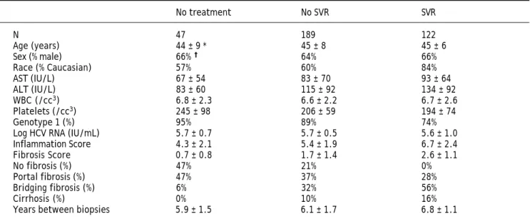
The demographic, biochemical, virologic and his-tologic features of the patients at baseline are sum-marized in table 1. Forty-seven patients did not receive HCV treatment, 189 were treated with inter-feron or peginterinter-feron with or without ribavirin but failed to achieve a SVR and 122 patients achieved a SVR. The mean age for all patients was 45 years, 65% were male and 65% where Caucasian. The time between the two liver biopsies for each of the three groups differed by less than 1 year.
Impact of interferon therapy on fibrosis progression
(p< 0.0001). In patients who received prior therapy the inflammation score also increased significantly from 5.4 ± 1.9 to 6.7 ± 2.5 (p < 0.0001). Although the mean change in inflammation scores were some-what greater in patients without prior treatment
(1.9 ± 2.7 vs. 1.3 ± 2.9), these differences were not
significant (p = 0.23). Fibrosis scores also increased significantly over time in both patient groups; from
0.9 ± 1.0 to 1.8 ± 1.7 (p = 0.0001) and 1.9 ± 1.4 to 2.5 ± 1.7 (p < 0.0001) respectively. The mean change in fibrosis scores were greater in patients without prior treatment compared to patients
with-out SVR (0.7 ± 1.1 vs. 0.4 ± 1.3 respectively).
How-ever, this difference was not significant (p = 0.15). Although the mean rate of fibrosis progression (Ishak units/year) was greater in patients without prior treatment this difference was also not
signifi-cant (0.13 ± 0.21 vs. 0.07 ± 0.27; p = 0.22).
The change in fibrosis over 5 years was as-sessed by comparing patients with the same de-gree of fibrosis at baseline (Figure 2). In this analysis patients were grouped into 4 categories based upon fibrosis scores; either none (Ishak score of 0), portal (score of 1 and 2), bridging (score of 3 and 4) or cirrhosis (score of 5 and 6). In patients with no fibrosis at baseline (Figure 2A) 59% of those without prior treatment had an increase in fibrosis; 50% progressed to portal fi-brosis, 5% bridging fibrosis and 4% to cirrhosis. A similar pattern of fibrosis progression was ob-served in patients who failed to achieve SVR; 48% progressed to portal fibrosis, 16% bridging fibrosis and 3% to cirrhosis.
The change in liver fibrosis over 5 years in pa-tients with portal fibrosis at baseline is illustrated in figure 2B. Fibrosis progression was also similar in patients who deferred treatment and in those who failed to achieve SVR; 32 and 25% of patients respec-tively progressed to bridging fibrosis or cirrhosis.
Table 1. Demographic, biochemical, hematologic, virologic and histologic findings at baseline.
No treatment No SVR SVR
N 47 189 122
Age (years) 44 ± 9 * 45 ± 8 45 ± 6
Sex (% male) 66% † 64% 66%
Race (% Caucasian) 57% 60% 84%
AST (IU/L) 67 ± 54 83 ± 70 93 ± 64
ALT (IU/L) 83 ± 60 115 ± 92 134 ± 92
WBC (/cc3) 6.8 ± 2.3 6.6 ± 2.2 6.7 ± 2.6
Platelets (/cc3) 245 ± 98 206 ± 59 194 ± 74
Genotype 1 (%) 95% 89% 74%
Log HCV RNA (IU/mL) 5.7 ± 0.7 5.7 ± 0.5 5.6 ± 1.0
Inflammation Score 4.3 ± 2.1 5.4 ± 1.9 6.7 ± 2.4
Fibrosis Score 0.7 ± 0.8 1.7 ± 1.4 2.6 ± 1.1
No fibrosis (%) 47% 21% 0%
Portal fibrosis (%) 47% 37% 28%
Bridging fibrosis (%) 6% 32% 56%
Cirrhosis (%) 0% 10% 16%
Years between biopsies 5.9 ± 1.5 6.1 ± 1.7 6.8 ± 1.1
*Values are given as mean ± standard deviation. † † † † † Values are given as percentage of patients in each group.
Figure 1. Change in mean inflammation score (left panel) and mean fibrosis score (right panel) over 5 years in patients with chronic HCV. Patients are grouped according to whether they received no treatment (No Tx) for chronic HCV or interfe-ron based therapy without SVR (No SVR). Vertical lines on the top of each bar represent standard deviation. An asterisk in-dicates the change between the initial and follow-up liver biopsies was statistically significant. P values are provided in the text.
Inflammation score
10
8
6
4
2
0
5
4
3
2
1
0
Fibrosis score
No Tx No SVR No Tx No SVR
*
*
*
*
a a a a a a a a
aaaaaaaaaaaaaaaaa a a a a a a a aaaaaaaaaaaaaaaa
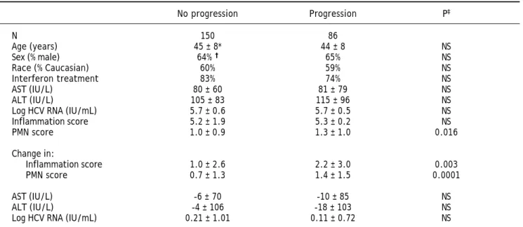
Table 2 compares various features from patients in the “No treatment” and “No SVR” groups based upon whether they developed fibrosis pro-gression or not. Propro-gression was observed in 86/ 236 patients (36%). Factors associated with fibro-sis progression on univariate analyfibro-sis included the baseline piecemeal necrosis score, and an in-crease in the total inflammation and piecemeal necrosis scores over time. Multiple logistic regres-sion analysis identified only an increase in the piecemeal necrosis and total inflammation scores as being independently associated with fibrosis progression.
Impact of SVR on fibrosis regression
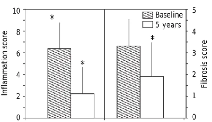
SVR was confirmed in 122 patients by assessing serum for HCV RNA once yearly for at least consec-utive 5 years after discontinuing treatment. All pa-tients were repeatedly HCV RNA undetectable. The mean serum AST and ALT in patients who achieved SVR declined significantly (p < 0.0001) from 93 ± 64 and 134 ± 92 to 31 ± 18 and 34 ± 16 respective-ly. Figure 3 illustrates the inflammation and fibro-sis scores at baseline and 5 years after achieving SVR. The inflammation score declined from 6.7 ± 2.4 to 2.2 ± 2.2 (p < 0.0001) and fibrosis score from
Figure 2. Percentage of patients with varying degrees of fibrosis on the second liver biopsy compared to the initial liver biop-sy. Patients either deferred treatment (No Tx) or received interferon therapy but failed to achieve a SVR (No SVR). All patients were followed for a minimum of 5 years before they underwent repeat liver biopsy. A. Patients with no fibrosis on the initial liver biopsy. B. Patients with portal fibrosis on the initial liver biopsy.
Table 2. Factors associated with fibrosis progression in patients with chronic HCV.
No progression Progression P‡
N 150 86
Age (years) 45 ± 8* 44 ± 8 NS
Sex (% male) 64% † 65% NS
Race (% Caucasian) 60% 59% NS
Interferon treatment 83% 74% NS
AST (IU/L) 80 ± 60 81 ± 79 NS
ALT (IU/L) 105 ± 83 115 ± 96 NS
Log HCV RNA (IU/mL) 5.7 ± 0.6 5.7 ± 0.5 NS
Inflammation score 5.2 ± 1.9 5.3 ± 0.2 NS
PMN score 1.0 ± 0.9 1.3 ± 1.0 0.016
Change in:
Inflammation score 1.0 ± 2.6 2.2 ± 3.0 0.003
PMN score 0.7 ± 1.3 1.4 ± 1.5 0.0001
AST (IU/L) -6 ± 70 -10 ± 85 NS
ALT (IU/L) -4 ± 106 -18 ± 103 NS
Log HCV RNA (IU/mL) 0.21 ± 1.01 0.11 ± 0.72 NS
* Values are given as mean ± standard deviation. † Values are given as percentage of patients in each group. ‡ P values > 0.05 are
recorded as being not significant (NS).
A B
Fibrosis on
second biopsy (%)
100
80
60
40
20
0
No Tx No SVR
Fibrosis on
second biopsy (%)
100
80
60
40
20
0
No Tx No SVR
CX BF PF None
a a aaaa a a a a a a a a a a
aaaaaaaaaaaaaaa aaaaaaaaaaaaaaa
a a a a a aaaaaaaaaaa aaaaaaaaaaa
aaaaaaaaaa aaaaaaaaaaa aaaaaaaaaaa aaaaaaaaaaa aaaaaaaaaaa aaaaaaaaaaa aaaaaaaaaaa aaaaaaaaaaa aaaaaaaaaaa aaaaaaaaaaa aaaaaaaaaaa aaaaaaaaaaa aaaaaaaaaaa aaaaaaaaaaa aaaaaaaaaaa aaaaaaaaaaa aaaaaaaaaaa aaaaaaaaaaa aaaaaaaaaaa
3.3 ± 1.3 to 1.8 ± 1.9 (p < 0.0001). Fibrosis regres-sion occurred at a mean of -0.23 Ishak units/year. Of the 122 patients with SVR 76 (63%) had regression in fibrosis by 1 or more stage, 40 (33%) had no change in fibrosis stage and 6 (1%) had more fibro-sis 5 years after they had achieved SVR. In the 34 patients with portal fibrosis at baseline 71% had no evidence of fibrosis 5 years after achieving SVR. In the 68 patients with bridging fibrosis at baseline 35% had no fibrosis and 26% portal fibrosis 5 years after achieving SVR. Of the 20 patients with cirrho-sis on the baseline biopsy, 2 had no fibrocirrho-sis, 2 portal fibrosis and 6 bridging fibrosis 5 years after achiev-ing SVR (Figure 4).
The relationship between the change in inflamma-tion and the change in fibrosis for patients with SVR is illustrated in figure 5. A decline in the inflammation score was associated with a linear de-cline in hepatic fibrosis. Seventeen percent (20/122) of patients either had no change or an increase of 1-4 points (6 patients) in the hepatic inflammation score after being HCV RNA undetectable in serum for at least 5 years. Only 4 of these 20 patients had a decline in the fibrosis score. No baseline features differentiated patients who failed to have a decline in hepatic inflammation from those that did have a decline in inflammation after achieving SVR. Specifi-cally, there were no significant differences in base-line serum ALT, serum HCV RNA level, the total inflammation score, piecemeal necrosis score or fi-brosis score. All patients who failed to have a decline in inflammation also remained persistently HCV RNA undetectable in serum by Taqman PCR.
Non-invasive markers of fibrosis
Table 3 compares markers of fibrosis in patients with fibrosis progression, no progression and in pa-tients with SVR. Papa-tients with fibrosis progression had a slight decline in the platelet count, patients with no change in fibrosis had no change in platelets and patients with SVR had a significant (p < 0.01) increase in the platelet count. Similar changes were noted in the APRI.
Figure 4. Percentage of patients with varying degrees of fibrosis on the second liver biopsy grouped according to the degree of fibrosis on the first biopsy. Patients were followed for 5 years after achieving a SVR and before the repeat liver biopsy was performed.
Figure 3. Change in mean inflammation score (left panel) and mean fibrosis score (right panel) over 5 years in patients who achieved an SVR following treatment with interferon ba-sed therapy. Vertical lines on the top of each bar represent standard deviation. An asterisk indicates that the change between the initial and follow-up liver biopsies was statisti-cally significant. P values are provided in the text.
Figure 5. Relationship between inflammation and fibrosis scores for patients who achieved an SVR. Patients were initially grouped according to the change in inflammation score (inflammation score on repeat liver biopsy - inflammation score on initial liver biopsy). Each point represents the mean change in fibrosis score for each change in inflammation score. The size of each point is proportional the number of patients with that degree of change in inflammation score. The line presents the calculated linear regression fit for the data. The equation of the regression line was: Y = 0.20X - 0.27 (p < 0.001).
Inflammation score
10
8
6
4
2
0
Fibrosis score
5
4
3
2
1
0
Fibrosis on
second biopsy (%)
100
80
60
40
20
0
Portal Bridging Cirrhosis
*
*
*
Baseline 5 years
1
0
-1
-2
-3
-4
Change in inflammation
-12 -10 -8 -6 -4 -2 0 2 4 6
C
hange in fibrosis
aaa aaaa
DISCUSSION
The results of this study have demonstrated that the vast majority of patients with chronic HCV who achieve SVR and remain HCV RNA undetectable in serum over many years have regression in fibrosis. Overall, about 40% of patients had complete loss of fibrosis and 50% no longer had evidence of cirrhosis 5 years after achieving SVR. Significant improve-ments in non-invasive markers of fibrosis, such as platelet count and APRI were also observed in pa-tients with SVR. The degree of fibrosis improvement was closely linked to changes in the inflammation score. A long term improvement in both inflamma-tion and fibrosis has also been observed in patients
who achieved SVR in a previous study.30
The current study also evaluated the impact of in-terferon based therapy in patients with chronic HCV who failed to achieve SVR. Previous studies suggest-ed that these patients also derive histologic benefit and this may reduce the rate of fibrosis
progres-sion.13,14,16,18,19 However, in many of these studies
the repeat liver biopsy was performed less than 1 year after discontinuing interferon treatment. None of the previous studies compared the histologic im-pact of interferon to a control group of untreated patients. In the HALT-C study, over 800 patients with advanced fibrosis or cirrhosis underwent 2-3
liver biopsies over 4-5 years.31,32 No significant
re-duction in fibrosis progression was observed even in patients who had profound virologic suppression during treatment with full-dose peginterferon dur-ing the lead-in phase and with low-dose
peginterfer-on maintenance therapy.32,33 Similar results were
observed when a more sensitive technique, morpho-metric analysis, was utilized to quantitate tissue
col-lagen.34 The unique aspect of the present study was
that changes in liver histology in patients who
failed to achieve SVR were compared to patients who deferred HCV treatment. The rate of fibrosis pro-gression observed in the NoTx group, 0.12 Ishak units/year, was very similar to that reported for
un-treated patients in previous studies,1,11,12,35 and
sim-ilar to that observed in patients with “No SVR”. Thus, although short term improvements in liver histology may be achieved following interferon based therapy in patients without SVR it is unlikely that a single course of HCV treatment significantly im-pacts the long term natural history of fibrosis pro-gression in the absence of SVR.
The primary limitation of this study was that pa-tients could not be matched for baseline characteris-tics. Patients with no fibrosis who achieved SVR could not be included because there was no reason for these patients to undergoing a repeat liver biopsy. In addition, patients with more fibrosis rarely deferred treatment or opted for retreatment if they failed to achieve an SVR. This explains why the “SVR” and “No SVR” groups had significantly high-er fibrosis scores at baseline than the “No Tx” group (Table 1). As a result, we only evaluated fi-brosis progression in patients with the same degree of fibrosis at baseline (Figure 2). Another limitation was that biopsies were not rescored and 2 different systems were utilized during the nearly 15 years re-quired to conduct this study. However, all biopsies were scored by the same pathologist during the en-tire study and the system utilized to convert Kn-odell to Ishak fibrosis scores was internally validated and tightly correlated.
Liver injury in chronic HCV is composed of two processes; inflammation and fibrosis. Several studies now strongly suggest that inflammation is the driv-ing force for fibrosis progression in patients with chronic HCV. Inflammation increases with
increas-ing fibrosis in chronic HCV11,12,36 and fibrosis
pro-Table 3. Impact of sustained virologic response on platelet count and APRI.
Baseline 5 years P
Platelet count
Progression 217 ± 54* 188 ± 75 0.0001
No progression 212 ± 78 212 ± 76
SVR 194 ± 74 230 ± 85
APRI index
Progression 0.80 ± 1.03 1.11 ± 1.20 0.0001
No progression 0.93 ± 0.22 0.96 ± 1.03
SVR 1.15 ± 1.02 0.33 ± 0.31
gression is strongly associated with higher
inflam-mation scores.11,12,36,37 The piecemeal necrosis
sub-score appears to be the most important part of the
inflammation score leading to fibrosis progression.12
Similar results were observed in the current study. Why inflammation increases in some patients with chronic HCV but remains stable in others remains undefined. Most likely, this is secondary to host
im-munologic factors and their response to HCV.38
Spe-cific genetic polymorphisms may be associated with fibrosis progression because they affect the host im-munologic response to HCV and the level of
inflam-mation.9,10 This possibility is worthy of additional
studies.
Previous studies in other liver disorders have demonstrated that reducing the degree of hepatic in-flammation can either lead to regression of fibrosis or delay fibrosis progression. Anti-viral therapy in
patients with chronic hepatitis B virus,39,40 and
sup-pressing the immune response in patients with
au-toimmune hepatitis41,42 are both associated with a
reduction in inflammation and fibrosis. Suppressing inflammation with maintenance interferon has been
shown to prevent fibrosis progression.43 Although
very few patients in the HALT-C trial had suppres-sion of HCV RNA with maintenance peginterferon, those patients in whom HCV RNA was suppressed to low or undetectable levels also had a reduction in inflammation scores and evidence of fibrosis
regres-sion.44 In the present study, a strong linear
relation-ship was observed between the decline in inflammation and fibrosis scores in patients who achieved SVR. Patients with SVR who had no im-provement in inflammation also had no improve-ment in fibrosis. Why some patients with SVR had no improvement in inflammation remains unclear at this time. This observation may explain why some patients who achieve SVR do not resolve cirrhosis and remain at risk for hepatocellular carcinoma.
In summary, the current study has demonstrated that in patients with chronic HCV a strong relation-ship exists between hepatic inflammation and fibrosis. In patients with ongoing chronic disease, increases in inflammation are associated with fibrosis progression. In contrast, achieving SVR and eradi-cating HCV, not just receiving interferon therapy, is associated with a marked reduction in inflammation and fibrosis regression. The future of HCV treat-ment is to suppress HCV with multiple oral anti-viral agents without interferon and/or ribavirin. This approach has already been demonstrated to achieve high rates of SVR with various
combina-tions of anti-viral agents.45-49 The observations of
the present study strongly suggest that fibrosis re-gression including resolution of cirrhosis will occur in the vast majority of patients who will achieve SVR with these future therapies.
ACKNOWLEDGEMENTS
The authors would like to acknowledge the as-sistance of our nursing staff (Charlotte Hofmann, Jennifer Salvatori, Denice Shelton, Paula Smith and Kim Williams), who assisted in the care of these pa-tients for more than a decade.
FINANCIAL DISCLOSURE
This study was not funded by any pharmaceutical company, private foundation or governmental agency.
The authors disclose the following relationships they have with various pharmaceutical companies and devise manufacturers which may be perceived as a conflict of interest with this study.
Dr. Shiffman is a consultant for GenProbe, Glaxo-Smithkline, Janssen, and Roche/Genentech; has attended advisor meetings with Achillion, Bay-er, Boehringer-Ingelheim, Bristol-Myers-Squibb, Gilead, Globeimmune, Janssen, Novartis, Roche/Ge-nentech, Merck and Vertex; has been a speaker for Boehringer-Ingelheim, GlaxoSmithKline, Gilead, Janssen, Roche/Genentech, Merck and Vertex; and has received research support from Abbvie, Anadys, Gilead, Bristol-Myers-Squibb, Globeimmune, Idenix, Intercept, Lumena, Merck, Roche/Genentech, Vertex.
Dr. Sterling has received research support from Abbvie, Boehringer-Ingelheim, Bayer, Bristol-Myers-Squibb, Merck, Roche/Genentech and Vertex; and has attended advisor meetings with Abbvie, Bayer, Bristol-Myers-Squibb, Gilead, Merck, Roche/Genen-tech, Salix, and Vertex.
Ms Hubbard, PA has no conflicts.
Ms Long, NP has attended advisor meetings with Boehringer-Ingelheim, Bristol-Myers-Squibb, Gilead, Merck and Vertex; and has been a speaker for Gile-ad, Merck and Vertex.
Dr. Luketic has received research support from Abbvie, Bristol-Myers-Squibb, GenFit, Gilead, Ide-nix, Intercept, Lumena, Novartis, Takeda and Vertex.
Drs. Contos, Fuchs and Stravitz report nothing to disclose.
REFERENCES
1. Poynard T, Bedossa P, Opolon P. Natural history of liver fi-brosis progression in patients with chronic hepatitis C. Lancet 1997; 349: 825-32.
2. Yano M, Kumada H, Kage M, Ikeda K, Shimamatsu K, Inoue O, Hashimoto E, et al. The long term pathological evolu-tion of chronic hepatitis C. Hepatology 1996; 23: 1334-40. 3. Puoti C, Castellacci R, Montagnese F, Zaltron S, Stornaiuo-lo G, Bergami N, Bellis L, et al. HistoStornaiuo-logical and viroStornaiuo-logical features and follow-up of hepatitis C virus carriers with normal aminotransferase levels: the Italian prospective study of the asymptomatic C carriers (ISACC). J Hepatol 2002; 37: 117-23.
4. Bialek SR, Terrault NA. The changing epidemiology and na-tural history of hepatitis C virus infection. Clin Liver Dis 2006; 10: 697-715.
5. Benhamou Y, DiMartino V, Bochet M, et al. Factors affec-ting liver fibrosis in human immunodeficiency virus- and hepatitis C virus-coinfected patients: impact of protease inhibitor therapy. Hepatology 2001; 34: 283-7.
6. Castéra L, Hézode C, Roudot-Thoraval F, Bastie A, Zafrani ES, Pawlotsky JM, Dhumeaux D. Worsening of steatosis is an independent factor of fibrosis progression in untrea-ted patients with chronic hepatitis C and paired liver biopsies. Gut 2003; 52: 288-92.
7. Moucari R, Asselah T, Cazals-Hatem D, Voitot H, Boyer N, Ripault MP, Sobesky R, et al. Insulin resistance in chronic hepatitis C: association with genotypes 1 and 4, serum HCV RNA level, and liver fibrosis. Gastroenterology 2008; 134(2): 416-23.
8. Peters MG, Terrault NA. Alcohol use and hepatitis C. Hepa-tology 2002; 36(Suppl. 1): S220-S225.
9. Huang H, Shiffman ML, Friedman S, Venkatesh R, Bzowej N, Abar OT, Rowland CM, et al. A 7 gene signature identifies the risk of developing cirrhosis in patients with chronic hepatitis C. Hepatology 2007; 46: 297-306.
10. Li Y, Chang M, Abar O, Garcia V, Rowland C, Catanese J, Ross D, et al. Multiple variants in toll-like receptor 4 gene modulate risk of liver fibrosis in Caucasians with chronic hepatitis C infection. J Hepatol 2009; 51: 750-7.
11. Colletta C, Smirne C, Fabris C, et al. Value of two noninva-sive methods to detect progression of fibrosis among HCV carriers with normal aminotransferases. Hepatology 2005; 42: 838-45.
12. Ghany MG, Kleiner DE, Alter H, Doo E, Khokar F, Promrat K, Herion D, et al. Progression of fibrosis in chronic hepatitis C. Gastroenterology 2003; 124: 97-104.
13. Shiffman ML, Hofmann CM, Contos MJ, Luketic VA, Sanyal AJ, Sterling RK, Ferreira-Gonzalez A, et al. A randomized, controlled trial of maintenance interferon for treatment of chronic hepatitis C non-responders. Gastroenterology 1999; 117: 1164-72.
14. McHutchison JG, Gordon SC, Schiff ER, Shiffman ML, Lee WM, Rustgi VK, Goodman ZD, et al. Interferon alfa-2b alo-ne or in combination with ribavirin as initial treatment for chronic hepatitis C. Hepatitis Interventional Therapy Group. N Engl J Med 1998; 339: 1485-92.
15. Fried MW, Shiffman ML, Reddy KR, Smith C, Marinos G, Gonçales FL Jr, Häussinger D, et al. Peginterferon alfa-2a plus ribavirin for chronic hepatitis C virus infection. N Engl J Med 2002; 347: 975-82.
16. Manns MP, McHutchison JG, Gordon SC, Rustgi VK, Shiff-man M, Reindollar R, GoodShiff-man ZD, et al. Peginterferon alfa-2b plus ribavirin compared with interferon alfa-alfa-2b plus ribavirin for initial treatment of chronic hepatitis C: a randomized trial. Lancet 2001; 358: 958-65.
17. Shiffman ML. Interferon-free regimens: the near future, the likely and the not so likely. Clin Liver Dis. 2011; 15:665-675.
18. Shiffman ML, Hofmann CM, Thompson EB, Ferreira-Gonza-lez A, Contos MJ, Koshy A, Luketic VA, et al. Relationship between biochemical, virologic and histologic response du-ring interferon treatment of chronic hepatitis C. Hepato-logy 1997; 26: 780-5.
19. Poynard T, McHutchison J, Manns M, Trepo C, Lindsay K, Goodman Z, Ling MH, et al. Impact of pegylated interfe-ron alfa-2b and ribavirin on liver fibrosis in patients with chronic hepatitis C. Gastroenterology 2002; 122: 1303-13.
20. Baran B, Gulluoglu M, Soyer OM, Ormeci AC, Gokturk S, Evirgen S, Yesil S, et al. Treatment failure may lead to ac-celerated fibrosis progression in patients with chronic he-patitis C. J Viral Hepatol 2014; 21: 111-20.
21. Maylin S, Martinot-Peignoux M, Moucari R, Boyer N, Ripault MP, Cazals-Hatem D, Giuily N, et al. Eradication of hepati-tis C virus in patients successfully treated for chronic he-patitis C. Gastroenterology 2008; 135: 821-9.
22. Reichard O, Glaumann H, Fryden A, Norkrans G, Wejstal R, Weiland O. Long-term follow-up of chronic hepatitis C pa-tients with sustained virological response to alpha-inter-feron. J Hepatol 1999; 30: 783-7.
23. Veldt BJ, Heathcote EJ, Wedemeyer H, Reichen J, Hof-mann P, Zeusem S, et al. Sustained virologic response and clinical outcomes in patients with chronic hepatitis C and advanced fibrosis. Ann Int Med 2007; 147:677-84. 24. Nishiguchi S, Kuroki T, Nakatani S, Morimoto H, Takeda T,
Nakajima S, Shiomi S, et al. Randomised trial of effects of interferon-alpha on incidence of hepatocellular carcinoma in chronic active hepatitis C with cirrhosis. Lancet 1995; 346: 1051-5.
25. Poynard T, Moussalli J, Munteanu M, Thabut D, Lebray P, Rudler M, Ngo Y, et al.; FibroFrance-GHPS group. Slow re-gression of liver fibrosis presumed by repeated biomar-kers after virological cure in patients with chronic hepatitis C. J Hepatol 2013; 59: 675-83.
26. Kleiner DE, Brunt EM, Van Natta M, Behling C, Contos MJ, Cummings OW, Ferrell LD, et al. Design and validation of a histological scoring system for nonalcoholic fatty liver di-sease. Hepatology 2005; 41: 1313-21.
27. Knodell RG, Ishak KG, Black WC, Chen TS, Craig R, Kaplo-witz N, Kiernan TW, et al. Formulation and application of a numerical scoring system for assessing histological activi-ty in asymptomatic chronic active hepatitis. Hepatology 1981; 1: 431-5.
28. Ishak K, Baptista A, Bianchi L, Callea F, De Groote J, Gu-dat F, Denk H, et al. Histological grading and staging of chronic hepatitis. J Hepatol 1995; 6: 696-9.
29. Wai CT, Greenson JK, Fontana RJ, Kalbfleisch JD, Marrero JA, Conjeevaram HS, Lok AS. A simple noninvasive index can predict both significant fibrosis and cirrhosis in pa-tients with chronic hepatitis C. Hepatology 2003; 38: 518-26.
31. Lee WM, Dienstag JL, Lindsay KL, Lok AS, Bonkovsky HL, Shiffman ML, Everson GT, et al. Evolution of the halt-c trial: Pegylated interferon as maintenance therapy for chronic hepatitis C in previous interferon nonresponders. Controlled Clinical Trials 2004; 25: 472-92.
32. Di Bisceglie AM, Shiffman ML, Everson GT, Lindsay KL, Everhart JE, Wright EC, Lee WM, et al. Prolonged therapy of advanced chronic hepatitis C with low-dose peginterfe-ron. N Engl J Med 2008; 359: 2429-41.
33. Shiffman ML, Morishima C, Dienstag JL, Lindsay KL, Hoefs JC, Lee WM, Wright EC, et al. Effect of HCV RNA Suppres-sion During Peginterferon Alfa-2a Maintenance Therapy on Clinical Outcomes in the Halt-C Trial. Gastroenterology 2009; 137: 1986-94.
34. Goodman ZD, Stoddard AM, Bonkovsky HL, Fontana RJ, Ghany MG, Morgan TR, Wright EC, et al. Fibrosis progres-sion in chronic hepatitis C: Morphometric image analysis in the HALT-C trial. Hepatology 2009; 50: 1738-49. 35. Wilson LE, Torbenson M, Astemborski J, et al. Progression
of liver fibrosis among injection drug users with chronic hepatitis C. Hepatology 2006; 43: 788-95.
36. Collier JD, Woodall T, Wight DG, et al. Predicting progres-sive hepatic fibrosis stage on subsequent liver biopsy in chronic hepatitis C virus infection. J Viral Hepat 2005; 12: 74-80.
37. Ryder S, Irving W, Jones D, et al. Trent Hepatitis C Study Group. Progression of hepatic fibrosis in patients with he-patitis C: a prospective repeat liver biopsy study. Gut 2004; 53: 451-5.
38. Rehermann B. Interaction between the hepatitis C virus and the immune system. Semin Liver Dis 2000; 20: 127-41. 39. Marcellin P, Chang TT, Lim SG, Tong MJ, Sievert W,
Shiff-man ML, Jeffers L, et al. Adefovir dipivoxil for the treat-ment of hepatitis B e antigen-positive chronic hepatitis B. N Engl J Med 2003; 348: 808-16.
40. Marcellin P, Heathcote EJ, Buti M, Gane E, de Man RA, Krastev Z, Germanidis G, et al. Tenofovir disoproxil fuma-rate versus adefovir dipivoxil for chronic hepatitis B. N Engl J Med 2008; 359: 2442-55.
41. Schvarcz R, Glaumann H, Weiland O. Survival and his-tological resolution of fibrosis in patients with au-toimmune chronic active hepatitis. J Hepatol 1993; 18: 15-23.
42. Cotler SJ, Jakate S, Jensen DM. Resolution of cirrhosis in autoimmune hepatitis with corticosteroid therapy. J Clin Gastroenterol 2001; 32: 428-30.
43. Shiffman ML, Hofmann CM, Contos MJ, Luketic VA, Sanyal AJ, Sterling RK, Ferreira-Gonzalez A, et al. A randomized, controlled trial of maintenance interferon for treatment of chronic hepatitis C non-responders. Gastroenterology 1999; 117: 1164-72.
44. Morishima C, Shiffman ML, Dienstag JL, Lindsay KL, Szabo G, Everson GT, Lok AS, et al. Reduction in Hepatic Inflam-mation Is Associated With Less Fibrosis Progression and Fewer Clinical Outcomes in Advanced Hepatitis C. Am J Gastroenterol 2012; 107(9): 1388-98.
45. Lawitz E, Mangia A, Wyles D, Rodriguez-Torres M, Hassa-nein T, Gordon SC, Schultz M, et al. Sofosbuvir for previo-usly untreated chronic hepatitis C infection. N Engl J Med 2013; 368: 1878-87.
46. Jacobson IM, Gordon SC, Kowdley KV, Yoshida EM, Ro-driguez-Torres M, Sulkowski MS, Shiffman ML, et al. So-fosbuvir for hepatitis C genotype 2 or 3 in patients without treatment options. N Engl J Med 2013; 368: 1867-77.
47. Suzuki Y, Ikeda K, Suzuki F, Toyota J, Karino Y, Chayama K, Kawakami Y, et al. Dual oral therapy with daclatasvir and asunaprevir for patients with HCV genotype 1b infec-tion and limited treatment opinfec-tions. J Hepatol 2013; 58: 655-62.
48. Poordad F, Lawitz E, Kowdley KV, Cohen DE, Podsadecki T, Siggelkow S, Heckaman M, et al. Exploratory study of oral combination antiviral therapy for hepatitis C. N Engl J Med 2013; 368: 45-53.