F Vizzutti et al. Non-invasive assessment of fibrosis in NAFLD 89
www.medigraphic.com
Annals of Hepatology 2009; 8(2): April-June: 89-94
Annals of Hepatology
Concise Review
Non-invasive assessment of fibrosis in non-alcoholic
fatty liver disease (NAFLD)
Francesco Vizzutti;1 Umberto Arena;1 Valerio Nobili;2 Roberto Tarquini;1 Marco Trappoliere;1 Giacomo Laffi;1 Fabio Marra;1,3 Massimo Pinzani1,3
1Department of Internal Medicine, University of Florence,
Firenze, Italy.
2Liver Unit, "Bambino Gesù" Children’s Hospital and Research
Institute, Rome, Italy.
3Center for Research, Higher Education and Transfer DENOThe,
University of Florence, Firenze, Italy.
Address for correspondence: Prof. Massimo Pinzani, M.D., Ph.D. Dipartimento di Medicina Interna Viale G.B. Morgagni, 85 50134 Firenze, ITALY Phone: +39 055 4296473 Fax: +39 055 417123 E-mail: [email protected]
Abbreviations:
NAFLD, non-alcoholic fatty liver disease; NASH, non-alcoholic steatohepatitis; CLD, chronic liver disease;
LSM, liver stiffness measurement; ALT, alanine aminotransferase; AST, aspartate aminotransferase; TE, transient elastography; BMI, body mass index; Z-BMI, Z-score of BMI; OGTT, oral glucose tolerance test
Manuscript received and accepted: March 31, 2009
Abstract
Non-alcoholic fatty liver disease (NAFLD) is the most common chronic liver disease in Western countries, and its prevalence is increasing worldwide. It currently affects approximately 30% of adults and 10% of chil-dren and adolescents. The resulting increase in the number of patients with NAFLD is expected to trans-late into increased numbers of patients with liver cir-rhosis, and hepatocellular carcinoma. In this context, it is particularly important to identify patients at risk for progressive chronic liver disease. Currently, liver bi-opsy is the gold standard to diagnose non-alcoholic ste-atohepatitis (NASH) and to establish the presence and stage of fibrosis. Due to the remarkable increase in the prevalence of NAFLD and the concomitant efforts in developing novel therapies for patients with NASH, invasive, simple, reproducible, and reliable non-invasive methodologies are needed. This paper
pro-vides a concise overview of the role of non-invasive di-agnostic tools for the determination of presence and ex-tent of fibrosis in NAFLD patients, with particular em-phasis on the methods currently available in clinical practice.
Key words: NAFLD, NASH, non-invasive, fibrosis.
Introduction
Non-alcoholic fatty liver disease (NAFLD) is the he-patic manifestation of the metabolic syndrome, a cluster of abnormalities related to insulin resistance, frequently associated with obesity. The high prevalence of NAFLD, and the likelihood of evolution to cirrhosis and its com-plications warrant increased attention toward this disor-der.1,2 Disease progression depends on the presence of
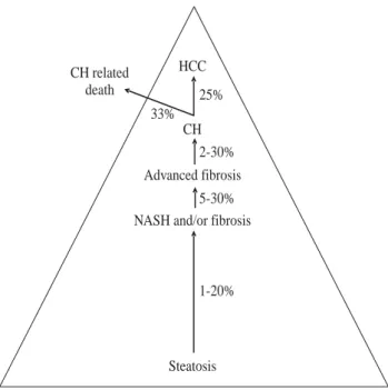
hepatocellular damage, inflammation and fibrogenesis which define a pathological entity called non-alcoholic steatohepatitis (NASH). The increasing number of pa-tients with NAFLD is expected to translate into increased prevalence of liver cirrhosis, and hepatocellular carcino-ma3 (Figure 1). Currently, histopathological analysis of
liver tissue represents the only mean to assess fibrosis in NAFLD4 and to disclose other histological finding
typi-cal of NASH. In the past decade, major efforts have been directed at identifying non-invasive methods for the as-sessment of liver fibrosis in different chronic liver diseas-es (CLD) including NAFLD. Several non-invasive ap-proaches — serum markers, transient elastography (i.e. liver stiffness measurement; LSM) and a re-visitation of classical imaging techniques — have been proposed as a replacement for, or to be used in combination with, histo-pathological analysis of liver biopsies.5 This article
pro-vides a concise overview on the non-invasive diagnostic methodologies proposed to differentiate simple fatty li-ver from possible fibrogenic evolution typical of NASH and to stage fibrosis in NASH. Given the considerable in-crease in the prevalence of overweight among children and adolescents in Western countries, NAFLD represents an emerging clinical problem affecting also a substantial proportion of these subjects (2.6 to 9.8%).6-9 Therefore, a
concise analysis of non-invasive approaches in the pedi-atric setting is also included.
Artemisa
www.medigraphic.com
Non-invasive methodologies: An overview
With the current epidemic of obesity, type 2 diabetes, and other abnormalities associated with the metabolic syn-drome, NAFLD has become the most frequent liver disease in Western countries, while its prevalence in the develop-ing world is increasdevelop-ing at a worrydevelop-ing pace. As already mentioned, NAFLD encompasses a spectrum of diseases ranging from simple steatosis with or without inflamma-tion, to a more severe entity, NASH that is associated with fibrosis and carries a significant risk to progress to cirrho-sis and its complications, including hepatocellular carci-noma (HCC).10 While simple steatosis will probably never
progress in the majority of patients,11 patients with NASH
and fibrosis require a close follow-up and enrolment in clinical trials for testing novel pharmacological approach-es, and may also have to undergo periodical screening for HCC. Thus, given the extremely high prevalence of this condition in the general population (up to 30% in West-ern countries)12 it is critical to define non-invasive
meth-ods that could allow to focus attention on those with the higher likelihood to progress.
It should be noted that fibrosis developing in the con-text of NASH shows several differences when compared to fibrosis developing in patients with chronic viral hep-atitis. First, fibrosis develops in a liver where hepato-cytes are laden with fat, an event associated with changes in the biochemical and biophysical properties of the tis-sue. Second, the pattern of fibrosis development in-volves predominantly zone 3, and leads to formation of
pericellular bundles (chicken-wire pattern). This aspect is also associated with the involvement of fibrogenic cells that are believed to be implicated in the process, with activated hepatic stellate cells playing a key role.13
The lack of a clear-cut biomarker that easily allows iden-tification of patients with NAFLD, together with the fact that metabolic abnormalities and cardiovascular disease may overshadow the hepatic disturbances and delay or prevent referral to a hepatologist, have delayed the rec-ognition of the factors associated with fibrosis. As a re-sult, sophisticated and extensively tested diagnostic al-gorithms have yet to be developed. Nonetheless, a num-ber of clinical studies that have cross-sectionally evaluated the patients with NASH and fibrosis have al-lowed the identification of factors associated with a greater risk of fibrosis.4,14-25 A list of the main clinical and
laboratory parameters related with advanced stages of disease is shown in Table I. In particular, the risk con-veyed by the clinical features of the metabolic syndrome indicates that the severity of the underlying pathophysi-ological abnormality has a significant impact on disease progression. Several factors, such as age, AST/ALT ratio, extracellular matrix proteins, and thrombocytopenia have been implicated in other types of liver diseases, in-cluding hepatitis C, indicating the role of matrix turnover and/or portal hypertension. Finally, the role of autoanti-bodies recognizing adducts with oxidative stress-related products recalls data previously described in alcoholic liver disease,26 while an increase in ferritin plasma levels
has been interpreted as a proxy of inflammatory activity rather than a marker of iron overload. Interestingly, age and insulin-resistance, that almost invariably emerge as risk factors in cross-sectional studies,14-16,19 appear to be
poorly correlated with fibrosis progression in longitudi-nal studies,4,18,26 reflecting the complexity of
understand-ing fibrosis dynamics in this disease.
A small number of studies have provided performance data for tests that identify fibrosis in patients with NAFLD (Table II). It is important to realize that only in few studies22,27-29 the results of the training set were
con-firmed in an independent, validation cohort. Moreover,
Abbreviations: NAFLD, non-acoholic fatty liver disease; NASH,
non-alcoholic steatohepatitis; CH, cirrhosis; HCC, hepatocellular carcinoma.
Figure 1. Natural course of NAFLD in 8-13 years (data from
re-ferences 1-4, 10, 12, 15, 18, 21, 26). Steatosis NASH and/or fibrosis
Advanced fibrosis CH HCC
1-20% 5-30% 2-30% 25% CH related
death
33%
Table I. Main predictive factors of advanced fibrosis in patients with
NASH (data from references 4, 14-25).
Clinically determinable factors Laboratory tests
• Older age • Low platelets count
• Gender • ALT, AST/ALT ratio
• Elevated BMI • Elevated ferritin levels
• Diabetes mellitus • Indexes of insulin resistance
• Visceral obesity (HOMA, QUICKI, OGIS)
• Metabolic syndrome • Elevated HA
• Systemic hypertension • Anti-MDA antibodies
Abbreviations: NASH, non-alcoholic steatohepatitis; BMI, body mass index; ALT,
www.medigraphic.com
interpretation of the available data is not always easy,particularly because these series often report on small numbers of patients. In addition, evaluation of fibrosis stage varies across studies. While the Brunt scoring sys-tem (or its recent modification)33 has been used more
of-ten, other studies have employed different systems, mak-ing comparisons difficult except for extreme stages. Ad-ditionally, the fact that performance of the tests varies based on the prevalence of the severity of fibrosis in the population tested makes difficult to extrapolate the re-sults in clinical practice. This is particularly important when considering that patients with NAFLD may be seen in settings (e.g. diabetes or obesity clinics) where the prevalence of advanced fibrosis is largely lower than in a Hepatology tertiary referral center.
It is interesting to note that the available non-invasive markers of fibrosis in NASH not only include single se-rum markers, or combination thereof, but algorithms that have been developed to accommodate clinical parame-ters, such as the presence or absence of diabetes.22,34 In
general, performance of these tests will allow identifying or excluding patients with severe fibrosis, although a sig-nificant proportion of the population is likely to fall in an undetermined area. Clearly, also in this case, a dynam-ic test that allows monitoring of changes in fibrosis or fi-brogenesis would greatly improve our ability to follow these patients.
Recently, transient elastography (TE) has been pro-posed also for the assessment of fibrosis in patients with different forms of chronic liver disease.5,35 TE is based on
a non-invasive medical device (Fibroscan®, Echosens
SA, Paris, France). This system has received a great
atten-tion in the past 5 years as a measurement of liver stiff-ness, which is considered a direct consequence of the fi-brotic evolution of CLD.36 The major limitation of this
technique in patients with NASH is represented by high prevalence of obesity, considering that a BMI ³ 28 is in-dependently associated with failure of TE examination.37
Moreover, the inter-observer agreement was found to be lower in the presence of moderate or severe steatosis.38
Yoneda et al39 recently reported that TE was successfully
used in 67 patients with NAFLD, demonstrating progres-sive increases in liver stiffness along the stages of fibro-sis, and excellent sensitivity and specificity in the iden-tification of patients with cirrhosis. An additional prob-lem that may arise in patients with NAFLD and metabolic syndrome is related to the possible presence of conges-tive heart failure that has been recently shown to influ-ence liver stiffness measurement.40
Although several tests (Table II) are sufficiently valid to identify patients with advanced fibrosis caused by NASH, a critical point is whether this is the most useful determination for a correct management of these patients. The sole recognition of bridging fibrosis and cirrhosis would overlook patients with lower degrees of fibrosis, which nonetheless are at risk to progress, especially if they are young. For this reason, a number of tests have been proposed to differentiate the presence of NASH from bland steatosis.41,42 Not surprisingly, some of the
biomarkers tested were similar to those used for the iden-tification of fibrosis.16,43 A combination of biochemical
markers was recently used to develop the NashTest, that was evaluated in a training set and a validation set.44
An-other interesting approach is represented by the
evalua-Table II. Serum markers of fibrosis in non-alcoholic fatty liver disease.
N. of Staging Fibrosis PPVa NPVa
Index pts system Factors stage Cut off (%) (%) AUC
BAAT15 93b METAVIR Age, BMI, serum ALT, triglycerides F ³ 2 vs £ 1 2 6 1 8 6 0.84
OELF27 61c Modified Age, HA, TIMP-1, PIIINP F ³ 3 vs £ 2 0.375 8 0 9 8 0.87
Scheuer 0.462 8 7 9 6
ELF plus simple 192c Kleiner HA, TIMP-1, PIIINP plus NFS F ³ 1 -5.002 8 6 6 6 0.84
clinical markers28 F ³ 2 -0.995 9 4 7 5 0.93
F ³ 3 -0.2826 7 7 9 9 0.98
HA 30 79 b Brunt HA F ³ 3 vs £ 2 46.1 5 1 9 6 0.92
NFS 22 480b Brunt Hyperglycemia, BMI, platelet F ³ 3 vs £ 2 <-1.455 5 6 9 3 0.88
253d count, albumin, AST:ALT, age > 0.676 9 0 8 5 0.82
NS39 112b Brunt Type IV collagen, HA F ³ 3 vs £ 2 Coll ³5 6 6 9 5 NS
or HA ³ 50
FT31,32 170b Brunt/ Total bilirubin, GGT, gender, age, F ³ 2 vs £ 1 0.30 5 4 9 0 0.81
97d Kleiner a2-macroglobulin, apolipoprotein 0.70 9 8 7 6
A1, haptoglobin F ³ 3 vs £ 2 0.30 7 1 9 8 0.88
0.70 9 7 8 9
BARD29 827b Kleiner Diabetes mellitus, BMI, ALT:AST F ³ 3 2-4 4 3 9 6 0.81
160d
a PPV and NPV vary based on the prevalence of the disease. b Training group. c Combination of training group and validation group. d Validation group.
Abbreviations: ALT, alanine aminotransferase; AST aspartate aminotransferase; GGT gamma-glutamyl transpeptidase; AUC, area under receiver operator curve; BAAT, BMI, ALT,
www.medigraphic.com
ESTE DOCUMENTO ES ELABORADO POR MEDI-GRAPHIC
tion of plasma caspase-3-generated cytokeratin-18 frag-ments, a biomarker of hepatocytes apoptosis.45 Levels of
cytokeratin-18 fragments were able to identify patients with NASH as compared to those with bland steatosis with remarkably high specificity and acceptable sensi-tivity. Larger studies are awaited to test the usefulness of this novel biomarker (for an extensive review see 41-42).
Other non-invasive test may be of use in patients with fatty liver. The SteatoTest, a derivation of FibroTest/Ac-tiTest46 has been proposed as a simple quantitative
esti-mate of liver steatosis. An algorithm based on body mass index (BMI), waist circumference, triglycerides and gam-ma-glutamyl transpeptidase was used to develop the fatty liver index47 that may be helpful in selecting subjects for
liver ultrasonography and lifestyle counseling. Finally, the ASH/NASH index (ANI) has been found to represent a useful tool for detecting alcohol abuse in patients with steatohepatis.48
The pediatric setting
Given the strong association of NAFLD with in-creased BMI and the considerable increase in the preva-lence of overweight among children and adolescents,49
NAFLD represents an emerging clinical problem affect-ing a substantial proportion of these subjects (2.6 to 9.8%),6,7 especially in the presence of obesity.8 Efforts in
identifying non-invasive methods for predicting fibrosis assume particular relevance in the pediatric setting, where the use of liver biopsy is perceived as bearing higher risks and is less acceptable than in adults.
Considering routine laboratory variables, the NASH Clinical Research Network recently50 failed to identify
tests with an adequate discriminating power to replace liver biopsy in evaluating NAFLD pattern and fibrosis se-verity in children and adolescents. However, other cross-sectional studies evaluating children with NASH and fi-brosis have allowed the identification of clinical and bio-chemical parameters associated with advanced stages of fibrosis in patients with NAFLD. Sartorio et al51 showed
that the Z-score of BMI (Z-BMI), ALT, uric acid, glucose during oral glucose tolerance test (OGTT), and insulin during OGTT are independent predictors of NAFLD in obese children, with most of the prediction explained by ALT and Z-BMI. Abdominal rather than generalized obe-sity contributes to liver fibrosis in children with NAFLD, and accordingly, waist circumference seems to be associ-ated with fibrosis. Therefore, the presence of abdominal obesity is an additional criterion for the selection of chil-dren and adolescents who should undergo extensive in-vestigation, including liver biopsy.52-53
We recently evaluated the diagnostic accuracy of TE54
and enhanced liver fibrosis test (ELF)55 in predicting
fi-brosis in a cohort of NAFLD pediatric patients. These au-thors demonstrated that TE is an accurate and reproduc-ible methodology to identify, in children and
adoles-cents affected by NAFLD, those without any degree of fi-brosis or significant fifi-brosis, or with advanced fifi-brosis. Similarly, the ELF test seems to predict fibrosis stages in pediatric NAFLD patients with a high degree of sensitivi-ty and specificisensitivi-ty, and, interestingly, the results were su-perior to those reported for adult patients with NAFLD.28
Information obtained through these methodologies may be relevant for identifying subjects with progressive fib-rogenic liver disease that require further histopathologi-cal analysis or therapeutic follow-up.
Conclusion
The large number of publications on non-invasive methodologies confirms the interest in, and need for, this type of innovation in the setting of NAFLD; however, the complexity of these surrogate non-invasive measures of disease progression need further investigation and guid-ance on their use and caution on results interpretation.
Some major considerations have arisen from the expe-rience accumulated so far. First, the majority of non-inva-sive methodologies have sufficient to excellent diagnos-tic accuracy for the detection (or exclusion) of advanced fibrosis and cirrhosis, but none allow a follow-up of the fibrogenic evolution of NAFLD in a stepwise fashion. In other words, due to the absence of a true gold standard, achieving 90% diagnostic accuracy remains a goal for the future. Therefore, non-invasive methodologies must be integrated with histology in indefinite cases and/or to confirm fibrotic evolution and discern other histological features of NASH.
Hopefully, new approaches employing high-through-put technologies including genomics, proteomics, me-tabolomics and glycomics may identify biomarkers that may help in categorizing patients, enhancing or replac-ing currently available non-invasive methodologies. Up to now, only single-center studies have employed these technologies in NAFLD.56,57 These types of studies must
be encouraged and clearly require a multicenter design in order to gather a large number of well-characterized cases and controls.
References
1 . Adams LA, Lymp JF, St. Sauver J, Sanderson SO, Lindor KD, Feldstein A, et al. The natural history of non-alcoholic fatty liver disease a population-based cohort study. Gastroenterology 2005; 129: 113-121.
2. Ekstedt M, Franzen LE, Mathiensen UI, Thorelius L, Holmqvist M, Bodemar G, et al. Long-term follow-up of patients with NAFLD and elevated liver enzymes. Hepatology 2006; 44: 865-873. 3. Bugianesi E, Leone N, Vanni E, Marchesini G, Brunello F, Carucci
P, et al. Expanding the natural history of non-alcoholic steatohepatitis: from cryptogenic cirrhosis to hepatocellular car-cinoma. Gastroenterology 2002; 123: 134-140.
www.medigraphic.com
5 . Pinzani M, Vizzutti F, Arena U, Marra F. Technology Insight: non-invasive assessment of liver fibrosis by biochemical scores and elastography. Nature Clinical Practice Gastroenterology &
Hepatology 2008; 5: 95-106.
6 . Tominaga K, Kurata JH, Chen YK, Fujimoto E, Miyagawa S, Abe I, et al. Prevalence of fatty liver in Japanese children and relationship to obesity. An epidemiological ultrasonographic sur-vey. Dig Dis Sci 1995; 40: 2002-2009.
7. Schwimmer JB, Deutsch R, Kahen T, Lavine JE, Stanley C, Behling C. Prevalence of fatty liver in children and adolescents.
Pediat-rics 2006; 118: 1388-1393.
8 . Chan DF, Li AM, Chu WC, Chan MH, Wong EM, Liu EK, et al. Hepatic steatosis in obese Chinese children. Int J Obes Relat
Metab Disord 2004; 28: 1257-1263.
9 . Kinugasa A, Tsunamoto K, Furukawa N, Sawada T, Kusunoki T, Shimada N. Fatty liver and its fibrous changes found in simple obesity of children. J Pediatr Gatroenterol Nutr 1984; 3: 408-414. 10. Angulo P. Nonalcoholic fatty liver disease. N Engl J Med 2002;
346: 1221-1231.
11. Teli MR, James OF, Burt AD, Bennett MK, Day CP. The natural history of nonalcoholic fatty liver: a follow-up study. Hepatology 1995; 22: 1714-1719.
12. McCullogh AJ. The epidemiology and risk factors of NASH. In: Farell GC, George J, De la MH, and McCollough AJ, eds. Fatty
liver disease: NASH and related disorders. Malden: MA: Blackwell
Publishing, 2005: 23-37.
13. Cassiman D, Libbrecht L, Desmet V, Denef C, Roskams T. He-patic stellate cell/myofibroblast subpopulations in fibrotic hu-man and rat livers. J Hepatol 2002; 36: 200-209.
14. Angulo P, Keach JC, Batts KP, Lindor KD. Independent predic-tors of liver fibrosis in patients with nonalcoholic steatohepatitis.
Hepatology 1999; 30: 1356–1362.
15. Ratziu V, Giral P, Charlotte F, Bruckert E, Thibault V, Theodorou I, et al. Liver fibrosis in overweight patients. Gastroenterology 2000; 118: 1117–1123.
16. Dixon JB, Bhathal PS, O’Brien PE. Nonalcoholic fatty liver disease: predictors of nonalcoholic steatohepatitis and liver fibrosis in the severely obese. Gastroenterology 2001; 121: 91–100.
17. Matteoni CA, Younossi ZM, Gramlich T, Boparai N, Liu YC, McCullough AJ. Nonalcoholic fatty liver disease: a spectrum of clinical and pathological severity. Gastroenterology 1999; 116: 1413-1419.
18. Fassio E, Álvarez E, Domínguez N, Landeira G, Longo C. Natu-ral history of non-alcoholic steatohepatitis: a longitudinal study of repeat liver biopsies. Hepatology 2004; 40: 820–826. 19. Albano E, Mottaran E, Vidali M, Reale E, Saksena S, Occhino G,
et al. Immune response towards lipid peroxidation products as a predictor of progression of non-alcoholic fatty liver disease to advanced fibrosis. Gut 2005; 54: 987–993.
20. Bugianesi E, Manzini P, D’Antico S, Vanni E, Longo F, Leone N, et al. Relative contribution of iron burden, HFE mutations, and insulin resistance to fibrosis in nonalcoholic fatty liver.
Hepatology 2004; 39: 179–187.
21. Marchesini G, Bugianesi E, Forlani G, Cerrelli F, Lenzi M, Manini R, et al. Nonalcoholic fatty liver, steatohepatitis, and the meta-bolic syndrome. Hepatology 2003; 37: 917–923.
22. Angulo P, Hui JM, Marchesini G, Bugianesi E, George J, Farrell GC, et al. The NAFLD fibrosis score: a non-invasive system that identifies liver fibrosis in patients with NAFLD. Hepatology 2007; 45: 846–854.
23. Tahan V, Canbakan B, Balci H, Dane F, Akin H, Can G, et al. Serum gamma glutamyltranspeptidase distinguishes non-alco-holic fatty liver disease at high risk. Hepatogastroenterology 2008; 55: 1433-1438.
24. Rodríguez-Hernández H, González JL, Márquez-Ramírez MD, Flores-Hernández M, Rodríguez-Morán M, Guerrero-Romero F. Risk factors associated with non-alcoholic fatty liver disease and its relationship with the hepatic histological changes. Eur J
Gastroenterol Hepatol 2008; 20: 399-403.
25. Miyaaki H, Ichikawa T, Nakao K, Yatsuhashi H, Furukawa R, Ohba K, et al. Clinicopathological study of nonalcoholic fatty liver disease in Japan: the risk factors for fibrosis. Liver Int 2008; 28: 519-524.
26. Harrison SA, Di Bisceglie AM. Advances in the understanding and treatment of nonalcoholic fatty liver disease. Drugs 2003; 63: 2379-2394.
27. Rosenberg WM, Voelker M, Thiel R, Becka M, Burt A, Schuppan D, et al. Serum markers detect the presence of liver fibrosis: a cohort study. Gastroenterology 2004; 127: 1704–1713. 28. Guha IN, Parkes P, Roderick P, Chattopadhyay D, Cross R,
Har-ris S, et al. Non-invasive Markers of Fibrosis in Nonalcoholic Fatty Liver Disease: Validating the European Liver Fibrosis Panel and Exploring Simple Markers. Hepatology 2008; 47: 455-460. 29. Harrison SA, Oliver D, Arnold HL, Gogia S, Neuschwander-Tetri BA. Development and validation of a simple NAFLD clini-cal scoring system for identifying patients without advanced dis-ease. Gut 2008; 57: 1441–1447.
30. Suzuki A, Angulo P, Lymp J, Li D, Satomura S, Lindor K. Hyaluronic acid, an accurate serum marker for severe hepatic fibrosis in patients with non-alcoholic fatty liver disease. Liver Int 2005; 25: 779-786.
31. Ratziu V, Massard J, Charlotte F, Messous D, Imbert-Bismut F, Bonyhay L, et al. Diagnostic value of biochemical markers (FibroTest-FibroSURE) for the prediction of liver fibrosis in pa-tients with non-alcoholic fatty liver disease. BMC Gastroenterol 2006; 6: 6.
32. Jacqueminet S, Lebray P, Morra R, Munteanu M, Devers L, Messous D, et al. Screening for liver fibrosis by using a noninvasive biomarker in patients with diabetes. Clin Gastroenterol Hepatol 2008; 6: 828-831.
33. Kleiner DE, Brunt EM, Van Natta M, Behling C, Contos MJ, Cummings OW, et al. Nonalcoholic Steatohepatitis Clinical Re-search Network. Design and validation of a histological scoring system for nonalcoholic fatty liver disease. Hepatology 2005; 41: 1313-1321.
34. Wong VW, Wong GL, Chim AM, Tse AM, Tsang SW, Hui AY, et al. Validation of the NAFLD fibrosis score in a Chinese popu-lation with low prevalence of advanced fibrosis. Am J
Gastroenterol 2008; 103: 1682-1688.
35. Rockey DC. Noninvasive assessment of liver fibrosis and portal hypertension with transient elastography. Gastroenterology 2008; 134: 8-14.
36. Sandrin L, Tanter M, Gennisson JL, Catheline S, Fink M. Shear elasticity probe for soft tissues with 1-D transient elastography.
IEEE Trans Ultrason Ferroelectr Freq Control 2002; 49: 436–
446.
37. Foucher J, Chanteloup E, Vergniol J, Castéra L, Le Bail B, Adhoute X, et al. Diagnosis of cirrhosis by transient elastography (Fibroscan): a prospective study. Gut 2006; 55: 403–408. 38. Fraquelli M, Rigamonti C, Casazza G, Conte D, Donato MF,
Ronchi G, et al. Reproducibility of transient elastography in the evaluation of liver fibrosis in patients with chronic liver disease.
Gut 2007; 56: 968–973.
39. Yoneda M, Yoneda M, Mawatari H, Fujita K, Endo H, Iida H, et al. Noninvasive assessment of liver fibrosis by measurement of stiffness in patients with non-alcoholic fatty liver disease (NAFLD). Dig Liver Dis 2008; 40: 371-378.
40. Lebray P, Varnous S, Charlotte F, Varaut A, Poynard T, Ratziu V. Liver stiffness is an unreliable marker of liver fibrosis in patients with cardiac insufficiency. Hepatology 2008; 48: 2089. 41. Wieckowska A, Feldstein AE. Diagnosis of non-alcoholic fatty liver disease: invasive versus non-invasive. Semin Liver Dis 2008; 28: 386-395.
42. Torres DM, Harrison SA. Diagnosis and therapy of nonalcoholic steatohepatitis. Gastroenterology 2008; 134: 1682-1698. 43. Sakugawa H, Nakayoshi T, Kobashigawa K, Yamashiro T,
www.medigraphic.com
44. Poynard T, Ratziu V, Charlotte F, Messous D, Munteanu M, Imbert-Bismut F, et al. Diagnostic value of biochemical markers (NashTest) for the prediction of non alcoholic steatohepatitis in patients with non-alcoholic fatty liver disease. BMC Gastroenterol 2006; 6: 34.
45. Wieckowska A, Zein NN, Yerian LM, Lopez AR, McCullough AJ, Feldstein AE. In vivo assessment of liver cell apoptosis as a novel biomarker of disease severity in nonalcoholic fatty liver disease. Hepatology 2006; 44: 27–33.
46. Poynard T, Ratziu V, Naveau S, Thabut D, Charlotte F, Messous D, et al. The diagnostic value of biomarkers (SteatoTest) for the prediction of liver steatosis. Comp Hepatol 2005; 23(4): 10. 47. Bedogni G, Bellentani S, Miglioli L, Masutti F, Passalacqua M,
Castiglione A, et al. The Fatty Liver Index: a simple and accurate predictor of hepatic steatosis in the general population. BMC
Gastroenterol 2006; 6: 33.
48. Dunn W, Angulo P, Sanderson S, Jamil LH, Stadheim L, Rosen C, et al. Utility of a new model to diagnose an alcohol basis for steatohepatitis. Gastroenterology 2006; 131: 1057–1063. 49. Hedley AA, Ogden CL, Johnson CL, Carroll MD, Curtin LR, Flegal
KM. Prevalence of overweight and obesity among US children, adolescents, and adults, 1999-2002. JAMA 2004; 291: 2847-2850. 50. Patton HM, Lavine JE, Van Natta ML, Schwimmer JB, Kleiner D, Molleston J; Nonalcoholic Steatohepatitis Clinical Research Net-work. Clinical correlates of histopathology in pediatric nonalco-holic steatohepatitis. Gastroenterology 2008; 135: 1961-1971.
51. Sartorio A, Del Col A, Agosti F, Mazzilli G, Bellentani S, Tiribelli C, et al. Predictors of non-alcoholic fatty liver disease in obese children. Eur J Clin Nutr 2007; 61: 877-883.
52. Manco M, Bedogni G, Marcellini M, Devito R, Ciampalini P, Sartorelli MR, et al. Waist circumference correlates with liver fibrosis in children with non-alcoholic steatohepatitis. Gut 2008; 57: 1283-1287.
53. Iacobellis A, Marcellini M, Andriulli A, Perri F, Leandro G, Devito R, et al. Non invasive evaluation of liver fibrosis in paedi-atric patients with nonalcoholic steatohepatitis. World J
Gastroenterol 2006; 12: 7821-7825.
54. Nobili V, Vizzutti F, Arena U, Abraldes JG, Marra F, Pietrobattista A, et al. Accuracy and reproducibility of transient elastography for the diagnosis of fibrosis in pediatric nonalcoholic steatohepatitis. Hepatology 2008; 48: 442-448.
55. Nobili V, Parkes J, Bottazzo G, Marcellini M, Cross R, Newman D, et al. Performance of ELF Serum Markers in Predicting Fibro-sis Stage in Pediatric Non-Alcoholic Fatty Liver Disease.
Gastro-enterology 2009; 136: 160-167.
56. Day CP. Genes or environment to determine alcoholic liver dis-ease and non-alcoholic fatty liver disdis-ease. Liver Int 2006; 26: 1021-1028.
57. Huang H, Shiffman ML, Cheung RC, Layden TJ, Friedman S, Abar OT, et al. Identification of two gene variants associated with risk of advanced fibrosis in patients with chronic hepatitis C.