Promotor methylation: Does it affect
response to therapy in chronic hepatitis C (G4) or fibrosis?
Abdel-Rahman N. Zekri,* Ahmed M. Raafat,* Suzan Elmasry,***Abeer A. Bahnassy,* Yasmin Saad,** Hamed A. Dabaon,**** Mohamed El-Kassas,** Hend I. Shousha,** Auhood A. Nassar,* Mohamed Ale EL-Dosouky,*** Nehal Hussein*
* Molecular Virology and Immunology Unit, Cancer Biology Department, National Cancer Institute, Cairo University, Cairo, Egypt. ** Department of Endemic Medicine and Hepatology, Faculty of Medicine, Cairo University, Cairo, Egypt.
*** Biochemistry Department, Faculty of Science, Cairo University, Cairo, Egypt. **** Organic Chemistry Department, Faculty of Science, Cairo University, Cairo, Egypt.
ABSTRACT
Background and aim. DNA methylation plays a critical role in the control of important cellular processes. The present study
assessed the impact of promoter methylation (PM) of some genes on the antiviral response to antiviral therapy and it’s rela-tion to the presence of fibrosis in HCV-4 infected patients from Egypt. Material and methods. Clinical, laboratory and histo-pathological data of 53 HCV-4 infected patients who were subjected to combined antiviral therapy were collected; patients were classified according to their response to treatment and the fibrosis status. The methylation profiles of the studied groups were determined using the following genes: APC, P14ARF, P73, DAPK, RASSF1A, and O6MGMT in patients’ plasma.
Re-sults. O6MGMT and P73 showed the highest methylation frequencies (64.2 and 50.9%) while P14 showed the lowest frequency
(34%). Sustained virological response (SVR) was 54.7%with no significant difference in clinico-pathological or laboratory fea-tures between the studied groups. PM of O6MGM was significantly higher in non-responders (p value 0.045) while DAPK showed high methylation levels in responders with no significance (p value: 0.09) andPM of RASSF1A was significantly related to mild fibrosis (p value: 0.019). No significant relations were reported between PM of any of the studied genes and patients’ features. Conclusion. PM of some Tumor Suppressor genes increases in chronic active HCV-4. However, only 06MGMT can be used as a predictor of antiviral response and RASSF1A as a marker of marked fibrosis in this small set of patients. An exten-ded study, including more patients is required to validate the results of this preliminary study.
Key words. HCV. Antiviral response. Promoter methylation.
Correspondence and reprint request: Abdel-Rahman N. Zekri M.Sc., Ph.D.
Virology and Immunology Unit, Cancer Biology Department, National Cancer Institute, Cairo University.
Kasr Al-Aini st., Fom El-Khaleg . Cairo, Egypt 11976 Tel.: +20101413521, Fax: +20223644720 E-mail: [email protected]
Manuscript received: December 23, 2013. Manuscript accepted: May 22, 2014.
INTRODUCTION
Hepatitis C virus (HCV) is the cause of a signifi-cant proportion of cases of chronic liver disease, hepatocellular carcinoma (HCC) and deaths from liver disease. Egypt has the highest prevalence of HCV worldwide (15%) and the highest prevalence of HCV-4, which accounts for almost 90% of the cases.1 Moreover a consistent increase of
seroposi-tivity for HCV antibodies with age was observed, with a peak level of 54.9% in all individuals for the age group 45-49 years.2 The goal of treatment is to
prevent complications of HCV infection, which is mainly achieved by elimination of the virus,
predict-ed by a sustainpredict-ed virological response (SVR).3 The
main predictors of SVR are the IL28B (IFN k3) pol-ymorphism, the HCV genotype, and the stage of fi-brosis. Other predictors of response include baseline HCV RNA levels, the dose and duration of therapy, host factors e.g. body mass index, age, insulin resistance, gender, and the characteristics of liver disease e.g. the levels of alanine aminotransferase (ALT), gamma glutamyltransferase (GGT), and the stage of fibrosis or co-infection with HIV or other hepatotropic virus.4
Epigenetic changes, including promoter methyla-tion (PM) of several genes play a critical role in the control of cellular processes through switching genes on or off leading to differential expression of the genes, which determines the expression of pro-teins.5 Some studies have shown that the presence
of hepatitis viruses, especially HCV, could play a role in accelerating the methylation process which is involved in HCC development, potentiate the pro-gression of HCV related liver disease and affect its response to treatment.6
p15, p16, and E-cadherin in tumor tissues and plas-ma obtained from 28 HCC patients from Egypt. The Promotor methylation frequency (PMF) ranged from 67.9% for p16 to 89.2% for p15 with a high concordance rate between plasma and tissue sam-ples.7 In another study, we found that PM of a
group of genes increases with disease progression from CH to HCC.8 In this study the PMF of p14,
p73, RASSF1A, CDH1 and O6MGMT was signifi-cantly higher in HCC and their ANT whereas PMF of APC was higher in CH and we were able to sug-gest a panel of 4 genes (APC, p73, p14, O6MGMT) that can independently classify cases into HCC and CH with high sensitivity and specificity.9 Then, it
may be valuable to assess whether PM of our previ-ously tested genes contribute to fibrogenesis and response to antiviral treatment in chronic HCV-4 related liver disease.
Therefore the current study was conducted to clarify the contribution of PM to: the development of fibrosis and the response to antiviral therapy using some genes that proved to be significant in our pre-viously studies (APC, P14ARF, P73, DAPK, RASSF1A and MGMT).
MATERIAL AND METHODS
Population samples
The study was conducted on 53 consecutive chronic HCV-4 patients from Egypt who were eligi-ble for treatment with pegylated interferon α and ribavirin. All patients fulfilled the standards of care, inclusion and exclusion criteria for interferon thera-py, which are applied on the national wide program controlled by the National Committee for Treatment of Viral Hepatitis. Informed consents were obtained from all the participants enrolled in the study, which was performed in accordance with the decla-ration of Helsinki, local and national laws (clinical trial NCT01758939).
For all patients height and weight were deter-mined at baseline and body mass index (BMI) was calculated (weight in kilograms divided by height in meters squared). Laboratory investigations includ-ing complete liver profile, kidney function, Alfa-Fe-toprotein (AFP), INR, and CBC were also done to justify suitability for therapy. HCV RNA was quan-tified in all patients’ sera using quantitative real time PCR at baseline, after 12, 24, 48 and 72 week of anti-viral therapy. Histological examination was done on core needle biopsies to determine the grade of necro-inflammation and the stage of fibrosis
ac-cording to the Metavir scoring system prior to treatment. Clinical and laboratory follow up were done for every patient to report any possible adverse side effects and the response to treatment according to IFN treatment guidelines.
Detection of promoter methylation
High molecular weight DNA was extracted from patient’s plasma samples collected before treat-ment, according to our previously published pro-tocol.7 Briefly, an equal volume of equilibrated
phenol (pH 7.0-7.5) was added to samples and vor-texed. The upper aqueous layer was removed and an equal volume of phenol/chloroform (1:1) was added and vortexed. The upper aqueous layer was removed again and an equal volume of chloro-form/isoamyl alcohol (24:1) was added and vor-texed. This was followed by the addition of 3 M Sodium acetate (pH 4.7-5.2), DNA precipitation by ice-cold ethanol and overnight incubation at -80 °C. The fluid was decanted and the DNA pellet was dissolved in sterile water.7
The extracted DNA was subjected to bisulfite treatment using EZ DNA methylation kit which uses 300 ng of the extracted Nucleic acid. This was followed by MSP using the primer sequences and the methylation- specific PCR conditions illus-trated in table 1. DNA methylation of CpG islands for p14, p73, APC, DAPK, RASSF1A and O6MGMT genes was determined using specific primers for methylated (M) and unmethylated (UM) DNA as previously described by.8 Negative
control samples (without DNA) were included in each PCR set. PCR products were analyzed on 4% ethidium bromide-stained agarose gel and visual-ized under ultraviolet illumination. The methyla-tion index (MI), defined as the ratio between the number of methylated genes and the total number of the studied genes for each sample was calculat-ed for all patients.8
Statistical Analysis
This was done using Statistical Package for So-cial Sciences, Version 17.0 (SPSS, Inc., Chicago, III., USA) for Windows. Continuous variables were analyzed as mean values ± standard devia-tion (SD) or median (range) as appropriate. Per-centages were calculated for categorical data. For categorical variables, differences were analyzed with χ2 (chi square) tests and Fisher’s exact test
variables with normal distribution were analyzed by Student’s T-test; comparison between three groups was done using Kruskelswallis test (non parametric analogue for ANOVA). P value of
≤ 0.05 was considered statistically significant.
RESULTS
Clinical and epidemiological data: Baseline de-mographic and laboratory features of all patients enrolled in the study in addition to the stage of fi-brosis are illustrated in table 2. Out of the 53 pa-tients assessed, 47 (89%) were males and 6 (11%) were females. Their ages ranged from 35 to 45 with a mean of 39.2. Forty patients (75.5%) showed mild to moderate fibrosis (F1/F2) and 13 (24.5%) showed marked fibrosis.
Out of 53 patients studied, 29 showed SVR (54.7%) and 24 showed either no response or relapse (45.3%). None of patients was excluded from the treatment due to emergence of any side effects and no patient received < 80% of the thera-peutic schedule. The demographic, laboratory and histopathological parameters of those patients are illustrated in table 3. No significant difference was observed between the two groups (responders and non-responders) regarding the age and sex, the
hematological parameters, liver profile, HCV viral load or different fibrosis stages.
Promotor methylation frequency (PMF)
The PMFs of all studied genes are illustrated in table 4. The 06MGMT showed the highest PMF (64.2%) followed by P73 (50.9%) and APC (49.1%) whereas P14 showed the lowest PMF (34%). Out of
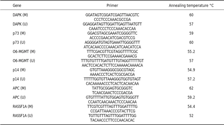
Table 1. Primers sequences and conditions of the methylation specific PCR (MSP).
Gene Primer Annealing temperature °C
DAPK (M) GGATAGTCGGATCGAGTTAACGTC 60
CCCTCCCAAACGCCGA
DAPK (U) GGAGGATAGTTGGATTGAGTTAATGTT 57
CAAATCCCTCCCAAACACCAA
p73 (M) GGACGTAGCGAAATCGGGGTTC 59
ACCCCGAACATCGACGTCCG
p73 (U) AGGGGATGTAGTGAAATTGGGGTTT 60
ATCACAACCCCAAACATCAACATCCA
O6-MGMT (M) TTTCGACGTTCGTAGGTTTTCGC 55.2
GCACTCTTCCGAAAACGAAACG
O6-MGMT (U) TTTGTGTTTTGATGTTTGTAGGTTTTTGT 57
AACTCCACACTCTTCCAAAAACAAAACA
p14 (M) GTGTTAAAGGGCGGCGTAGC 54.9
AAAACCCTCACTCGCGACGA
p14 (U) TTTTTGGTGTTAAAGGGTGGTGTAGT 57.2
CACAAAAACCCTCACTCACAACAA
APC (M) TATTGCGGAGTGCGGGTC 62
TCAACGAACTCCCGACGA
APC (U) GTGTTTTATTGTGGAGTGTGGGTT 59.2
CCAATCAACAAACTCCCAACAA
RASSF1A (M) TTCGTCGTTTAGTTTGGATTTTG 54.4
CCGATTAAACCCGTACTTCG
RASSF1A (U) TGTTGTTTAGTTTGGATTTTGG 52
TACAACCCTTCCCAACACAC
Table 2. Clinico-pathological features of the studied patients.
Variables Patients n = 53
Age (years) 39.2 ± 8.9
Sex M/F 47/6
BMI27.2 ± 4.2
Platelets/mm3 197.7 ± 64.8 Total bilirubin mg/dl 0.8 ± 0.3
ALT IU/L 69.6 ± 44.2
AFP ng/ml 7.2 ± 10.4
HCV viral load IU/ml 371.785 ± 762.2 Fibrosis as n (%):
Mild to moderate (F1& F2) 40 (75.5%) Marked (F3& F4) 13 (24.5%)
the six genes assessed for PM, only RASSF1Agene methylation was significantly related to the pres-ence of mild fibrosis (p value: 0.019, and 06MGMT was significantly correlated with patient’s response to therapy (p value: 0.045). On the other hand, PM of DAPK was higher in responders than in non re-sponders, however, the difference between the two groups did not reach a statistically insignificant lev-el (p value: 0.097) (Tables 5 and 6)(Figure 1).
Promotor methylation index (PMI)
There is no significant difference between IFN responders and IFN non-responders regarding the methylation index (2.76 ± 1.4 and 2.83 ± 1.46; p = 0.851) respectively as well as no significant dif-ference in methylation index between mild fibrosis (F1 and F2) and marked fibrosis (F3 and F4) (Table 5).
DISCUSSION
Aberrant methylation in the promoter regions of tumor suppressor genes (TSGs) is a crucial
epige-netic alterations that contribute to deregulation of many cellular processes leading finally to the initia-tion and progression of human cancers.9,10 Several
studies revealed that different types of cancer, in-cluding HCC, show distinct DNA methylation pro-files; suggesting the existence of cancer- type specific methylation signatures.11,12 Other studies have
men-tioned a possible HCV- induced HCC methylation profile.6,8 Such epigenetic defects have also been
ob-served in non-cancerous liver tissues of HCC pa-tients, which are usually show evidence of chronic inflammation.13,14
Though Egypt has the highest prevalence of HCV infection in the world, DNA methylation profiles for HCV has not been well studied yet and there are only few studies in this context. In an early case control study by our group,7 we were able to detect a
high frequency of APC, FHIT, p15, p16 and E-cad-herin-PM (range 67.9-89.2%) in the plasma and tis-sues of 28 chronic HCV and/or HBV- associated
Table 3. Clinico-pathological features of the responder and non- responder patients
Variables Responders (n = 29) Non-responders (n = 24) p-value
Age (years) 38.6 ± 8.8 39.8 ± 9.0 0.646
Sex M/F 26/3 21/3 1
BMI26.3±3.8 28.3 ± 4.5 0.081
Platelets/mm3 188.1± 63.6 209.3 ± 65.7 0.241
ALT IU/L 64.4 ± 47.4 75.9 ± 40.2 0.231
AFP ng/mL 5.3 ± 7.4 9.5 ± 12.9 0.15
HCV viral load IU/mL 500.000 ± 24.8 30.000 ± 300 0.368
Fibrosis as n (%):
Mild to moderate (F1& F2) 23 (79.3%) 17 (70.8%) 0.264
Marked (F3& F4) 6(20.7%) 7 (29.2%)
Data are represented by Mean ± SD. P-value > 0.05 is not significant. AFP: α-fetoprotein. ALT: alanine aminotransferase. BMI: body mass index. F: female. HCV: hepatitis C virus. M: male.
Table 4. Promotor Methylation Frequency of in the studied
genes.
Studied genes Methylated Un-methylateds
genes gene
n (%) n (%)
O6MGMT 34 (64.2) 19 (35.8)
APC 26 (49.1) 27 (50.9)
RASSF1A 22 (41.5) 31 (58.5)
DAP-kinase 22 (41.5) 31 (58.5)
P73 27 (50.9) 26 (49.1)
P14 18 (34) 35 (66)
Numbers are represented as n (%).
Figure 1. The significant relation between the methylation status of RASSF1A and fibrosis stages (p value: 0.019).
1 2 3 4
Fibrosis
Percent of positive cases
100 90 80 70 60 50 40 30 20 10 0
HCC patients, with a high concordance for all stud-ied genes. However, no significant association was found, in this study, between the methylation status of any gene and the presence of hepatitis virus infec-tion. This was partially attributed to the small sam-ple size in this study. Then, we assessed the contribution of methylation status to the develop-ment and progression of HCV- associated HCC and CH in Egyptian patients using a specific panel of genes (APC, FHIT, p15, p73, p14, p16, DAPK1, CDH1, RARb, RASSF1A, O6MGMT).8 We found that
HCV infection may contribute to hepatocarcinogene-sis through enhancing PM of certain genes. A panel of 4 genes (APC, p73, p14, O6MGMT) out of 11 test-ed genes successfully classifitest-ed cases into HCC or CH with high accuracy (89.9%), sensitivity (83.9%) and specificity (94.7%).
A more extended confirmatory study, including 516 Egyptian patients with HCV-related liver disease (208 HCC, 108 liver cirrhosis, 100 CHC and 100 con-trols), was then performed to detect PM of P14, P15, P73 and Mismatch repair gene (O6MGMT) in pa-tient’s plasma by using EpiTect Methyl qPCR Array technology.15 This study provided evidence that PM
of the studied genes is an early event in hepatocar-cinogenesis and showed specific DNA methylation
signatures associated with the potential clinical ap-plications in diagnosis and prognosis of HCC.15
The current study was then conducted to determine the impact of PM of our specified panel of genes on the degree of fibrosis in chronic HCV infected patients and their response to combined antiviral therapy.
Our results regarding the correlation between PM of the tested genes and patients’ response to an-tiviral therapy differ from previously published data in Western countries or USA. We found that only O6MGMT PM significantly affected patients’ response to antiviral therapy (p value, 0.045), being significantly higher in non-responders than in responders. This is explainable since some previous studies have shown that; 06MGM plays an impor-tant role in cytoprotection through preventing DNA damage and triggering DNA repair mechanisms. Therefore, PM of 06MGM is frequently detected in chronic hepatitis patients.16
On the other hand, PM of DAPK showed a tenden-cy to affect patients’ response to treatment though this did not reach a statistically significant level (p = 0.097). This could be attributed, at least partially, to the small sample size. None of the other tested genes was significantly associated with response to antiviral treatment.
Table 5. The methylation status of the studied group in relation to IFN response.
Methylated gene Responders Non-Responders p-value
N = 29 N = 24
O 6MGMT 15 (51.7%) 19 (79.2%) 0.045*
APC 13 (44.8%) 13 (54.2%) 0.473
RASSF1A 11 (37.9%) 11 (45.8%) 0.540
DAP-kinase 14 (48.3%) 8 (33.3%) 0.097
P73 12 (41.4%) 15 (62.5%) 0.328
P14 9 (31%) 9 (37.5%) 0.930
Methylation Index 2.76 ± 1.41 2.83 ± 1.46 0.851
* P-value > 0.05 is not significant.
Table 6. Correlation between promoter methylation of the studied genes and degree of fibrosis.
Methylated gene Mild Fibrosis (F1&F2) Marked fibrosis (F3&F4) p-value
n = 40 n = 13
O6MGMT 26 (65.0%) 8 (61.5%) 0.543
APC 20 (50.0%) 6 (46.2%) 0.687
RASSF1A 20 (50.0%) 2 (15.4%) 0.019*
DAP-kinase 18 (45.0%) 4 (30.8%) 0.366
P73 21 (52.5%) 6 (46.2%) 0.691
P14 15 (37.5%) 3 (23.1%) 0.340
In the current study 06MGMT has the highest pretreatment PM frequency among HCV infected pa-tients (64.2%) followed by P73 (50.9%), APC (49.1%), RASSF1A/DAP-kinase (41.5%) and P14 (34%). Our data in this regard is different from what has been previously reported by our group in chronic HCV and/or HBV-associated HCC)8 or by
Gioia, et al.17 These two studies showed that PM of
the RASSF1A gene was the most frequently detected with progression from regenerative conditions to cirrhosis. The variability in results of different stud-ies regarding the frequency of PM of the assessed genes could be attributed to several factors includ-ing: the differences in the CpG sites tested, environ-mental factors, HCV genotype present as well as geographical and racial differences. However, some previous reports have also shown significant associ-ation between O6MGMT-PM and HCV infection in-cluding the study of Matsukura, et al.18
The p73 is the second most frequently methylated gene in our tested group. Data regarding PM of the p73 gene in chronic active hepatitis patients are still immature and the few available reports in literature show a low PM frequency in CH patients.19 This
contradicts with our results since PM of the p73 was detected in 50.9% of the studied patients.
Data regarding PM of the APC gene varied signif-icantly in different studies. In the current study, APC-PM was relatively high in CH patients (49.1%), confirming the data obtained from our previous study on the Egyptian population.8 However,
Nomoto, et al.20 reported a much lower frequency of
APC- PM (21.6%) in CH patients with cirrhosis. One possible explanation for the difference between the results of the two studies could be attributed either to different HCV genotypes, the presence of fibrosis and/or environmental factors in Egyptian population.
Within our studied panel of genes, p14 showed the lowest frequency of PM among HCV patients (34%). Similar findings were reported by Anzola,21
who showed that p14 PM was associated with the pathogenesis of HCC and suggested that inactiva-tion of p14 through PM could be an important mechanism for HCV-induced HCC.8,21 To the best of
our knowledge, data regarding the impact of PM on the response to antiviral therapy in HCV-associated CH patients are still preliminary. Thus, further studies including larger number of patients are still needed to evaluate and validate the already available small studies including ours.
Only few studies have addressed the association between gene methylation status and the
develop-ment of fibrosis in hepatitis patients. Murphy, et al.22 confirmed in their study the implication of
methylation status of some genes in the progression of mild NAFLD (Non-alcoholic fatty liver disease) into advanced NAFLD, steato-hepatitis, fibrosis and carcinogenesis. Their study confirmed the presence of functionally relevant differences in the methyla-tion patterns, which can distinguish a mild from an advanced disease. However no similar studies have been done in chronic hepatitis patients, except for the current study, which shows that only PM of the RASSF1A gene was significantly associated with mild fibrosis in the studied patients (p = 0.0.019). This provides an evidence for the role of an intact RASSF1A gene in the induction of fibrogenesis in chronic HCV patients.
We conclude that, promoter methylation of some genes increases in HCV genotype 4-associated chronic active hepatitis. However, only O6MGMT methylation can significantly affect patients’ re-sponse to antiviral treatment, whereas RASSF1A is involved in the regulating the process of fibrogenesis and therefore, it could be helpful in predicting the stage of fibrosis or in the differentiation between mild and marked fibrosis in those patients.
ACKNOWLEDGMENT
We would like to thank Prof. Waleed M. Sief, professor of Virology and Immunology, Cancer Biol-ogy Department, National Cancer Institute for help-ing in statistical analyses of the data.
ABBREVIATIONS
• AFP: Alfa-Fetoprotein.
• ALT: Alanine aminotransferase. • BMI: body mass index.
• GGT: gamma glutamyltransferase. • HCC: hepatocellular carcinoma. • HCV: hepatitis C virus.
• HCVG4: hepatitis C virus genotype 4. • M: methylated DNA.
• MI: methylation index.
• PMF: The Promotor methylation frequency. • SVR: Sustained virological response. • TSCs: tumor suppressor genes. • UM: unmethylated DNA.
REFERENCES
2. Elkady A, Tanaka Y, Kurbanov F, Sugauchi F, Sugiyama F, Khan M, Sayed A, et al. Genetic Variability of Hepatitis C Virus in South Egypt and its Possible Clinical Implication. J Med Virol 2009; 1023: 1015-23.
3. Strader DB, Wright T, Thomas DL, Seeff LB. Diagnosis, management, and treatment of hepatitis C. Hepatology (Baltimore, Md) 2004; 39: 1147-71.
4. Manns MP, Wedemeyer H, Cornberg M. Treating viral he-patitis C: efficacy, side effects, and complications. Gut 2006; 55:1350-9.
5. Robertson KD. DNA methylation and human disease. Natu-re Reviews Genetics 2005; 6: 597-610.
6. Okamoto Y, Shinjo K, Shimizu Y, Tsuyoshi S, Yamao K, Wentao G, Makiko F, et al. Hepatitis virus infection affects DNA methylation in mice with humanized livers. Gastroenterology 2014; 146: 562–72.
7. Iyer P, Zekri AR, Hung CW, Schiefelbein E, Ismail K, Hablas A, Seifeldin I, et al. Concordance of DNA methylation pat-tern in plasma and tumor DNA of Egyptian hepatocellular carcinoma patients. Experimental and Molecular Patho-logy 2010; 88: 107–11.
8. Zekri ARN, Bahnasy A , Shoeab FEM, Elzahraa F, Mohamed W, El-Dahshan D, Ali F, et al. Methylation of multiple genes in hepatitis C virus associated hepatocellular carcinoma. Journal of Advanced Research 2014; 5: 27-40.
9. Baylin SB, Ohm JE. Epigenetic gene silencing in cancer -a mech-anism for e-arly oncogenic p-athw-ay -addiction? N-a- Na-ture Reviews Cancer 2006; 6: 107–16.
10. Issa J. CpG island methylator phenotype in cancer. Na-ture Reviews Cancer 2004; 4: 988-93.
11. Yang B, Guo M, Herman JG, Clark DP. Aberrant Promo-ter Methylation Profiles of Tumor Suppressor Genes in Hepatocellular Carcinoma Materials and Methods. Am J Pathol 2003;163: 1101-7.
12. Yu J, Ni M, Xu J, Zhang H, Gao B, Gu J, Chen J, et al. Me-thylation profiling of twenty promoter-CpG islands of genes which may contribute to hepatocellular carcino-genesis. BMC Cancer 2002; 2: 29.
13. Braakhuis BJM, Tabor MP, Kummer JA, Brakenhoff RH. A Genetic Explanation of Slaughter’s Concept of Field Cancerization: Evidence and Clinical Implications A
Ge-netic Explanation of Slaughter’s Concept of Field Cance-rization: Evidence and clinical implications. Cancer Re-search 2003; 63: 1727-30.
14. Shen L, Kondo Y, Rosner GL, Xiao L, Hernandez N, Vila-ythong J, Houlihan P, et al. MGMT promoter methylation and field defect in sporadic colorectal cancer. Journal of the National Cancer Institute 2005; 97: 1330-8. 15. Zekri AE-RN, Nassar AA-M, El-Din El-Rouby MN, Shousha
H, Barakat A, El-Desouky E, Zayed N, et al. Disease pro-gression from chronic hepatitis C to cirrhosis and hepa-tocellular carcinoma is associated with increasing DNA promoter methylation. Asian Pacific Journal of Cancer Prevention. APJCP 2013; 14: 6721-6.
16. Li Z, Zhang H, Yang J, Hao T, Li S. Promoter hypermethyla-tion of DNA damage response genes in hepatocellular carci-noma. Cell Biology International 2012; 36: 427-32. 17. Di Gioia S, Bianchi P, Destro A, Grizzi F, Malesci A, Luigi
L, Massimo L, et al. Quantitative evaluation of RASSF1A methylation in the non-lesional, regenerative and neo-plastic liver. BMC Cancer 2006;6:89.
18. Matsukura S, Soejima H, Nakagawachi T,Yakushiji H, Ogawa A, Fukuhara M, Miyazaki K, et al. CpG methyla-tion of MGMT and hMLH1 promoter in hepatocellular carcinoma associated with hepatitis viral infection. Bri-tish Journal of Cancer 2003; 88: 521-9.
19. Matsumura T, Makino R, Mitamura K. Frequent Down-Re-gulation of E-cadherin by Genetic and Epigenetic Chan-ges in the Malignant Progression of Hepatocellular Carcinomas. Clinical Cancer Research 2001; 7: 594-9.
20. Nomoto S, Kinoshita T, Kato K, Otani S, Kasuya H, Takeda S, Kanazumi N, et al. Hypermethylation of mul-tiple genes as clonal markers in multicentric hepatoce-llular carcinoma. British Journal of Cancer 2007; 97: 1260-5.
21. Saiz A, Anzola M, Cuevas N, Lo M, Martý M, Jose J. p14 ARF gene alterations in human hepatocellular carcino-ma. Eur J Gastroenterol Hepatol 2004; 16: 19-26. 22. Murphy SK, Yang H, Moylan CA, Pang H, Dellinger A,