Rev Electron Biomed / Electron J Biomed 2012;1:1.
ISSN: 1697-090X
Inicio Home
Comité Editorial Editorial Board
Comité Científico Scientific Committee
Normas para los autores
Instruction to Authors
Derechos de autor / Copyright
Contacto/Contact:
Rev Electron Biomed / Electron J Biomed 2012;1:1-68 Enero - Abril 2012 / January - April 2012
EDITORIAL /EDITORIAL
3-6.- Q FEVER AND PREGNANCY
7-10.- FIEBRE Q Y EMBARAZO Santiago González Quijada MD, PhD
Servicio de Medicina Interna. Complejo Asistencial Universitario de Burgos. Burgos. España
11-15.- THIOSEMICARBAZONES AS NEW ANTITUMOR COMPOUNDS
16-20.- TIOSEMICARBAZONAS, NUEVOS COMPUESTOS CONTRA EL CÁNCER Prof. Javier García Tojal, PhD.
Facultad de Ciencias. Universidad de Burgos. Burgos. España
ORIGINALS / ORIGINALES
21-25 PROPUESTA DE UN MÉTODO DIAGNÓSTICO-PRONÓSTICO PARA EL ANÁLISIS Y EL SEGUIMIENTO DE LA HEPATOPATÍA ALCOHÓLICA CRÓNICA
Juan Llor Baños, Luis Fernando De La Fuente Crespo
Medicina Interna, Hospital de León y Universidad de León. León, España
26-36.- EFFECT OF BROMINE ATOMS NUMBER ON THE CYTOTOXICITY OF TWO 2-FURYLETHYLENE DERIVATIVE SUBSTANCES IN NORMAL AND TUMORAL CELL LINES
Oscar Hernández, Yudiska Martinez, Héctor Pimentel, Llanetsy Llanes, Giselle Pérez, Nayadis Vazquez, Sandra Fernández, Quesada Lidice, Aime Debesa.
Centro de Inmunología y Productos Biológicos (CENIPBI), Universidad Médica y Centro Provincial de Genética Médica, Camagüey. Centro de Bioactivos Químicos (CBQ), Universidad Central de Las Villas, Villa Clara. Cuba.
37-40.- EFECTO DEL TRATAMIENTO CON OXALIPLATINO SOBRE LOS NIVELES DE TIORREDOXINA Y DE LA MOLÉCULA IDO (INDOLAMINA 2,3 DIOXIGENASA) EN LINEAS CELULARES DE CÁNCER
COLORRECTAL.
Mónica Cavia, Susana Gonzalez-Mateo, Carlos García Giron, Pilar Muñiz PhD
Unidad de Investigación del Hospital General Yagüe. Complejo Asistencial Universitario de Burgos. Departamento de Bioquímica y Biología Molecular. Universidad de Burgos. Burgos. España
41-48.-EFECTOS ADVERSOS DERMATOLÓGICOS POR ANTIBACTERIANOS. SISTEMA CUBANO DE FARMACOVIGILANCIA, 2007-2009.
Ismary Alfonso Orta, Giset Jimenez Lopez
Unidad Coordinadora Nacional de Farmacovigilancia, Dirección de medicamentos y Tecnología. Departamento Nacional de Farmacoepidemiología. MINSAP. Cuba
CASE REPORTS / CASOS CLÍNICOS
49-59.- MICROSCOPIA ELECTRONICA DE BARRIDO DE LA VELLOSIDAD PLACENTARIA CORRESPONDIENTE A FETO CON MÚLTIPLES MALFORMACIONES CONGÉNITAS Olivar C Castejón S. Prof. Titular en Biología Celular. Director del CIADANA.
Laboratorio de Microscopía Electrónica. Facultad de Ciencias de la Salud. Universidad de Carabobo - Núcleo Aragua.
Centro de Investigación y Análisis Docente Asistencial del Núcleo Aragua (CIADANA). Maracay, Edo. Aragua.
http://biomed.uninet.edu/2012/n1/index.html
Rev Electron Biomed / Electron J Biomed 2012;1:2.
Venezuela.
INTERNET REVIEWS / REVISIONES EN INTERNET
60-65.- IMPACTO DE LA METODOLOGÍA MALDI-TOF EN LA IDENTIFICACIÓN CLÍNICA DE AGENTES INFECCIOSOS.
Gabriel A. March, José María Eiros.
Hospital Clínico Universitario y Facultad de Medicina de Valladolid. Valladolid. España
LETTERS TO THE EDITOR / CARTAS AL EDITOR
66-68.-¿PUEDE SER LA OCTOPAMINA UN MARCADOR BIOLÓGICO DE LA ENFERMEDAD DE PARKINSON?.
Esther Cubo Delgado MD, PhD.
Servicio Neurologia. Complejo Asistencial Universitario de Burgos. Burgos. España.
http://biomed.uninet.edu/2012/n1/index.html
Rev Electron Biomed / Electron J Biomed 2012;1:3. Editorial: Q FEVER AND PREGNANCY
ISSN: 1697-090X
Inicio Home
Indice del volumen Volume index
Comité Editorial Editorial Board
Comité Científico Scientific
Committee
Normas para los autores
Instruction to Authors
Derechos de autor Copyright
Contacto/Contact:
Rev Electron Biomed / Electron J Biomed 2012;1:3-6.
Editorial:
Q FEVER AND PREGNANCY
Santiago González Quijada MD, PhD
Department of Internal Medicine
Complejo Asistencial Universitario de Burgos Burgos. Spain
sgq @ hgy.es
Version en español
Q fever (QF) is a worldwide zoonosis caused by Coxiella Burnetti. People are mainly infected due to inhalation of contaminated aerosols
1,3. It is generally treated as an occupational disease, although isolated cases and outbreaks in people with indirect contact with infected animals have been reported
1. Acute QF has a wide clinical spectrum that includes isolated febrile syndrome, pneumonia and hepatitis, although the majority of cases are asymptomatic
4. Chronic infection mainly involves endocarditis, although it can affect vascular implants and bones
1,4. Serology continues to be the diagnostic method of reference. Domestic animals and farm ungulates are the main reservoirs, and cause abortions and various obstetric complications in these.
Q fever is usually asymptomatic during pregnancy, although there is a higher risk of it becoming chronic
2-6. Furthermore, it has been associated with diverse obstetric complications, such as miscarriages, intrauterine death, prematurity, oligoamnios and delayed intrauterine growth
2-3. However, it is highly unlikely, except in epidemic situations, that the primary form of this infection is contracted during pregnancy.
As we will see below, something different could happen as regards the
http://biomed.uninet.edu/2012/n1/editorial-en.html
Rev Electron Biomed / Electron J Biomed 2012;1:4. Editorial: Q FEVER AND PREGNANCY
secondary or recurrence of the infection.
This microorganism can persist in the uterus, breast, bone marrow, and other body tissues for years, and be reactivated later in situations of
immunodepression or pregnancy
3,8-10. This fact, known for a long time in animals, can also be a common cause of obstetrics complications in humans.
Our group has recently demonstrated a strong association between serology titers compatible with active or recent Q fever infections and miscarriage in the province of Burgos (Spain)
9. According to our data, up to 12% of
miscarriages could be associated with this infection, probably due to
reactivation of a previous infection. To understand the scope of these findings, it should be remembered that miscarriage is by far the most frequent
obstetric complication (it explains up to 20% of fetal mortality), and up to the present time, a significant relationship between miscarriage and other
infections has been unable to be shown.
It may be worth noting that the mortality and morbidity associated with toxoplasmosis during pregnancy is 1-4 per 1000 births, and for congenital rubella it is approximately 0.2 per 10,000 births
5.
Other groups that have work on the subject have obtained disparate and contradictory results, perhaps due to the limited seroprevalence observed and the deficient methodology applied
4-8, 10,11. Something similar occurs with the more delayed obstetrics complications (those that take place in the second or third trimester of the pregnancy) where the discrepancies observed are even greater. As regards this last aspect, we do not know what happens in our area, a traditionally endemic Q fever region.
We conclude by pointing out the need to perform serological screening of C.
burnetti in all pregnant women who are seen in our clinics.
It should be remembered that the primary form of the infection has been successfully treated in pregnant women, and that there are effective vaccines that could prevent the infection.
Lastly, and given the strong relationship between serology compatible with active Q fever and miscarriage, a clinical trial should be carried out to
demonstrate whether treatment with specific antibiotics could alter the risk of suffering from it. This should be done at the beginning of the pregnancy, since 50% of our pregnant women miscarriage before seeing the doctor. In fact we should know if there has been previous exposure to the microorganism before the pregnancy, when the preventive measures against a possible reactivation of the infection would be more effective.
http://biomed.uninet.edu/2012/n1/editorial-en.html
Rev Electron Biomed / Electron J Biomed 2012;1:5. Editorial: Q FEVER AND PREGNANCY
REFERENCES
1.- Raoult D, Tissot-Dupont H, Foucault C, et al. Q fever 1985-1998.
Clinical and epidemiologic features of 1,383 infections. Medicine (Baltimore) 2000;79:109-123.
2.- Tissot-Dupont H, Vaillant V, Rey S, Raoult D. Role of sex, age, previous valve lesion, and pregnancy in the clinical expression and outcome of Q fever after a large outbreak. Clin Infect Dis 2007;44:232- 237.
3.- Vaidya VM, Malik SV, Kaur S, Kumar S, Barbuddhe SB.
Comparison of PCR, immunofluorescence assay, and pathogen isolation for diagnosis of q fever in humans with spontaneous abortions. J Clin Microbiol 2008;46:2038-2044.
4.- Dupont HT, Thirion X, Raoult D. Q fever serology: cutoff determination for microimmunofluorescence. Clin Diagn Lab Immunol 1994;1:189-196.
5.- Raoult D, Fenollar F, Stein A. Q fever during pregnancy:
diagnosis, treatment, and follow-up. Arch Intern Med 2002;162:701- 704.
6.- Stein A; Raoult D. Q fever during pregnancy: a public health problem in southern France. Clin Infect Dis 1998;27:592-596.
7.- Carcopino X, Raoult D, Bretelle F, Boubli L, Stein A. Managing Q fever during pregnancy: the benefits of long-term cotrimoxazole therapy. Clin Infect Dis 2007;45:548-555.
8.- Langley JM, Marrie TJ, Leblanc JC, Almudevar A, Resch L, Raoult D. Coxiella burnetii seropositivity in parturient women is associated with adverse pregnancy outcomes. Am J Obstet Gynecol 2003;189:228 -232.
9.- Santiago González Quijada, Beatriz Manzanedo Terán, Patricia Siesto Murias, Ainhoa Aregita Anitua, José Luis Barba Cermeño, and Arturo Bodega Frías. Q fever and spontaneous abortion. Clin
Microbiol Infect 2012; 10.1111/j.1469-0691.201.
10.- Harris RJ, Storm PA, Lloyd A, Arens M, Marmion BP. Long- term persistence of Coxiella burnetii in the host after primary Q fever. Epidemiol Infect 2000;124:543-549.
11.- Wim van der Hoek1, Jamie CE Meekelenkamp, Alexander CAP Leenders, Nancy Wijers1, Daan W Notermans, Chantal WPM
fhttp://biomed.uninet.edu/2012/n1/editorial-en.html
Rev Electron Biomed / Electron J Biomed 2012;1:6. Editorial: Q FEVER AND PREGNANCY
Hukkelhoven. Antibodies against Coxiella burnetii and pregnancy outcome during the 2007-2008 Q fever outbreaks in the Netherlands.
BMC Infectious Diseases 2011;11:44-49.
CORRESPONDENCE:
Santiago González Quijada MD, PhD Servicio de Medicina Interna
Complejo Asistencial Universitario de Burgos Burgos. España
Mail: sgq @ hgy.es
http://biomed.uninet.edu/2012/n1/editorial-en.html
Rev Electron Biomed / Electron J Biomed 2012;1:7. Editorial: FIEBRE Q Y EMBARAZO
ISSN: 1697-090X
Inicio Home
Indice del volumen Volume index
Comité Editorial Editorial Board
Comité Científico Scientific
Committee
Normas para los autores
Instruction to Authors
Derechos de autor Copyright
Contacto/Contact:
Rev Electron Biomed / Electron J Biomed 2012;1:7-10.
Editorial:
FIEBRE Q Y EMBARAZO
Santiago González Quijada MD, PhD
Servicio de Medicina Interna
Complejo Asistencial Universitario de Burgos Burgos. España
sgq @ hgy.es
English Version
La fiebre Q (FQ) es una zoonosis de distribución mundial causada por Coxiella Burnetti. Las personas se infectan principalmente por la inhalación de aerosoles contaminados
1,3. En general, se trata de una enfermedad ocupacional, aunque ocasionalmente se describen casos aislados y brotes en personas en contacto indirecto con animales infectados
1. La FQ aguda ofrece un amplio espectro clínico, que incluye síndrome febril aislado, neumonía y hepatitis, aunque la mayoría de los casos son asintomáticos
4. La infección crónica cursa principalmente con endocarditis, aunque también puede afectar a prótesis vasculares y huesos
1,4. La serología sigue siendo el método
diagnóstico de referencia. Los animales domésticos y ungulados de granja son los principales reservorios, y en ellos la infección causa abortos y diversas complicaciones obstétricas.
En embarazadas, la fiebre Q suele cursar de forma asintomática, aunque presenta mayor riesgo de cronificación
2-6. Además, se ha puesto en relación con diversas complicaciones obstétricas, como el aborto espontáneo, muerte intraútero, prematuridad, oligoamnios y retraso del crecimiento intrauterino
2-3
. Sin embargo, excepto en situaciones de epidemia, es poco probable que una embarazada contraiga la forma primaria de esta infección. Como veremos a continuación, algo diferente podría ocurrir con respecto a la infección
http://biomed.uninet.edu/2012/n1/editorial-es.html
Rev Electron Biomed / Electron J Biomed 2012;1:8. Editorial: FIEBRE Q Y EMBARAZO
secundaria o recidiva de la infección.
Este microorganismo puede persistir en el útero, mamas, médula ósea y otros tejidos corporales durante años, y reactivarse posteriormente en situaciones de inmunodepresión o embarazo
3,8-10. Este hecho, conocido desde hace
tiempo en animales, puede ser también una causa frecuente de complicaciones obstétricas en humanos.
Recientemente nuestro grupo ha demostrado una fuerte asociación entre títulos serológicos compatibles con infección activa o reciente de fiebre Q y aborto espontaneo en la provincia de Burgos (España)
9. Según nuestros datos, hasta el 12% de los abortos podrían estar relacionados con esta infección, probablemente por reactivación de una infección previa. Para comprender el alcance de estos hallazgos, es necesario recordar que el aborto es con mucho la complicación obstétrica más frecuente (explica hasta el 20% de la
mortalidad fetal), y que hasta el momento actual no se había podido demostrar una asociación importante entre aborto y otras infecciones.
Para que valga de comparación, la morbilidad y mortalidad asociadas a la Toxoplasmosis durante el embarazo es de 1-4 por 1000 nacimientos, y para la Rubeola congénita de aproximadamente 0.2 por 10.000 nacimientos
5.
Otros grupos que han trabajado en el tema han obtenido resultados dispares y contradictorios, quizás en relación con la escasa seroprevalencia observada y la deficiente metodología aplicada
4-8, 10,11. Algo similar ocurre con las complicaciones obstétricas más tardías (aquellas que tienen lugar en el segundo y tercer trimestre del embarazo, dónde las discrepancias observadas son aún mayores. En cuanto a este último aspecto, desconocemos lo que ocurre en nuestro medio, una región tradicionalmente endémica de fiebre Q.
Concluimos señalando la necesidad de realizar un screening serológico de C.
burnetti a todas las mujeres embarazadas que acudan a nuestra consulta.
Hay que recordar que la forma primaria de la infección ha sido tratada con éxito en las embarazadas, y que existen vacunas eficaces que podrían prevenir la infección.
Por último, y dado la fuerte asociación que hemos encontrado entre serología compatible con fiebre Q activa y aborto, habría que realizar un ensayo clínico para demostrar si el tratamiento con antibioterapia específica podría
modificar el riesgo de padecerlo. Para ello deberíamos conocer el status serológico en los primeros momentos del embarazo, ya que el 50% de nuestras gestantes abortan antes de haber acudido al médico. Incluso, sería necesario saber si ha habido exposición previa al germen antes del embarazo, cuando las medidas preventivas contra una posible reactivación infecciosa podrían ser más eficaces.
http://biomed.uninet.edu/2012/n1/editorial-es.html
Rev Electron Biomed / Electron J Biomed 2012;1:9. Editorial: FIEBRE Q Y EMBARAZO
REFERENCIAS
1.- Raoult D, Tissot-Dupont H, Foucault C, et al. Q fever 1985-1998.
Clinical and epidemiologic features of 1,383 infections. Medicine (Baltimore) 2000;79:109-123.
2.- Tissot-Dupont H, Vaillant V, Rey S, Raoult D. Role of sex, age, previous valve lesion, and pregnancy in the clinical expression and outcome of Q fever after a large outbreak. Clin Infect Dis 2007;44:232- 237.
3.- Vaidya VM, Malik SV, Kaur S, Kumar S, Barbuddhe SB.
Comparison of PCR, immunofluorescence assay, and pathogen isolation for diagnosis of q fever in humans with spontaneous abortions. J Clin Microbiol 2008;46:2038-2044.
4.- Dupont HT, Thirion X, Raoult D. Q fever serology: cutoff determination for microimmunofluorescence. Clin Diagn Lab Immunol 1994;1:189-196.
5.- Raoult D, Fenollar F, Stein A. Q fever during pregnancy:
diagnosis, treatment, and follow-up. Arch Intern Med 2002;162:701- 704.
6.- Stein A; Raoult D. Q fever during pregnancy: a public health problem in southern France. Clin Infect Dis 1998;27:592-596.
7.- Carcopino X, Raoult D, Bretelle F, Boubli L, Stein A. Managing Q fever during pregnancy: the benefits of long-term cotrimoxazole therapy. Clin Infect Dis 2007;45:548-555.
8.- Langley JM, Marrie TJ, Leblanc JC, Almudevar A, Resch L, Raoult D. Coxiella burnetii seropositivity in parturient women is associated with adverse pregnancy outcomes. Am J Obstet Gynecol 2003;189:228 -232.
9.- Santiago González Quijada, Beatriz Manzanedo Terán, Patricia Siesto Murias, Ainhoa Aregita Anitua, José Luis Barba Cermeño, and Arturo Bodega Frías. Q fever and spontaneous abortion. Clin
Microbiol Infect 2012; 10.1111/j.1469-0691.201.
10.- Harris RJ, Storm PA, Lloyd A, Arens M, Marmion BP. Long- term persistence of Coxiella burnetii in the host after primary Q fever. Epidemiol Infect 2000;124:543-549.
11.- Wim van der Hoek1, Jamie CE Meekelenkamp, Alexander CAP Leenders, Nancy Wijers1, Daan W Notermans, Chantal WPM
http://biomed.uninet.edu/2012/n1/editorial-es.html
Rev Electron Biomed / Electron J Biomed 2012;1:10. Editorial: FIEBRE Q Y EMBARAZO
Hukkelhoven. Antibodies against Coxiella burnetii and pregnancy outcome during the 2007-2008 Q fever outbreaks in the Netherlands.
BMC Infectious Diseases 2011;11:44-49.
CORRESPONDENCIA:
Santiago González Quijada MD, PhD Servicio de Medicina Interna
Complejo Asistencial Universitario de Burgos Burgos. España
Mail: sgq @ hgy.es
http://biomed.uninet.edu/2012/n1/editorial-es.html
Rev Electron Biomed / Electron J Biomed 2012;1:11. Editorial: THIOSEMICARBAZONES AS NEW ANTITUMOR COMPOUNDS
ISSN: 1697-090X
Inicio Home
Indice del volumen Volume index
Comité Editorial Editorial Board
Comité Científico Scientific
Committee
Normas para los autores
Instruction to Authors
Derechos de autor Copyright
Contacto/Contact:
Rev Electron Biomed / Electron J Biomed 2012;1:11-15.
Editorial:
THIOSEMICARBAZONES AS NEW ANTITUMOR COMPOUNDS
Javier García Tojal, PhD
Profesor Titular de Química Inorgánica.
Facultad de Ciencias. Universidad de Burgos.
Burgos. España
qipgatoj @ ubu.es
Version en español
The search of new compounds with medicinal applications is promoting important therapeutic advances and generates a new scientific
multidisciplinary field where chemistry, pharmacy and biomedicine overlap together with other specialities. Thiosemicarbazones are some of the studied compounds. These sulfur-containing organic substances exhibit an interesting biological activity, which has been studied for more than fifty years
1. In this sense, it is worth mentioning the efforts carried out in the cancer research.
During the middle 1960s some of these derivatives showed high antitumor activity in different assays
2-3. This fact encouraged to perform the first clinical trials, early stopped due to the unexpected low activity in humans
4. In addition, cellular targets of these substances were identified, mainly redox cellular processes and several enzymatic systems as topoisomerases,
dehydrogenases, polymerases and nucleoside kinases
5-8. Among the last ones, it is remarkable the inhibition of ribonucleotide reductases (RNRs), enzymes
http://biomed.uninet.edu/2012/n1/editorial-jgt-en.html
Rev Electron Biomed / Electron J Biomed 2012;1:12. Editorial: THIOSEMICARBAZONES AS NEW ANTITUMOR COMPOUNDS
that transform ribonucleoside diphosphates into deoxyribonucleoside diphospates to give the basic constituents of DNA
9.
The synthesis of new more selective and less toxic compounds led to the attainment of a novel drug (3-aminopyridine-2-carboxaldehyde
thiosemicarbazone, ATSC), which is currently involved in phase I and II clinical trials
10-13. In parallel, other compounds exhibiting even better
biological activity than ATSC have been developed
14. Among them, there are some metal-organic derivatives. Binding of such organic molecules to metal ions gives rise to very stable coordination compounds. It also allows to modify the physicochemical properties of the thiosemicarbazones. Thus, their usually low water solubility drastically increases upon coordination. Moreover, a fine selection of the metal ion leads to a control of the stability of these substances against decomposition reactions.
Finally, the coordination induces changes in the acid-base behavior and the redox properties as in the metal ions as in the thiosemicarbazones. These physicochemical transformations affect the biological activity. For instance, it has recently been suggested that inhibition of RNRs is actually carried out by thiosemicarbazoneiron complexes
15, it has also been demonstrated that copper derivatives activate lysosomal apoptosis pathway
16and, finally, both ions-containing compounds are responsible for the redox activity shown by thiosemicarbazones inside the cell
17.
On the other hand, thiosemicarbazonecopper compounds are yielding quite promising findings in diagnosis. In this regard,
64Cu-based
thiosemicarbazone radiopharmaceuticals are being explored to be used in PET (positron emission tomography) because of the hypoxia-selective tissue uptake, and they have been approved for use in clinical trials in patients with cervical cancer
18.
In summary, the interesting results obtained in therapy and diagnosis become the thiosemicarbazones and their metal complexes into very attractive
systems to be studied with a great applicative prospect. However, it is necessary a deeper research of these compounds in order to diminish their toxicity and increase the effectiveness in humans.
To achieve these goals, the preparation of new compounds, studies on structure-properties relationships and the reactivity against biomolecules, which are in an early stage, should be carried out. The advances in this field could be very useful to interpret the biological properties, as the interactions and bonding of thiosemicarbazone compounds to biological targets, and virtually to increase their therapeutic possibilities.
REFERENCES
1.- Brockman RW, Thomson JR, Bell MJ, Skipper HE. Observations
http://biomed.uninet.edu/2012/n1/editorial-jgt-en.html
Rev Electron Biomed / Electron J Biomed 2012;1:13. Editorial: THIOSEMICARBAZONES AS NEW ANTITUMOR COMPOUNDS
on the antileukemic activity of pyridine-2-carboxaldehyde
thiosemicarbazone and thiocarbohydrazone. Cancer Res. 1956, 16:
167-170.
2.- French FA, Blanz EJ. Carcinostatic activity of thiosemicarbazones of formyl heteroaromatic compounds. III. Prymary correlation. J Med Chem. 1966, 9: 585-589.
3.- Blanz EJ, French FA, DoAmaral JR, French DA. Carcinostatic activity of thiosemicarbazone of formyl heteroaromatic compounds.
VII. 2-formylpyridine derivatives bearing additional ring substituents. J Med Chem. 1970, 13: 1124-1130.
4.- DeConti RC, Toftness BR, Agrawal KC, Tomchick R, Mead JAR, Bertino JR, Sartorelli AC, Creasey WA. Clinical and pharmacological studies with 5-hydroxy-2-formylpyridine thiosemicarbazone. Cancer Res. 1972, 32: 1455-1462.
5.- Chan-Stier CH, Minkel D, Petering DH. Reactions of bis thiosemicarbazonato copper(II) complexes with tumor-cells and mitochondria. Bioinorg Chem. 1976, 6: 203-217.
6.- Kaska W, Carrano C, Michalowski J, Jackson J, Levinson W.
Inhibition of RNA dependent RNA-polymerase and malignant transforming ability of rous-sarcoma virus by thiosemicarbazone transition, metal complexes. Bioinorg Chem. 1978, 8: 225-236.
7.- Miller III MC, Stineman CN, Vance JR, West DX, Hall IH. The cytotoxicity of copper(II) complexes of 2-acetyl-pyridyl-N-4-
substituted thiosemicarbazones. Anticancer Res. 1998, 18: 4131-4140.
8.- Miller III MC, Bastow KF, Stineman CN, Vance JR, Song SC, West DX, Hall IH. The cytotoxicity of 2-formyl and 2-acetyl-(6- picolyl)-N-4-substituted thiosemicarbazones and their copper(II) complexes. Arch Pharm Pharm Med Chem. 1998, 331: 121-127.
9.- Moore EC, Zedeck MS, Agrawal KC, Sartorelli AC. Inhibition of ribonucleoside diphosphate reductase by 1-formylisoquinoline thiosemicarbazone and related compounds. Biochemistry. 1970, 9:
4492-4498.
10.- Finch RA, Liu MC, Cory AH, Cory JG, Sartorelli AC. Triapine (3-aminopyridine-2-carboxaldehyde thiosemicarbazone, 3-AP), an inhibitor of ribonucleotide reductase with antineoplastic activity.
Advan Enzyme Regul. 1999, 39: 3-12.
11.- Feun L, Modiano M, Lee K, Mao J, Marini A, Savaraj N, Plezia P, Almassian B, Colacino E, Fischer J, MacDonald S. Phase I and
http://biomed.uninet.edu/2012/n1/editorial-jgt-en.html
Rev Electron Biomed / Electron J Biomed 2012;1:14. Editorial: THIOSEMICARBAZONES AS NEW ANTITUMOR COMPOUNDS
pharmacokinetic study of 3-aminopyridine-2-carboxaldehyde thiosemicarbazone (3-AP) using a single intravenous dose schedule.
Cancer Chemother Pharmacol. 2002, 50: 223-229.
12.- Karp JE, Giles FJ, Gojo I, Morris L, Greer J, Johnson B, Thein M, Sznol M, Low J. A Phase I study of the novel ribonucleotide reductase inhibitor 3-aminopyridine-2-carboxaldehyde
thiosemicarbazone (3-AP, Triapine®) in combination with the nucleoside analog fludarabine for patients with refractory acute leukemias and aggressive myeloproliferative disorders. Leuk Res.
2008, 32: 71-77.
13.- Traynor AM, Lee JW, Bayer GK, Tate JM, Thomas SP,
Mazurczak M, Graham DL, Kolesar JM, Schiller JH. A phase II trial of Triapine® (NSC# 663249) and gemcitabine as second line
treatment of advanced non-small cell lung cancer: Eastern
Cooperative Oncology Group Study 1503. Invest. New Drugs. 2010, 28: 91-97.
14.- Yuan J, Lovejoy DB, Richardson DR. Novel di-2-pyridyl-derived iron chelators with marked and selective antitumor activity: in vitro and in vivo assessment. Blood. 2004, 104: 1450-1458.
15.- Shao J, Zhou B, Di Bilio AJ, Zhu L, Wang T, Qi C, Shih J, Yen Y. A ferrous-Triapine complex mediates formation of reactive oxygen species that inactivate human ribonucleotide reductase. Mol Cancer Ther. 2006, 5: 586-592.
16.- Lovejoy DB, Janson PJ, Brunk UT, Wong J, Ponka P,
Richardson DR. Antitumor activity of metal-chelating compound Dp44mT is mediated by formation of a redox-active copper complex that accumulates in lysosomes. Cancer Res. 2011, 71: 5871-5780.
17.- Yu Y, Rahmanto YS, Hawkins CL, Richardson DR. The potent and novel thiosemicarbazone chelators di-2-pyridylketone-4,4-
dimethyl-3-thiosemicarbazone and 2-benzoylpyridine-4,4-dimethyl-3- thiosemicarbazone affect crucial thiol systems required for
ribonucleotide reductase activity. Mol Pharmacol. 2011, 79: 921-931.
18.- Holland JP, Barnard, PJ, Collison D, Dilworth JR, Edge R, Green JC, McInnes EJL. Spectroelectrochemical and computational studies on the mechanism of hypoxia selectivity of copper
rediopharmaceuticals. Chem Eur J. 2008; 14: 5890-5907.
CORRESPONDENCE:
Prof. Javier García Tojal
Profesor Titular de Química Inorgánica.
http://biomed.uninet.edu/2012/n1/editorial-jgt-en.html
Rev Electron Biomed / Electron J Biomed 2012;1:15. Editorial: THIOSEMICARBAZONES AS NEW ANTITUMOR COMPOUNDS
Facultad de Ciencias.
Universidad de Burgos Plaza Misael Bañuelos s/n 09001. Burgos. España Mail: qipgatoj @ ubu.es
http://biomed.uninet.edu/2012/n1/editorial-jgt-en.html
Rev Electron Biomed / Electron J Biomed 2012;1:16. Editorial: TIOSEMICARBAZONAS, NUEVOS COMPUESTOS CONTRA EL CÁNCER
ISSN: 1697-090X
Inicio Home
Indice del volumen Volume index
Comité Editorial Editorial Board
Comité Científico Scientific
Committee
Normas para los autores
Instruction to Authors
Derechos de autor Copyright
Contacto/Contact:
Rev Electron Biomed / Electron J Biomed 2012;1:16-20.
Editorial:
TIOSEMICARBAZONAS, NUEVOS COMPUESTOS CONTRA EL
CÁNCER
Javier García Tojal, PhD
Profesor Titular de Química Inorgánica.
Facultad de Ciencias. Universidad de Burgos.
Burgos. España
qipgatoj @ ubu.es
English Version
La búsqueda de nuevos compuestos con aplicaciones en medicina está impulsando importantes avances terapéuticos y genera una nueva área científica multidisciplinar donde confluyen química, farmacia y biomedicina, aparte de otras especialidades. Entre las familias de compuestos investigados se encuentran las tiosemicarbazonas. Éstas son sustancias orgánicas que contienen azufre y exhiben una interesante actividad biológica que viene siendo estudiada desde hace unos cincuenta años
1. Destacan, en este sentido, los trabajos desarrollados en la lucha contra el cáncer. Ya a mediados de los años sesenta algunos de estos derivados mostraron una importante actividad antitumoral en ensayos celulares y también en otros realizados sobre
organismos superiores (ratones y perros, principalmente)
2-3. Ello animó a efectuar los primeras pruebas clínicas, que fueron interrumpidas al no rendir los resultados esperados
4.
http://biomed.uninet.edu/2012/n1/editorial-jgt-es.html
Rev Electron Biomed / Electron J Biomed 2012;1:17. Editorial: TIOSEMICARBAZONAS, NUEVOS COMPUESTOS CONTRA EL CÁNCER
También se identificaron los blancos celulares sobre los que actúan estas sustancias: fundamentalmente, procesos celulares de oxidación-reducción y múltiples sistemas enzimáticos (topoisomeras, deshidrogenasas, polimerasas, nucleósido kinasas)
5-8. Entre estos últimos destaca la inhibición de las
ribonucleótido reductasas (RNRs), familia de enzimas que cataliza la
conversión de ribonucleósido difosfatos en desoxirribonucleósido difosfatos, paso previo a la formación de las unidades constituyentes del ADN
9.
La síntesis de nuevos compuestos, cada vez más selectivos y menos tóxicos, condujo a la obtención de una nueva droga (3-amino-2-piridinacarbaldehído tiosemicarbazona, ATSC) sujeta, actualmente, a nuevos ensayos en fases clínicas I y II
10-13. Paralelamente, se han ido introduciendo mejoras que permiten disponer, a día de hoy, de compuestos que dan aún mejores resultados que la ATSC en los ensayos biológicos
14. Entre ellos, algunos derivados metalo-orgánicos. Y es que la unión de estas sustancias orgánicas a iones metálicos da lugar a complejos o compuestos de coordinación
habitualmente muy estables, y permite modificar varias de las propiedades físico-químicas de las tiosemicarbazonas. Así, su solubilidad en agua, normalmente baja, puede aumentar considerablemente. Además, una cuidadosa selección del metal permite modular la estabilidad de estas moléculas frente a distintas reacciones de descomposición.
Finalmente, la coordinación cambia drásticamente la acidez y las propiedades óxido-reductoras tanto de los iones metálicos como de las propias
tiosemicarbazonas. Estas modificaciones físico-químicas influyen
notablemente en la actividad biológica. Por ejemplo, en los últimos años se ha sugerido que la inhibición de las RNRs se debe, en realidad, a complejos tiosemicarbazona-hierro
15, además se ha demostrado que los derivados con cobre activan procesos en los lisosomas que inducen la muerte celular
16y, en definitiva, que los complejos con ambos iones son los responsables de la actividad óxido-reductora que muestran las tiosemicarbazonas dentro de las células
17.
Por otro lado, sistemas tiosemicarbazona-cobre están proporcionando resultados muy prometedores en el capo de la diagnosis clínica. En este sentido, se están explorando radiofármacos basados en núcleos de
64Cu para su uso en PET (tomografía por emisión de positrones), pues han demostrado ser selectivos para la detección de hipoxia y están siendo sometidos a pruebas clínicas para detección de cáncer de cuello de útero
18.
En resumen, los interesantes resultados obtenidos en los campos de la terapia y diagnosis clínica convierten a las tiosemicarbazonas y sus complejos
metálicos en sistemas muy atractivos para su estudio y de gran proyección aplicativa. Sin embargo, queda mucho por investigar en estos compuestos con el fin de disminuir su toxicidad y aumentar su efectividad en humanos.
Es necesario profundizar aún más en la preparación de nuevos compuestos, en el establecimiento de relaciones estructura-propiedades y el conocimiento de la reactividad de estas sustancias frente biomoléculas. Todo ello permitirá
http://biomed.uninet.edu/2012/n1/editorial-jgt-es.html
Rev Electron Biomed / Electron J Biomed 2012;1:18. Editorial: TIOSEMICARBAZONAS, NUEVOS COMPUESTOS CONTRA EL CÁNCER
interpretar mejor su actividad biológica y usar estos conocimientos en el diseño de fármacos con mayores posibilidades terapéuticas.
REFERENCIAS
1.- Brockman RW, Thomson JR, Bell MJ, Skipper HE. Observations on the antileukemic activity of pyridine-2-carboxaldehyde
thiosemicarbazone and thiocarbohydrazone. Cancer Res. 1956, 16:
167-170.
2.- French FA, Blanz EJ. Carcinostatic activity of thiosemicarbazones of formyl heteroaromatic compounds. III. Prymary correlation. J Med Chem. 1966, 9: 585-589.
3.- Blanz EJ, French FA, DoAmaral JR, French DA. Carcinostatic activity of thiosemicarbazone of formyl heteroaromatic compounds.
VII. 2-formylpyridine derivatives bearing additional ring substituents. J Med Chem. 1970, 13: 1124-1130.
4.- DeConti RC, Toftness BR, Agrawal KC, Tomchick R, Mead JAR, Bertino JR, Sartorelli AC, Creasey WA. Clinical and pharmacological studies with 5-hydroxy-2-formylpyridine thiosemicarbazone. Cancer Res. 1972, 32: 1455-1462.
5.- Chan-Stier CH, Minkel D, Petering DH. Reactions of bis thiosemicarbazonato copper(II) complexes with tumor-cells and mitochondria. Bioinorg Chem. 1976, 6: 203-217.
6.- Kaska W, Carrano C, Michalowski J, Jackson J, Levinson W.
Inhibition of RNA dependent RNA-polymerase and malignant transforming ability of rous-sarcoma virus by thiosemicarbazone transition, metal complexes. Bioinorg Chem. 1978, 8: 225-236.
7.- Miller III MC, Stineman CN, Vance JR, West DX, Hall IH. The cytotoxicity of copper(II) complexes of 2-acetyl-pyridyl-N-4-
substituted thiosemicarbazones. Anticancer Res. 1998, 18: 4131-4140.
8.- Miller III MC, Bastow KF, Stineman CN, Vance JR, Song SC, West DX, Hall IH. The cytotoxicity of 2-formyl and 2-acetyl-(6- picolyl)-N-4-substituted thiosemicarbazones and their copper(II) complexes. Arch Pharm Pharm Med Chem. 1998, 331: 121-127.
9.- Moore EC, Zedeck MS, Agrawal KC, Sartorelli AC. Inhibition of ribonucleoside diphosphate reductase by 1-formylisoquinoline thiosemicarbazone and related compounds. Biochemistry. 1970, 9:
4492-4498.
http://biomed.uninet.edu/2012/n1/editorial-jgt-es.html
Rev Electron Biomed / Electron J Biomed 2012;1:19. Editorial: TIOSEMICARBAZONAS, NUEVOS COMPUESTOS CONTRA EL CÁNCER
10.- Finch RA, Liu MC, Cory AH, Cory JG, Sartorelli AC. Triapine (3-aminopyridine-2-carboxaldehyde thiosemicarbazone, 3-AP), an inhibitor of ribonucleotide reductase with antineoplastic activity.
Advan Enzyme Regul. 1999, 39: 3-12.
11.- Feun L, Modiano M, Lee K, Mao J, Marini A, Savaraj N, Plezia P, Almassian B, Colacino E, Fischer J, MacDonald S. Phase I and pharmacokinetic study of 3-aminopyridine-2-carboxaldehyde thiosemicarbazone (3-AP) using a single intravenous dose schedule.
Cancer Chemother Pharmacol. 2002, 50: 223-229.
12.- Karp JE, Giles FJ, Gojo I, Morris L, Greer J, Johnson B, Thein M, Sznol M, Low J. A Phase I study of the novel ribonucleotide reductase inhibitor 3-aminopyridine-2-carboxaldehyde
thiosemicarbazone (3-AP, Triapine®) in combination with the nucleoside analog fludarabine for patients with refractory acute leukemias and aggressive myeloproliferative disorders. Leuk Res.
2008, 32: 71-77.
13.- Traynor AM, Lee JW, Bayer GK, Tate JM, Thomas SP,
Mazurczak M, Graham DL, Kolesar JM, Schiller JH. A phase II trial of Triapine® (NSC# 663249) and gemcitabine as second line
treatment of advanced non-small cell lung cancer: Eastern
Cooperative Oncology Group Study 1503. Invest. New Drugs. 2010, 28: 91-97.
14.- Yuan J, Lovejoy DB, Richardson DR. Novel di-2-pyridyl-derived iron chelators with marked and selective antitumor activity: in vitro and in vivo assessment. Blood. 2004, 104: 1450-1458.
15.- Shao J, Zhou B, Di Bilio AJ, Zhu L, Wang T, Qi C, Shih J, Yen Y. A ferrous-Triapine complex mediates formation of reactive oxygen species that inactivate human ribonucleotide reductase. Mol Cancer Ther. 2006, 5: 586-592.
16.- Lovejoy DB, Janson PJ, Brunk UT, Wong J, Ponka P,
Richardson DR. Antitumor activity of metal-chelating compound Dp44mT is mediated by formation of a redox-active copper complex that accumulates in lysosomes. Cancer Res. 2011, 71: 5871-5780.
17.- Yu Y, Rahmanto YS, Hawkins CL, Richardson DR. The potent and novel thiosemicarbazone chelators di-2-pyridylketone-4,4-
dimethyl-3-thiosemicarbazone and 2-benzoylpyridine-4,4-dimethyl-3- thiosemicarbazone affect crucial thiol systems required for
ribonucleotide reductase activity. Mol Pharmacol. 2011, 79: 921-931.
18.- Holland JP, Barnard, PJ, Collison D, Dilworth JR, Edge R, Green JC, McInnes EJL. Spectroelectrochemical and computational
http://biomed.uninet.edu/2012/n1/editorial-jgt-es.html
Rev Electron Biomed / Electron J Biomed 2012;1:20. Editorial: TIOSEMICARBAZONAS, NUEVOS COMPUESTOS CONTRA EL CÁNCER
studies on the mechanism of hypoxia selectivity of copper rediopharmaceuticals. Chem Eur J. 2008; 14: 5890-5907.
CORRESPONDENCIA:
Prof. Javier García Tojal
Profesor Titular de Química Inorgánica.
Facultad de Ciencias.
Universidad de Burgos Plaza Misael Bañuelos s/n 09001. Burgos. España Mail: qipgatoj @ ubu.es
http://biomed.uninet.edu/2012/n1/editorial-jgt-es.html
Electron J Biomed 2012;1:21. Llor y col. PROPUESTA DE UN MÉTODO DIAGNÓSTICO-PRONÓSTICO...
ISSN: 1697-090X
Inicio Home
Indice del volumen Volume index
Comité Editorial Editorial Board
Comité Científico Scientific Committee
Normas para los autores Instruction to Authors
Derechos de autor Copyright
Contacto/Contact:
PROPUESTA DE UN MÉTODO DIAGNÓSTICO-PRONÓSTICO PARA EL ANÁLISIS Y EL SEGUIMIENTO DE LA HEPATOPATÍA
ALCOHÓLICA CRÓNICA
Juan Llor Baños
1, Luis Fernando De La Fuente Crespo
21
Medicina Interna. Hospital de León.
2Universidad de León.
León, España
juan.llor.b @ gmail.com
Rev Electron Biomed / Electron J Biomed 2012;1:21-25
Comentario del revisor Dr. Alberto Enrique D'Ottavio PhD. Profesor e Investigador, Facultad de Ciencias Médicas, Universidad Nacional de Rosario, Rosario. Argentina.
Comentario de la revisora Dra. Larisa Ivón Carrera, PhD. Profesora e Investigadora, Facultad de Ciencias Médicas, Universidad Nacional del Litoral, Santa Fe. Argentina
RESUMEN:
El alcoholismo crónico es enfermedad que requiere constante vigilancia y control. Basado en la importante alteración orgánica que causa la toxicidad alcohólica, se ideó el "método diagnóstico - pronóstico" aplicándolo a pacientes con ingesta alcohólica de riesgo y con alteración hepática por el alcohol.
Se han seguido 116 pacientes con ingesta alcohólica de riesgo, 97 hombres y 19 mujeres, y con signos constatados de hepatopatía alcohólica, procediendo al seguimiento periódico para informar al paciente de la evolución de su hepatopatía y actualización de su pronóstico.
Se comprobó que la aplicación de dicho método conduce hacia una muy significativa reducción y resolución de la ingesta alcohólica, con una mayor eficacia en las mujeres que en los hombres, y permitió concluir que aplicar en esos pacientes el "método diagnóstico - pronóstico" facilita una supresión prolongada de la ingesta, y refuerza la decisión de permanecer abstemio.
PALABRAS CLAVE: Método. Hepatopatía crónica. Alcohol
SUMMARY: PROPOSAL OF A DIAGNOSTIC-PROGNOSTIC METHOD FOR ANALYZING AND FOLLOWING-UP CHRONIC ALCOHOLIC HEPATOPATHY
Chronic alcoholism is a disease requiring constant monitoring and control. Taking into account the important organic alteration caused by alcoholic toxicity, the "diagnostic- prognostic method" has been conceived and applied to patients with risky alcohol consumption and alcohol- induced liver alterations.
116 patients - 97 males and 19 females - with a history of risky alcohol ingestion and showing alcoholic liver disease signs have been followed up on a periodic basis in order to inform them of both their liver disease evolution and updated prediction.
It has been proved that applying the new "diagnosis - prediction" method leads to a very significant reduction and resolution of alcohol ingestion, with higher effectiveness in females than in males; at the same time, it facilitates a long term suppression and reinforces the patient's decision to remain abstemious
KEYWORDS: Method. Chronic hepatopathy. Alcohol
http://biomed.uninet.edu/2012/n1/llor.html
Electron J Biomed 2012;1:22. Llor y col. PROPUESTA DE UN MÉTODO DIAGNÓSTICO-PRONÓSTICO...
INTRODUCCIÓN
El alcoholismo crónico es una enfermedad que requiere una constante vigilancia y control en los pacientes y, también, de su entorno. El problema sanitario tiene dimensiones enormes. En España, que al respecto ocupa puesto medio en la Comunidad Europea, entre las personas que beben (11,1%), se llega a una ingesta de riesgo (a partir de 40 gr./día de alcohol en el hombre, y 24 gr./día en la mujer) en el 5,3% de la población (7,4% de los hombres y 3,1 de las mujeres)1. Los datos que disponemos hablan de forma elocuente de la gran importancia que tiene prestar la máxima atención a esa patología y reducir su fuerte carga lesiva: la ingesta de 40 gr/día acarrea ya un riesgo anual de fallecer del 7,5% por enfermedad ocasionada por el alcohol, llegando al 10% cuando se ingiere los 80 gr/día, las muertes por cirrosis hepática enólica constituyen el 10,5% de la población, y el coste social del alcoholismo en España supone los 6.428 millones de euros, y un coste sanitario de 1.785 millones de euros2
Muchos pacientes alcohólicos, en un estado de enfermedad crónica, tendrán repetidos periodos de abstemias y recaídas. Con intención de vencer ese ciclo vicioso en el alcohólico crónico se han invertido muchas energías con el fin de controlar y solucionar con eficacia el estado de inestabilidad casi permanente en que se encuentran dichos enfermos. Uno de los métodos con resultados más prometedores los ha proporcionado el conocido método de la intervención breve3, que aplicado a los pacientes alcohólicos con ingesta de riesgo, sin estado de dependencia, y a través de un tratamiento de conducta individualizado, con consejos breves, evaluación y educación para el cambio, y un seguimiento periódico a lo largo de un año4, logra que dichos pacientes reduzcan la ingesta y los problemas relacionados con el alcohol en un 20%3. Sin embargo, sigue siendo un desafío estabilizar la situación de los pacientes con el fin de que reduzcan las frecuentes recaídas, y disminuyan los tratamientos terapéuticos5.
Sabido es que el alcoholismo es responsable de forma directa de 60 entidades nosológicas6. Entre ellas, ocupa un papel especialmente relevante la hepatopatía alcohólica, que llega a constituir la mayor causa de enfermedad crónica hepática en el mundo occidental7, y genera un amplio espectro de alteraciones en ese órgano. Partiendo de tal específica característica e intentando controlar y comprometer al paciente, hemos concebido un método designado "diagnóstico - pronóstico", que sirviéndose de los distintos estadios de afectación clínica hepática generados por el alcohol puede ser utilizado para una más ajustada valoración de su situación y de su pronóstico a mediano y/o largo plazo.
El presente trabajo describe, aplica y evalúa el aludido método.
MATERIAL Y METODOS
116 pacientes (97 hombres y 19 mujeres) con ingesta excesiva enólica (más de 5 bebidas/día) y con signos de hepatopatía alcohólica fueron monitoreados, una vez descartada otro tipo de hepatopatía subyacente.
En cada uno de ellos fueron analizados: a) marcadores biológicos hepáticos (las alanino y aspartato-transaminasas - ALT y AST-, la gamma glutamil transpeptidasa -GGT-, bilirrubina, actividad protrombina y tiempo de cefalina); (b) valores hematológicos relacionados con la
hipertensión portal (contaje de las tres series y volumen corpuscular medio -VCM)- y (c) pruebas de imagen hepáticas: ecografía, tomografía axial computarizada, y ocasional resonancia magnética nuclear.
A todos los pacientes, sin que recibieran pauta de tratamiento farmacológico por su alcoholismo, se les aplicó el "método diagnóstico-pronóstico"
que consistió en: 1, informar al paciente y a su entorno familiar de su actual alteración hepática cuanto de su pronóstico a mediano y a largo plazo, de persistir ingiriendo alcohol, y 2, proceder a un seguimiento trimestral, en el que se ofrecía una nueva valoración actualizada del diagnóstico y pronóstico según la evolución de los marcadores biológicos del momento y de las periódicas pruebas de imagen.
En todo momento fueron respetadas las normas éticas vigentes para este tipo de proceder. El análisis estadístico consistió en contrastar la frecuencia obtenida mediante la aplicación del "método diagnóstico - pronóstico" con la estándar esperada (estimada en el 20%)8.
Complementariamente, se recurrió al Ji cuadrado 2) a fin de justipreciar diferencias sexuales vinculadas con la eficacia de aplicación del método.
RESULTADOS
De los 116 pacientes con hepatopatía alcohólica crónica confirmada se registró, tras la aplicación del "método diagnóstico - pronóstico" que, durante más de un año, dejaron de beber 82 (70,6%), y continuaron bebiendo 34 (29,3%) (Figura 1).
http://biomed.uninet.edu/2012/n1/llor.html
Electron J Biomed 2012;1:23. Llor y col. PROPUESTA DE UN MÉTODO DIAGNÓSTICO-PRONÓSTICO...
Figura 1.- Pacientes que dejan y siguen con ingesta alcohólica
También, se estableció que, con dicha aplicación, no fueron reveladas diferencias notorias entre quienes no presentaban cirrosis (el 71% dejó de beber y el 29% prosiguió bebiendo), y los que la presentaban (el 70% abandonó la bebida y el 30% perseveró en ella) (Figura 2).
Figura 2.- Aplicación en pacientes no cirróticos y cirróticos
La eficacia del método reveló una muy alta significación (p<0,0001) en la resolución y reducción prolongada de la ingesta alcohólica crónica cuando se cotejaron a los que se les aplicó (reducción de la ingesta 81.8%: 82 dejan de beber y 13 beben menos) con quienes no lo siguieron.
La eficacia de "método diagnóstico - pronóstico" fue mayor en las mujeres (del 87,47%) frente a los hombres (del 67,01%) (X2 3,87, p<0,05) (Fig.
3).
Asimismo, fue puesta en evidencia similar eficacia en las mujeres (87,47%) respecto de la mostrada en los hombres (67,01%) (p<0,05) (Figura 3).
http://biomed.uninet.edu/2012/n1/llor.html
Electron J Biomed 2012;1:24. Llor y col. PROPUESTA DE UN MÉTODO DIAGNÓSTICO-PRONÓSTICO...
Figura 3.- Aplicación en hombres y mujeres
DISCUSIÓN
La alta significación en la resolución y reducción prolongada de la ingesta alcohólica crónica a través del "método diagnóstico-pronóstico", también significativa en relación con el conocido "método de intervención breve" (índice de reducción media en la ingesta alcohólica del 20%)3,
probablemente pueda atribuirse a la introducción de una sucesiva y evolutiva valoración de la referida hepatopatía, al actualizar sucesivamente el diagnóstico y pronóstico, que sirve no sólo al paciente sino igualmente a su entorno familiar. Ello permite comprobar una mejoría o empeoramiento lo más ajustada a la realidad clínica según se suprima, reduzca o se persista en la ingesta alcohólica.
Asimismo, cabría constatar que la mejoría clínica - orgánica en caso de supresión o reducción de la bebida, actuaría como potente refuerzo positivo en la conveniencia de continuar en la decisión de reducir y prescindir de la ingesta.
Asimismo, y excediendo lo antedicho dable es constatar mejoría clínica - orgánica en caso de supresión o reducción de la bebida, lo que actuaría como potente refuerzo positivo en la conveniencia de continuar en la decisión de reducir y prescindir de la ingesta.
La diferencia sexual de la eficacia del método no tiene explicación clara, aunque, al menos, lleva a suponer que el hábito alcohólico adquiere distintas características en uno y otro sexo. Es posible que en la mujer sea de índole más circunstancial y que, por lo tanto, ellas sean más sensibles a percatarse del deterioro clínico orgánico a las que se halla sometiéndolas el alcohol sin aportar eficaz solución alguna. En contraste, el alcoholismo masculino adquiriría un cariz más constitucional, no obedecería a una determinada causa, y, por ende, resultaría más difícil de erradicar, llegando a hacerlo recién al constatárseles un un evidente y claro deterioro orgánico.
Por otra parte, el método no muestra diferenciación en su eficacia entre pacientes cirróticos y no cirróticos, posiblemente por hacer desarrollar en unos y otros similar responsabilidad en sus decisiones de cara al pronóstico.
En suma, puede concluirse que adoptar el "método diagnóstico - pronóstico" en la hepatopatía crónica alcohólica, teniendo como punto de partida la diferencia intersexual precitada, bien podría: 1) facilitar una supresión prolongada de la ingesta de manera significativa, y 2) permitir suministrar al paciente datos objetivos actualizados que contribuyeran a reforzar su decisión en superar su hábito alcohólico.
REFERENCIAS
1.- Ministerio de Sanidad y Consumo. Prevención de los Problemas Derivados del Alcohol. Primera Conferencia de Prevención y Promoción de la Salud en la Práctica Clínica en España. 2008; 19
2.- European Status Report on Alcohol and Health. 2010; 319
3.- Fleming MF, Barry KL, Manwell LB, Johnson K, London R. Brief Physician Advice for Problem Alcohol Drinkers: A Randomized Trial in Community-Based Primary Care Practices. Journal of the American Medical Association 1997; 277: 1039-1045
4.- Organización Panamericana de la Salud. Informaciones Clínicas Básicas para la Identificación y el Manejo de Riesgos y Problemas.
Efectividad de las Intervenciones Breves 2008; 91
5.- McKay JR, Hiller Sturmhöfel S. Treating Alcoholism As a Chronic Disease. Approaches to Long Term Continuing Care Alcohol Research & Health 2011; 33: 356-370.
http://biomed.uninet.edu/2012/n1/llor.html
Electron J Biomed 2012;1:25. Llor y col. PROPUESTA DE UN MÉTODO DIAGNÓSTICO-PRONÓSTICO...
6.- World Health Organization Global Status Report on Alcohol and Health. Global Alcohol-Attributable Deaths as a Percentage of Total Deaths by Disease or Injury, 25; 2011.
7.- Gao B, Bataller R. Alcoholic Liver Disease: Pathogenesis and New Terapeutic Targets. Gastroenterology 2011; 141: 1572-1585 8.- National Institute on Alcohol Abuse Alcoholism (NIAAA). Alcohol Researchers Prove Success with Primary Care Interventions. 1997, April 1.
ADVERTENCIA:
Referencias elementales en contexto divulgativo del presente trabajo aparecieron en la prensa (Diario de León, 27.XII.2011) en forma de entrevista sobre los trabajos que estaban desarrollando al respecto en el Servicio de Medicina Interna del Hospital Universitario de León (España).
CORRESPONDENCIA:
Dr. Juan Llor Baños
Medicina Interna. Hospital de León.
León, España
juan.llor.b @ gmail.com
Comentario del revisor Dr. Alberto Enrique D'Ottavio PhD.Profesor e Investigador, Facultad de Ciencias Médicas, Universidad Nacional de Rosario, Rosario. Argentina.
La propuesta de un singular método diagnóstico-pronóstico para el análisis y el seguimiento de la hepatopatía alcohólica crónica ofrecen determinados resultados que, además de invitar a su profundización, pueden, a la par, resultar de interés para quienes incursionan actualmente en el manejo de esta patología.
Comentario de la revisora Dra. Larisa Ivón Carrera, PhD. Profesora e Investigadora, Facultad de Ciencias Médicas, Universidad Nacional del Litoral, Santa Fe. Argentina
El control y el monitoreo diacrónico de la hepatopatía crónica producida por ingesta alcohólica prolongada requiere, para una evolución satisfactoria, especial compromiso por parte no sólo del paciente sino de su entorno familiar.
Este método diagnóstico-pronóstico, propuesto por los autores, se halla en sintonía con tal requerimiento y pone de relieve definidos resultados de viable aplicación.
Recibido, 18 de diciembre de 2011. Recibido revisado, 13 de febrero de 2012 Publicado, 22 de abril de 2012
http://biomed.uninet.edu/2012/n1/llor.html
Electron J Biomed 2012;1:26. Hernandez et al. EFFECT OF BROMINE ATOMS NUMBER ON THE CYTOTOXICITY...
ISSN: 1697-090X Inicio Home
Indice del volumen Volume index
Comité Editorial Editorial Board
Comité Científico Scientific Committee
Normas para los autores Instruction to Authors
Derechos de autor Copyright
Contacto/Contact:
EFFECT OF BROMINE ATOMS NUMBER ON THE CYTOTOXICITY OF TWO 2-FURYLETHYLENE
DERIVATIVE SUBSTANCES
IN NORMAL AND TUMORAL CELL LINES.
1 Oscar Hernández PhD, 1 Yudiska Martinez MSc, 2 Héctor Pimentel MSc,
1 Llanetsy Llanes, 3 Giselle Pérez PhD, 1 Nayadis Vazquez, 1 Sandra Fernández,
1 Quesada Lidice, 1 Aime Debesa
1 Centro de Inmunología y Productos Biológicos (CENIPBI), Universidad Médica. Camagüey.
2 Centro Provincial de Genética Médica. Camagüey.
3 Centro de Bioactivos Químicos (CBQ).
Universidad Central de Las Villas. Villa Clara.
Cuba
oscar.hernandez1964 @ yahoo.es
Rev Electron Biomed / Electron J Biomed 2012;1:26-36
Comment of the reviewer Prof. Pilar Muñiz Rodríguez PhD. Titular del Área de Bioquímica y Biología Molecular de la Facultad de Ciencias de la Universidad de Burgos. España.
Comment of the reviewer Prof. Francisco Abad Santos Clinical Pharmacology. Hospital la Princesa. Departamento de Farmacología y Terapéutica de la Facultad de Medicina. Universidad Autónoma de Madrid. España
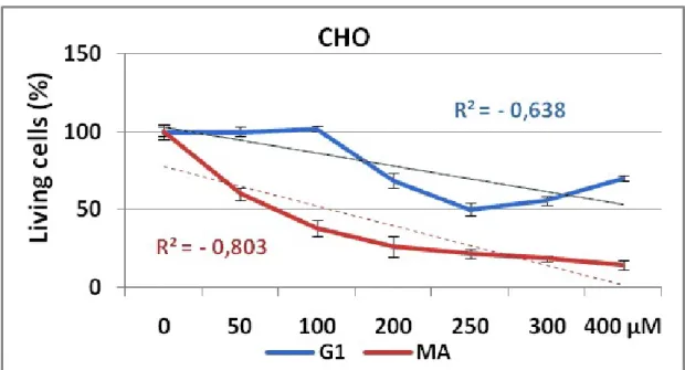
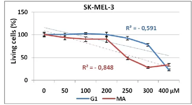
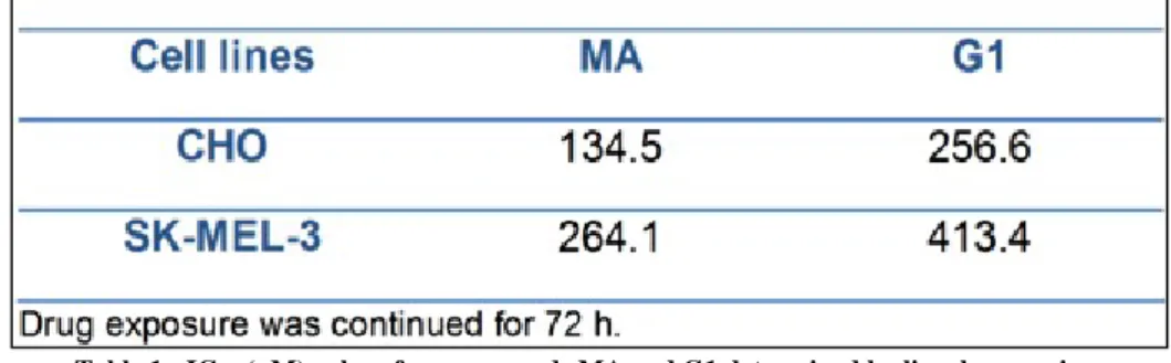
SUMMARY: The study was performed to investigate the effect of bromine atoms number present in two tested substances derivatives of 2-furylethylene on cell proliferation. The substances carrying one or two Br atoms were coded as MA and G1 respectively. The neutral red uptake (NRU) assay and mitotic index (MI) were used for this purpose.
The presence of two bromine atoms on the molecule of G1 inhibited markedly the cytotoxicity of this composite. For CHO cell line, the IC50 values were 256.6 µM for G1 and 134.5 µM for MA; whereas in SK MEL-3 (human melanoma) cell line, the IC50 were 413.4 µM and 264.1 µM for G1 and MA respectively. The IC50 values obtained in both cell lines were higher than 100 µM and showed no specificity for tumoral cells.
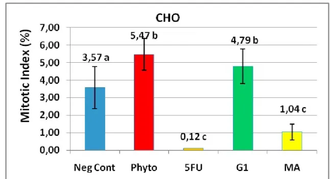
The MI obtained with the G1 composite showed no significant differences with phytohaemoglutinine used as positive control.
The anti-proliferative effect and MI were related with the number of bromine atoms on the molecules assayed. Another experiment was conducted with the MA product to obtain information about the acute oral toxicity class methods. The tested compound was classified in the 3th toxicity class with a fixed LD (50) cut-off value of 200 mg/kg of body weight.
KEYWORDS: Bromine, Cytotoxicity, Acute toxicitybromine, Mitotic index. Furylethylene, CHO.
http://biomed.uninet.edu/2012/n1/hernandez.html
Electron J Biomed 2012;1:27. Hernandez et al. EFFECT OF BROMINE ATOMS NUMBER ON THE CYTOTOXICITY...
RESUMEN: EFECTO DEL NÚMERO DE ÁTOMOS DE BROMO EN DERIVADOS DEL 2-FURILETILENO SOBRE LA CITOTOXICIDAD DE LÍNEAS CELULARES NORMALES Y TUMORALES.
El objetivo del estudio fue investigar el efecto que tiene el número de átomos de bromo presente en dos sustancias derivadas del 2- furiletileno sobre la proliferación celular. Las sustancias portando uno o dos átomos de bromo en su molécula se codificaron como MA y G1 respectivamente y su efecto sobre las líneas celulares se evaluó mediante las técnicas de captación de rojo neutro (CRN) y del indice mitótico (IM). La presencia de dos átomos de bromo en la molécula del G1 inhibió marcadamente la
citotoxicidad de este compuesto, para la línea celular CHO (ovario de hámster chino) los valores de IC50 fueron 256.6 µM para el G1 y 134.5 µM para el MA; mientras que en la línea SK MEL-3 (melanoma human), los valores de IC50 fueron 413.4 µM y 264.1 µM para el G1 y MA respectivamente. Los valores de IC50 obtenidos por estos compuestos en ambas líneas celulares fueron superiores a 100 µM y no mostraron especificidad por la línea tumoral.
El IM obtenido con el compuesto G1 no mostró diferencias significativas con la fitohaemoglutinina, empleada como control positivo del ensayo. El efecto anti-proliferativo y el IM estuvieron relacionado con el número de átomos de bromo en las moléculas ensayadas. Por otra parte un estudio llevado a cabo con el compuesto MA brindó información sobre la toxicidad aguda del mismo utilizando el método de las clases, clasificándose este producto en la clase 3 con un valor de corte LD (50) de 200 mg/kg de peso corporal.
PALABRAS CLAVE: Bromo. Citotoxicidad. Toxicidad aguda. Índice mitótico. Furiletileno. CHO.
INTRODUCTION
Since the 1990's, 2-furylethylene derivatives have been regarded as an interesting range of biological active compounds. They are one of the useful classes of synthetic agents with a wide range of biological activities including, antibacterial, antifungus and antiprotozoal properties1-4. The 2-furylethylenes, also known as vinylfurans or ethenylfurans, are derivatives of the ethene where a furan ring is attached to one of its carbon atoms (the ? carbon). The presence of a nitro group at position 5 of the furan ring in most of the 2 furylethylenes reported in literature until the 1980's, limited the use of these compounds because the aromatic nitro compounds in general, and nitrofurans in particular, have mutagenic and carcinogenic properties5-6. Subsequent studies opened a novel opportunity for these compounds, because it demonstrated that the presence of a nitro group at position 5 of the furan ring is not a necessary condition for the development of the antimicrobial activity of such chemicals7-8.
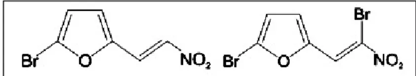
The (1-(5-bromofur-2-yl) nitroethene) and (1 (5 bromofur 2 yl) 2 bromo 2 nitroethene) coded for this paper as MA and G1 respectively are a synthetic 2-furylethylene derivatives. They have one or two bromine atoms added into the molecule and were used in this study in order to compare their effect on the biological activity changes. The addition of bromine atoms in different combinations could cause diverse responses due to the atomic mass of this element (79,904). The position and number of the bromine atoms on the structure of several natural9-11 and synthesized12-13 compounds has attracted the attention of many biomedical research in order to use these compounds in future human health treatments. Some of these works show that bromination and the position of this atom over those substances increased the cytotoxicity of the compounds tested14-15. Other results have shown that chemical modifications like a substitution of bromine or iodine has a significant effect on the binding properties of the product to DNA16-17.
There has been recently an increasing interest in controlling macromolecular conformations and interactions through the adding of halogen bonding to sensitized compounds. Halogen bonds are favorable electrostatic interactions between polarized,
electropositive chlorine, bromine or iodine atoms. An halogen bond is a mainly an electrostatic interaction between a classic hydrogen bond acceptor, such as the electronegative O, N or S18. This sort of bond seems to play a role in binding and recognition similar to that in the thyroid-related hormones, a diversity of short X···O interactions (X = halogens) that are involved in protein/ligand recognition19.
The in vitro systems are ideally suited for investigations of the molecular, cellular and physiological mechanisms of chemically induced toxicity; the main justification for developing in vitro toxicity tests is that they will make toxicology a more scientifically based practice. It is even more obvious that the development and incorporation of stepwise testing strategies, combining experimental data from a range of alternative methods (metabolic and kinetic modeling, quantitative structure-activity relationships - QSAR, and in vitro tests), provide the most advanced way to predict toxicity, reducing at the same time the number of laboratory animals used for testing.
Moreover, the costs of assessing potential health effects of around 200,000 substances per year that are newly identified or synthesized require alternatives methods of analysis. But we must be sure that the obtained results with these alternative methods are reliable in order to substitute or reduce the number of tests in animals. In this investigation we used an in vitro system as an alternative method to evaluate the bromine atoms number effect on cellular toxicity of these 2-furylethylene derivatives using normal and tumoral cell lines.
MATERIAL AND METHODS.
http://biomed.uninet.edu/2012/n1/hernandez.html
Electron J Biomed 2012;1:28. Hernandez et al. EFFECT OF BROMINE ATOMS NUMBER ON THE CYTOTOXICITY...
The experiments were carried out at the Molecular and Cell Biology Department and at the Animal Housing Laboratory of the Centro de Inmunología y Productos Biológicos (CENIPBI) at the Universidad de Ciencias Médicas "Carlos J. Finlay" in Camagüey, Cuba.
1.-Synthetic product used in the trail
The substances tested were coded as MA 1-(5-bromofur-2-yl) nitroethene and G1 (1 (5-bromofur-2-yl)-2-bromo-2-nitroethene) a 2-furylethylene derivatives synthesized and gently offered by the group of chemical synthesis of the Centro de Bioactivos Químicos (CBQ), at the Universidad Central de Las Villas, Santa Clara, Cuba. Its global formulas are: C6H4BrO3N and C6H3Br2O3N for MA and G1 respectively. Both of them consisted of a yellow crystalline powder and its purity was certified by analytical testing to be higher than 99.0% in both cases. The molecular formulas of these composites are showed below:
2.-Cell lines and culture conditions
To assess the cytotoxic effect of the synthesized composite we used CHO-K1 cell line (Chinese Hamster Ovary), purchased from ECACC (Nº85050302), and SK-MEL-3 (ATCC NºHTB-69) a human melanoma cell line, kindly given by the Centro de Inmunología Molecular, Havana, Cuba. RPMI 1640 medium (Gibco 31800-014) supplemented with 10% Fetal Bovine Serum (Gibco-BRL, Grand Island, NY, USA) and 2 mM glutamine piruvate without antibiotic was used for this cell line.
The cellular suspensions were obtained from a 25 cm2 flask at 70-80% final confluence. The detached cells by tripsine solution treatment (0.125%, 53 mM EDTA in PBS 1X) during 3 5 minutes at 37°, were counted in a Neubauer camera using the trypan blue dye exclusion test.
The viability was higher than 90% in all assays. The cells density used varied between 20 and 30×103 cells/well in 96 wells plates (COSTAR N°-402037310) seeded at 100 µl final volume. The plates were incubated in 100% humidity and 5% CO2 atmosphere at 37°C, during 24 hours before the treatment application to permit the cellular adhesion to the bottom plate.
3.- Animals
Wistar Rats, SPF from Laboratory Animals Breeding Center, CENPALAB, (Cuba). Age: 35 - 41 days, weight 101-125 g.
Microbiological status: free from ecto and endoparasites, mycoplasms and pasteurellae. 30 females were supplied by airplane in rats boxes and acclimatized (22 ± 3 °C; 50 ± 10%) for 1 week in the Animal Housing Laboratory (CENIPBI).
During the experiment, animals were housed in polycarbonate cages Type 4 (3 rats/cage) on sterilized wood shavings as a bedding. Room temperature and humidity were regulated at 22 ± 3 °C and 50 ± 10%). Lighting conditions: Fluorescents lamps, with a sequence of 12 hours of light and 12 hours of dark. Air flow rate 0,5 m/s above cages.
Feeding: A commercial diet (8 mm pellet) manufactured by CENPALAB, Havana, Cuba (ISO 9001 Certified Laboratory) was heated for 20 min at 110 °C, and available ad libitum. Drinking water: acidic sterilized water, ad libitum.
Experiment performed from July 7 to July 22, 2010 using 9 rats/experiment.
The behavioral parameters were measured as indicated by OECD 423 guideline.
4.-Experimental Design
4.1.-Cytotoxic Test
The neutral red uptake (NRU)20 was selected from the different in vitro tests described to evaluate the cytotoxicity. Prior to each experiment the test substances were dissolved in a (DMSO/ETOH) (v/v) mix and immediately diluted into medium. During drug exposure the concentrations of DMSO/ETOH mix, in treated and control wells, were kept below 0.1%. We prepared a 500 mM main solution of the product to then make the following doses: 400, 300, 250, 200, 100 and 50 µM afterwards, the doses and control solutions were filtered using 0.45 and 0.22 µM membranes. The negative control was the RPMI medium with 0.1% of the diluent used (DMSO/ETOH) without the test substances. As a positive control 10 ?g/ml of 5-fluorouracile (5-FU), was used. The tests were carried out in triplicate (3 wells/dose) during 72 hours. To keep the optimal concentrations and cultural conditions 100 µl of the medium were changed daily, containing the adequate doses, including controls. The optical density was recorded in an ELISA reader plate equipment (Lab Systems MULTISKAN Plus) at 540 nm wavelength. A lineal regression method was used to determine the IC50 value21-22, using Prism; GraphPad Software (5.02 version).
4.2.-Determination of Mitotic Index (MI)
http://biomed.uninet.edu/2012/n1/hernandez.html
Electron J Biomed 2012;1: